|
Pemphigoid
Pemphigoid is a group of rare autoimmune blistering diseases of the skin, and mucous membranes. As its name indicates, pemphigoid is similar in general appearance to pemphigus, but, unlike pemphigus, pemphigoid does not feature acantholysis, a loss of connections between skin cells. Pemphigoid is more common than pemphigus, and is slightly more common in women than in men. It is also more common in people aged over 70 years than it is in younger people. Classification IgG The forms of pemphigoid are considered to be connective tissue autoimmune skin diseases. There are several types: * Gestational pemphigoid (PG) (formerly called ''Herpes gestationis'') * Bullous pemphigoid (BP) Rarely affects the mouth * Mucous membrane pemphigoid (MMP) or (cicatricial pemphigoid), (No skin involvement) Bullous and mucous membrane pemphigoid usually affect persons who are over age 60. Gestational pemphigoid occurs during pregnancy, typically in the second or third trimester, or immediate ... [...More Info...] [...Related Items...] OR: [Wikipedia] [Google] [Baidu] |
Bullous Pemphigoid
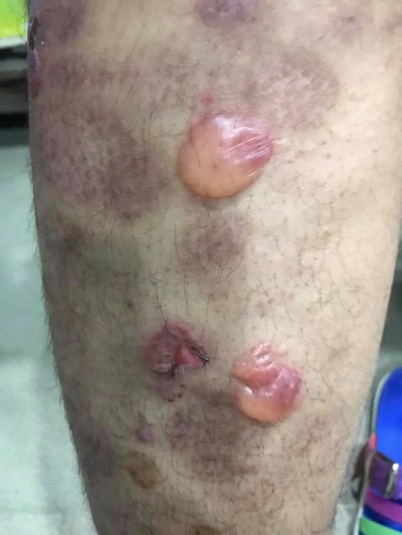
Bullous pemphigoid (type of pemphigoid) is an autoimmune pruritic skin disease which typically occurs in people aged over 60, that may involve the formation of blisters ( bullae) in the space between the epidermal and dermal skin layers. It is classified as a type II hypersensitivity reaction, which involves formation of anti- hemidesmosome antibodies, causing a loss of keratinocytes to basement membrane adhesion. Signs and symptoms Clinically, the earliest lesions may appear as a hives-like red raised rash, but could also appear dermatitic, targetoid, lichenoid, nodular, or even without a rash ( essential pruritus). Tense bullae eventually erupt, most commonly at the inner thighs and upper arms, but the trunk and extremities are frequently both involved. Any part of the skin surface can be involved. Oral lesions are present in a minority of cases. The disease may be acute, but can last from months to years with periods of exacerbation and remission. Several other skin ... [...More Info...] [...Related Items...] OR: [Wikipedia] [Google] [Baidu] |
Mucous Membrane Pemphigoid
Mucous membrane pemphigoid is a rare chronic autoimmune subepithelial blistering disease characterized by erosive lesions of the mucous membranes and skin. It is one of the pemphigoid diseases that can result in scarring. Signs and symptoms The autoimmune reaction most commonly affects the oral mucosa in the mouth, causing lesions in the gums (gingiva), known as desquamative gingivitis. More severe cases can also affect areas of mucous membrane elsewhere in the body, such as the sinuses, genitals, anus, and cornea. When the cornea of the eye is affected, repeated scarring may result in blindness. ''Brunsting–Perry cicatricial pemphigoid'' is a rare variant of mucous membrane pemphigoid involving the scalp and the neck without mucosal involvement. It is proposed by some authors that this be called a variant of epidermolysis bullosa acquisita. Nikolsky's sign (gentle lateral pressure) on unaffected mucosa or skin raises a bulla. If no lesions are present on examination it ma ... [...More Info...] [...Related Items...] OR: [Wikipedia] [Google] [Baidu] |
List Of Target Antigens In Pemphigoid
Circulating auto-antibodies in the human body can target normal parts of the skin leading to disease. This is a list of antigens in the skin that may become targets of circulating auto-antibodies leading to the various types of pemphigoid. Of note, there are also several other diseases that are caused by auto-antibodies that target the same anatomic area of the skin which is termed the basement membrane zone. These diseases include: Footnotes See also * List of target antigens in pemphigus * List of immunofluorescence findings for autoimmune bullous conditions * List of cutaneous conditions * List of genes mutated in cutaneous conditions * List of histologic stains that aid in diagnosis of cutaneous conditions References * * {{DEFAULTSORT:Target antigens in pemphigoid Cutaneous conditions Dermatology-related lists ... [...More Info...] [...Related Items...] OR: [Wikipedia] [Google] [Baidu] |
List Of Cutaneous Conditions
Many skin conditions affect the human integumentary system—the organ system covering the entire surface of the body and composed of skin, hair, nails, and related muscle and glands. The major function of this system is as a barrier against the external environment. The skin weighs an average of four kilograms, covers an area of two square metres, and is made of three distinct layers: the epidermis, dermis, and subcutaneous tissue. The two main types of human skin are: glabrous skin, the hairless skin on the palms and soles (also referred to as the "palmoplantar" surfaces), and hair-bearing skin.Burns, Tony; ''et al''. (2006) ''Rook's Textbook of Dermatology CD-ROM''. Wiley-Blackwell. . Within the latter type, the hairs occur in structures called pilosebaceous units, each with hair follicle, sebaceous gland, and associated arrector pili muscle. In the embryo, the epidermis, hair, and glands form from the ectoderm, which is chemically influenced by the underlying meso ... [...More Info...] [...Related Items...] OR: [Wikipedia] [Google] [Baidu] |
Gestational Pemphigoid
Gestational pemphigoid (GP) is a rare autoimmune variant of the skin disease bullous pemphigoid, and first appears in pregnancy. It presents with tense blisters, small bumps, hives and intense itching, usually starting around the navel before spreading to limbs in mid-pregnancy or shortly after delivery. The head, face and mouth are not usually affected. It may flare after delivery before resolving around three to six months after the pregnancy. It can be triggered by subsequent pregnancies, menstrual periods and oral contraceptive pill. A molar pregnancy and choriocarcinoma can provoke it. In some people, it persists long-term. It is associated with premature delivery of a small baby, a few who may be born with blisters and urticaria, which generally resolves within six weeks. It does not spread from one person to another, and does not run in families. Diagnosis is by visulaization, biopsy and immunofluorescence. It can resemble pruritic urticarial papules and plaques of ... [...More Info...] [...Related Items...] OR: [Wikipedia] [Google] [Baidu] |
Desquamative Gingivitis
Desquamative gingivitis is an erythematous (red), desquamatous (shedding) and ulcerated appearance of the gums. It is a descriptive term and can be caused by several different disorders. Signs and symptoms Desquamative gingivitis involves lesions of the free and attached gingiva. Unlike plaque-induced inflammation of the gums (normal marginal gingivitis), desquamative gingivitis extends beyond the marginal gingiva, involving the full width of the gingiva and sometimes the alveolar mucosa. The term "full width gingivitis" usually refers to the oral lesions of orofacial granulomatosis however. The color is another dissimilarity between typical marginal gingivitis and desquamative gingivitis, in the latter it is dusky red. Plasma cell gingivitis is another form of gingivitis which affects both the attached and free gingiva. Cause Caused by various autoimmune diseases as well as allergies. Erosive lichen planus, mucous membrane pemphigoid, pemphigus vulgaris, erythema exsudativum m ... [...More Info...] [...Related Items...] OR: [Wikipedia] [Google] [Baidu] |
Hemidesmosome
Hemidesmosomes are very small stud-like structures found in keratinocytes of the epidermis of skin that attach to the extracellular matrix. They are similar in form to desmosomes when visualized by electron microscopy, however, desmosomes attach to adjacent cells. Hemidesmosomes are also comparable to focal adhesions, as they both attach cells to the extracellular matrix. Instead of desmogleins and desmocollins in the extracellular space, hemidesmosomes utilize integrins. Hemidesmosomes are found in epithelial cells connecting the basal epithelial cells to the lamina lucida, which is part of the basal lamina. Hemidesmosomes are also involved in signaling pathways, such as keratinocyte migration or carcinoma cell intrusion. Structure Hemidesmosomes can be categorized into two types based on their protein constituents. Type 1 hemidesmosomes are found in stratified and pseudo-stratified epithelium. Type 1 hemidesmosomes have five main elements: integrin α6 β4, plectin in its ... [...More Info...] [...Related Items...] OR: [Wikipedia] [Google] [Baidu] |
Symblepharon
A symblepharon is a partial or complete adhesion of the palpebral conjunctiva of the eyelid to the bulbar conjunctiva of the eyeball. It results either from disease (conjunctival sequelae of trachoma) or trauma. Cicatricial pemphigoid and, in severe cases, rosacea Rosacea is a long-term skin condition that typically affects the face. It results in redness, pimples, swelling, and small and superficial dilated blood vessels. Often, the nose, cheeks, forehead, and chin are most involved. A red, enlarge ... may cause symblepharon. It is rarely congenital. Its treatment is ''symblepharectomy''. See also * Ankyloblepharon References Further reading * Disorders of conjunctiva {{Symptom-stub ... [...More Info...] [...Related Items...] OR: [Wikipedia] [Google] [Baidu] |
Pemphigus
Pemphigus ( or ) is a rare group of blistering autoimmune diseases that affect the skin and mucous membranes. The name is derived from the Greek root ''pemphix'', meaning "pustule". In pemphigus, autoantibodies form against desmoglein, which forms the "glue" that attaches adjacent epidermal cells via attachment points called desmosomes. When autoantibodies attack desmogleins, the cells become separated from each other and the epidermis becomes detached, a phenomenon called acantholysis. This causes blisters that slough off and turn into sores. In some cases, these blisters can cover a large area of the skin. Originally, the cause of this disease was unknown, and "pemphigus" was used to refer to any blistering disease of the skin and mucosa. In 1964, researchers found that the blood of patients with pemphigus contained antibodies to the layers of skin that separate to form the blisters. In 1971, an article investigating the autoimmune nature of this disease was published ... [...More Info...] [...Related Items...] OR: [Wikipedia] [Google] [Baidu] |
Acantholysis
Acantholysis is the loss of intercellular connections, such as desmosomes, resulting in loss of cohesion between keratinocytes,Kumar, Vinay; Fausto, Nelso; Abbas, Abul (2004) ''Robbins & Cotran Pathologic Basis of Disease'' (7th ed.). Saunders. Page 1230. . seen in diseases such as pemphigus vulgaris. It is absent in bullous pemphigoid, making it useful for differential diagnosis. This histological feature is also seen in herpes simplex infections (HSV 1 and 2) and varicella zoster infections (chicken pox and shingles). See also * Skin lesion * Skin disease * List of cutaneous conditions Many skin conditions affect the human integumentary system—the organ system covering the entire surface of the body and composed of skin, hair, nails, and related muscle and glands. The major function of this system is as a barrier agai ... References External links Dermatologic terminology {{dermatology-stub ... [...More Info...] [...Related Items...] OR: [Wikipedia] [Google] [Baidu] |
Blister
A blister is a small pocket of body fluid ( lymph, serum, plasma, blood, or pus) within the upper layers of the skin, usually caused by forceful rubbing ( friction), burning, freezing, chemical exposure or infection. Most blisters are filled with a clear fluid, either serum or plasma. However, blisters can be filled with blood (known as " blood blisters") or with pus (for instance, if they become infected). The word "blister" entered English in the 14th century. It came from the Middle Dutch and was a modification of the Old French , which meant a leprous nodule—a rise in the skin due to leprosy. In dermatology today, the words ''vesicle'' and ''bulla'' refer to blisters of smaller or greater size, respectively. To heal properly, a blister should not be popped unless medically necessary. If popped, the bacteria can spread. The excess skin should not be removed because the skin underneath needs the top layer to heal properly. Causes A blister may form when the skin ... [...More Info...] [...Related Items...] OR: [Wikipedia] [Google] [Baidu] |


