|
Posterior Gray Column
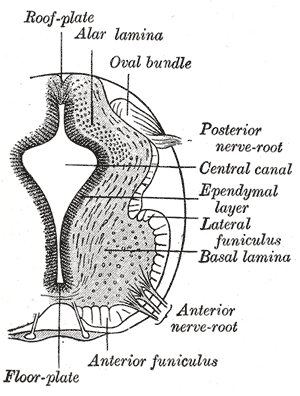
The grey columns are three regions of the somewhat ridge-shaped mass of grey matter in the spinal cord. These regions present as three columns: the anterior grey column, the posterior grey column, and the lateral grey column, all of which are visible in cross-section of the spinal cord. The anterior grey column is made up of alpha motor neurons, gamma motor neurons, and small neurons thought to be interneurons. It affects the skeletal muscles. The posterior grey column receives several types of sensory information regarding touch and sensation from receptors in the skin, bones, and joints, including fine touch, proprioception, and vibration. It contains the cell bodies of second-order sensory neurons and their synapses with the pseudounipolar first-order sensory neurons (whose cell bodies are located within the sensory ganglia (a.k.a. dorsal root ganglia)). The lateral grey column is only present in the thoracic region and upper lumbar segments (T1-L2). It contains preg ... [...More Info...] [...Related Items...] OR: [Wikipedia] [Google] [Baidu] |
Grey Columns
The grey columns are three regions of the somewhat ridge-shaped mass of grey matter in the spinal cord. These regions present as three columns: the anterior grey column, the posterior grey column, and the lateral grey column, all of which are visible in cross-section of the spinal cord. The anterior grey column is made up of alpha motor neurons, gamma motor neurons, and small neurons thought to be interneurons. It affects the skeletal muscles. The posterior grey column receives several types of sensory information regarding touch and sensation from receptors in the skin, bones, and joints, including fine touch, proprioception, and Oscillation, vibration. It contains the cell bodies of Second-order neuron, second-order sensory neurons and their synapses with the Pseudounipolar neuron, pseudounipolar First-order neuron, first-order sensory neurons (whose cell bodies are located within the Sensory ganglia, sensory ganglia (a.k.a. dorsal root ganglia)). The lateral grey colum ... [...More Info...] [...Related Items...] OR: [Wikipedia] [Google] [Baidu] |
Sensory Ganglia
A dorsal root ganglion (or spinal ganglion; also known as a posterior root ganglion) is a cluster of neurons (a ganglion) in a dorsal root of a spinal nerve. The cell bodies of sensory neurons known as first-order neurons are located in the dorsal root ganglia. The axons of dorsal root ganglion neurons are known as afferents. In the peripheral nervous system, afferents refer to the axons that relay sensory information into the central nervous system (i.e. the brain and the spinal cord). Structure The neurons comprising the dorsal root ganglion are of the pseudo-unipolar type, meaning they have a cell body (soma) with two branches that act as a single axon, often referred to as a ''distal process'' and a ''proximal process''. Unlike the majority of neurons found in the central nervous system, an action potential in posterior root ganglion neuron may initiate in the ''distal process'' in the periphery, bypass the cell body, and continue to propagate along the ''proximal proces ... [...More Info...] [...Related Items...] OR: [Wikipedia] [Google] [Baidu] |
Muscle Spindles
Muscle spindles are stretch receptors within the body of a skeletal muscle that primarily detect changes in the length of the muscle. They convey length information to the central nervous system via afferent nerve fibers. This information can be processed by the brain as proprioception. The responses of muscle spindles to changes in length also play an important role in regulating the contraction of muscles, for example, by activating motor neurons via the stretch reflex to resist muscle stretch. The muscle spindle has both sensory and motor components. * Sensory information conveyed by primary type Ia sensory fibers which spiral around muscle fibres within the spindle, and secondary type II sensory fibers * Activation of muscle fibres within the spindle by up to a dozen gamma motor neurons and to a lesser extent by one or two beta motor neurons ''.'' Structure Muscle spindles are found within the belly of a skeletal muscle. Muscle spindles are fusiform (spindle-shaped), a ... [...More Info...] [...Related Items...] OR: [Wikipedia] [Google] [Baidu] |
Intrafusal Muscle Fibers
Intrafusal muscle fibers are skeletal muscle fibers that serve as specialized sensory organs (proprioceptors). They detect the amount and rate of change in length of a muscle.Casagrand, Janet (2008) ''Action and Movement: Spinal Control of Motor Units and Spinal Reflexes.'' University of Colorado, Boulder. They constitute the muscle spindle, and are innervated by both sensory (afferent) and motor (efferent) fibers. Intrafusal muscle fibers are not to be confused with extrafusal muscle fibers, which contract, generating skeletal movement and are innervated by alpha motor neurons. Structure Types There are two types of intrafusal muscle fibers: nuclear bag fibers and nuclear chain fibers. They bear two types of sensory ending, known as annulospiral and flower-spray endings. Both ends of these fibers contract, but the central region only stretches and does not contract. Intrafusal muscle fibers are walled off from the rest of the muscle by an outer connective tissue sheat ... [...More Info...] [...Related Items...] OR: [Wikipedia] [Google] [Baidu] |
Muscle Contraction
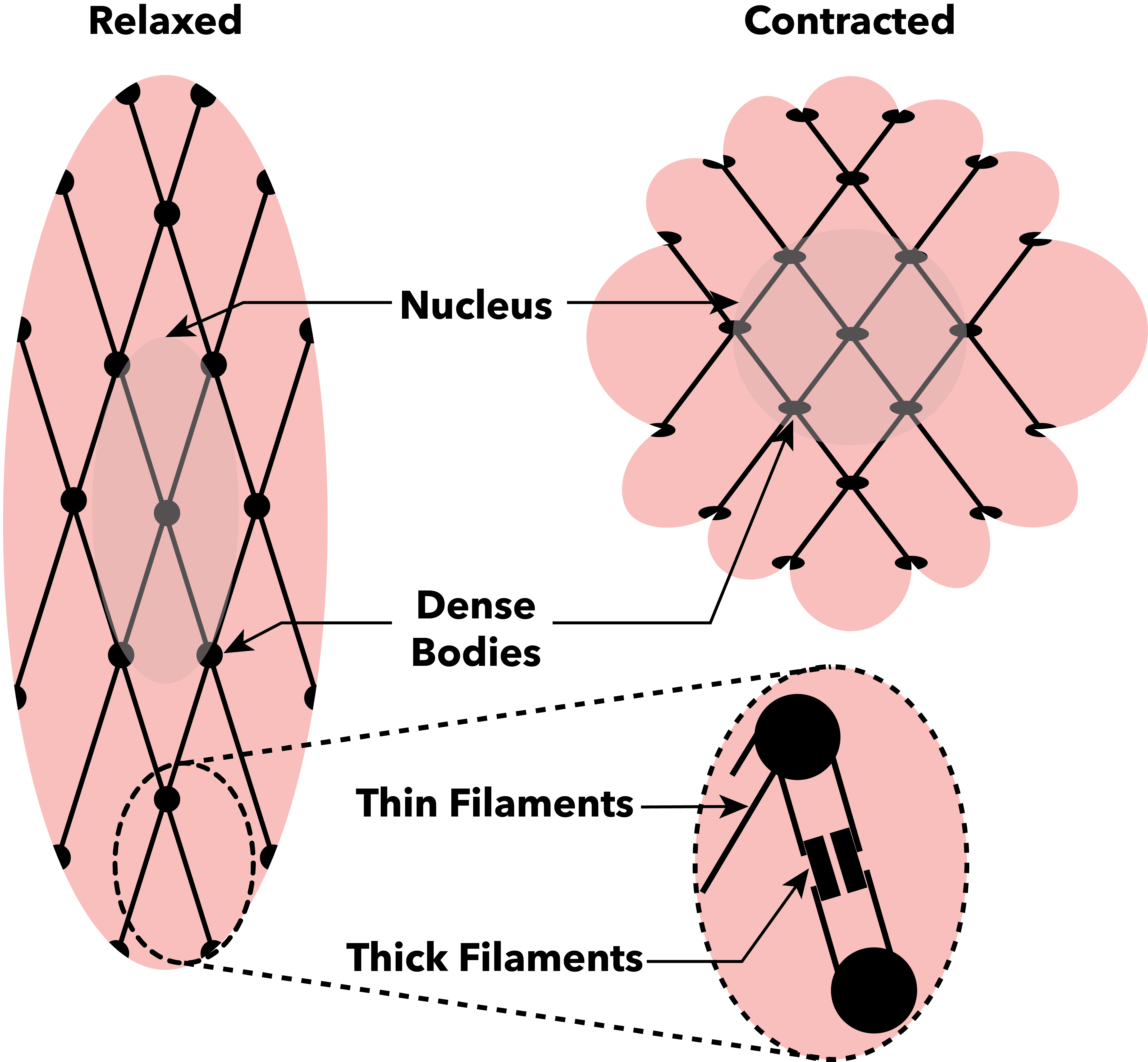
Muscle contraction is the activation of Tension (physics), tension-generating sites within muscle cells. In physiology, muscle contraction does not necessarily mean muscle shortening because muscle tension can be produced without changes in muscle length, such as when holding something heavy in the same position. The termination of muscle contraction is followed by muscle relaxation, which is a return of the muscle fibers to their low tension-generating state. For the contractions to happen, the muscle cells must rely on the change in action of two types of Myofilament, filaments: thin and thick filaments. The major constituent of thin filaments is a chain formed by helical coiling of two strands of actin, and thick filaments dominantly consist of chains of the Motor protein, motor-protein myosin. Together, these two filaments form myofibrils - the basic functional organelles in the skeletal muscle system. In vertebrates, Muscle cell#Muscle contraction in striated muscle, skele ... [...More Info...] [...Related Items...] OR: [Wikipedia] [Google] [Baidu] |
Neuromuscular Junctions
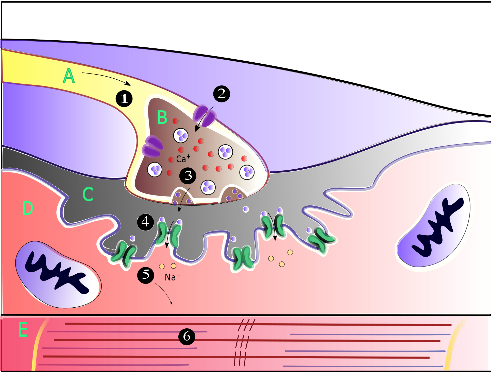
A neuromuscular junction (or myoneural junction) is a chemical synapse between a motor neuron and a muscle fiber. It allows the motor neuron to transmit a signal to the muscle fiber, causing muscle contraction. Muscles require innervation to function—and even just to maintain muscle tone, avoiding atrophy. In the neuromuscular system, nerves from the central nervous system and the peripheral nervous system are linked and work together with muscles. Synaptic transmission at the neuromuscular junction begins when an action potential reaches the presynaptic terminal of a motor neuron, which activates voltage-gated calcium channels to allow calcium ions to enter the neuron. Calcium ions bind to sensor proteins (synaptotagmins) on synaptic vesicles, triggering vesicle fusion with the cell membrane and subsequent neurotransmitter release from the motor neuron into the synaptic cleft. In vertebrates, motor neurons release acetylcholine (ACh), a small molecule neurotransmitter, which ... [...More Info...] [...Related Items...] OR: [Wikipedia] [Google] [Baidu] |
Extrafusal Muscle Fibers
Extrafusal muscle fibers are the standard skeletal muscle fibers that are innervated by alpha motor neurons and generate tension by contracting, thereby allowing for skeletal movement. They make up the large mass of skeletal striated muscle tissue and are attached to bone by fibrous tissue extensions (tendons). Each alpha motor neuron and the extrafusal muscle fibers innervated by it make up a motor unit. The connection between the alpha motor neuron and the extrafusal muscle fiber is a neuromuscular junction, where the neuron's signal, the action potential, is transduced to the muscle fiber by the neurotransmitter acetylcholine. Extrafusal muscle fibers are not to be confused with intrafusal muscle fibers, which are innervated by sensory nerve endings in central noncontractile parts and by gamma motor neurons in contractile ends and thus serve as a sensory proprioceptor. Extrafusal muscle fibers can be generated in vitro (in a dish) from pluripotent stem cells through dir ... [...More Info...] [...Related Items...] OR: [Wikipedia] [Google] [Baidu] |
Neuromorphology
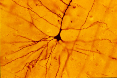
Neuromorphology (from Greek νεῦρον, neuron, "nerve"; μορφή, morphé, "form"; -λογία, -logia, “study of”) is the study of nervous system form, shape, and structure. The study involves looking at a particular part of the nervous system from a molecular and cellular level and connecting it to a physiological and anatomical point of view. The field also explores the communications and interactions within and between each specialized section of the nervous system. Morphology is distinct from morphogenesis. Morphology is the study of the shape and structure of biological organisms, while morphogenesis is the study of the biological development of the shape and structure of organisms. Therefore, neuromorphology focuses on the specifics of the structure of the nervous system and not the process by which the structure was developed. Neuromorphology and morphogenesis, while two different entities, are nonetheless closely linked. History Progress in defining the morpho ... [...More Info...] [...Related Items...] OR: [Wikipedia] [Google] [Baidu] |
Interneurons
Interneurons (also called internuncial neurons, association neurons, connector neurons, or intermediate neurons) are neurons that are not specifically motor neurons or sensory neurons. Interneurons are the central nodes of neural circuits, enabling communication between sensory or motor neurons and the central nervous system (CNS). They play vital roles in reflexes, neuronal oscillations, and neurogenesis in the adult mammalian brain. Interneurons can be further broken down into two groups: local interneurons and relay interneurons. Local interneurons have short axons and form circuits with nearby neurons to analyze small pieces of information. Relay interneurons have long axons and connect circuits of neurons in one region of the brain with those in other regions. However, interneurons are generally considered to operate mainly within local brain areas. The interaction between interneurons allows the brain to perform complex functions such as learning and decision-making. S ... [...More Info...] [...Related Items...] OR: [Wikipedia] [Google] [Baidu] |
Gamma Motor Neurons
A gamma motor neuron (γ motor neuron), also called gamma motoneuron, or fusimotor neuron, is a type of lower motor neuron that takes part in the process of muscle contraction, and represents about 30% of ( Aγ) fibers going to the muscle. Like alpha motor neurons, their cell bodies are located in the anterior grey column of the spinal cord. They receive input from the reticular formation of the pons in the brainstem. Their axons are smaller than those of the alpha motor neurons, with a diameter of only 5 μm. Unlike the alpha motor neurons, gamma motor neurons do not directly adjust the lengthening or shortening of muscles. However, their role is important in keeping muscle spindles taut, thereby allowing the continued firing of alpha neurons, leading to muscle contraction. These neurons also play a role in adjusting the sensitivity of muscle spindles. The presence of myelination in gamma motor neurons allows a conduction velocity of 4 to 24 meters per second, signifi ... [...More Info...] [...Related Items...] OR: [Wikipedia] [Google] [Baidu] |
Alpha Motor Neurons
Alpha (α) motor neurons (also called alpha motoneurons), are large, multipolar lower motor neurons of the brainstem and spinal cord. They innervate extrafusal muscle fibers of skeletal muscle and are directly responsible for initiating their contraction. Alpha motor neurons are distinct from gamma motor neurons, which innervate intrafusal muscle fibers of muscle spindles. While their cell bodies are found in the central nervous system (CNS), α motor neurons are also considered part of the somatic nervous system—a branch of the peripheral nervous system (PNS)—because their axons extend into the periphery to innervate skeletal muscles. An alpha motor neuron and the muscle fibers it innervates comprise a motor unit. A motor neuron pool contains the cell bodies of all the alpha motor neurons involved in contracting a single muscle. Location Alpha motor neurons (α-MNs) innervating the head and neck are found in the brainstem; the remaining α-MNs innervate the rest of the b ... [...More Info...] [...Related Items...] OR: [Wikipedia] [Google] [Baidu] |
Lower Motor Neuron
Lower motor neurons (LMNs) are motor neurons located in either the anterior grey column, anterior nerve roots (spinal lower motor neurons) or the cranial nerve nuclei of the brainstem and cranial nerves with motor function (cranial nerve lower motor neurons). Many voluntary movements rely on spinal lower motor neurons, which innervate skeletal muscle fibers and act as a link between upper motor neurons and muscles. Cranial nerve lower motor neurons also control some voluntary movements of the eyes, face and tongue, and contribute to chewing, swallowing and vocalization. Damage to lower motor neurons often leads to hypotonia, hyporeflexia, flaccid paralysis as well as muscle atrophy and fasciculations. Classification Lower motor neurons are classified based on the type of muscle fiber they innervate: * Alpha motor neurons (α-MNs) innervate extrafusal muscle fibers, the most numerous type of muscle fiber and the one involved in muscle contraction. * Beta motor neurons (β-M ... [...More Info...] [...Related Items...] OR: [Wikipedia] [Google] [Baidu] |


