|
Posterior Communicating Artery
In human anatomy, the left and right posterior communicating arteries are small arteries at the base of the brain that form part of the circle of Willis. Anteriorly, it unites with the internal carotid artery (ICA) (prior to the terminal bifurcation of the ICA into the anterior cerebral artery and middle cerebral artery); posteriorly, it unites with the posterior cerebral artery. With the anterior communicating artery, the posterior communicating arteries establish a system of collateral circulation in cerebral circulation. Anatomy The arteries contribute to the blood supply of the optic tract. The two posterior communicating arteries often differ in size. Relations Each posterior communicating artery is situated within the interpeduncular cistern, superolateral to the pituitary gland. Each are is situated upon the medial surface of the ipsilateral cerebral peduncle and adjacent to the anterior perforated substance. The ipsilateral oculomotor nerve (CN III) passes inferol ... [...More Info...] [...Related Items...] OR: [Wikipedia] [Google] [Baidu] |
Circle Of Willis
The circle of Willis (also called Willis' circle, loop of Willis, cerebral arterial circle, and Willis polygon) is a circulatory anastomosis that supplies blood to the brain and surrounding structures in reptiles, birds and mammals, including humans. It is named after Thomas Willis (1621–1675), an English physician. Structure The circle of Willis is a part of the cerebral circulation and is composed of the following arteries: * Anterior cerebral artery (left and right) at their A1 segments * Anterior communicating artery * Internal carotid artery (left and right) at its distal tip (carotid terminus) * Posterior cerebral artery (left and right) at their P1 segments * Posterior communicating artery (left and right) The middle cerebral arteries, supplying the brain, are also considered part of the Circle of Willis Origin of arteries The left and right internal carotid arteries arise from the left and right common carotid arteries. The posterior communicating artery is given ... [...More Info...] [...Related Items...] OR: [Wikipedia] [Google] [Baidu] |
Collateral Circulation
Collateral circulation is the alternate Circulatory system, circulation around a blocked blood vessel, artery or vein via another path, such as nearby minor vessels. It may occur via preexisting vascular redundancy (analogous to redundancy (engineering), engineered redundancy), as in the circle of Willis in the brain, or it may occur via new branches formed between adjacent blood vessels (neovascularization), as in the eye after a retinal embolism or in the brain when an instance of arterial constriction occurs due to Moyamoya disease. Its formation may be related by pathological conditions such as high vascular resistance or ischaemia. It is occasionally also known as accessory circulation, auxiliary circulation, or secondary circulation. It has surgery, surgically created analogues in which shunt (medical), shunts or circulatory anastomosis, anastomoses are constructed to bypass circulatory problems. An example of the usefulness of collateral circulation is a systemic thromboem ... [...More Info...] [...Related Items...] OR: [Wikipedia] [Google] [Baidu] |
Oculomotor Nerve
The oculomotor nerve, also known as the third cranial nerve, cranial nerve III, or simply CN III, is a cranial nerve that enters the orbit through the superior orbital fissure and innervates extraocular muscles that enable most movements of the eye and that raise the eyelid. The nerve also contains fibers that innervate the intrinsic eye muscles that enable pupillary constriction and accommodation (ability to focus on near objects as in reading). The oculomotor nerve is derived from the basal plate of the embryonic midbrain. Cranial nerves IV and VI also participate in control of eye movement. Structure The oculomotor nerve originates from the third nerve nucleus at the level of the superior colliculus in the midbrain. The third nerve nucleus is located ventral to the cerebral aqueduct, on the pre-aqueductal grey matter. The fibers from the two third nerve nuclei located laterally on either side of the cerebral aqueduct then pass through the red nucleus. From the ... [...More Info...] [...Related Items...] OR: [Wikipedia] [Google] [Baidu] |
Aneurysm
An aneurysm is an outward :wikt:bulge, bulging, likened to a bubble or balloon, caused by a localized, abnormal, weak spot on a blood vessel wall. Aneurysms may be a result of a hereditary condition or an acquired disease. Aneurysms can also be a wikt:Special:Search/nidus, nidus (starting point) for clot formation (thrombosis) and Embolism, embolization. As an aneurysm increases in size, the risk of rupture increases, which could lead to uncontrolled bleeding. Although they may occur in any blood vessel, particularly lethal examples include aneurysms of the circle of Willis in the brain, aortic aneurysms affecting the thoracic aorta, and abdominal aortic aneurysms. Aneurysms can arise in the heart itself following a Myocardial infarction, heart attack, including both Ventricular aneurysm, ventricular and atrial septal aneurysms. There are congenital atrial septal defect, atrial septal aneurysms, a rare heart defect. Etymology The word is from Greek language, Greek: ἀνεύρ� ... [...More Info...] [...Related Items...] OR: [Wikipedia] [Google] [Baidu] |
Cerebral Aneurysm
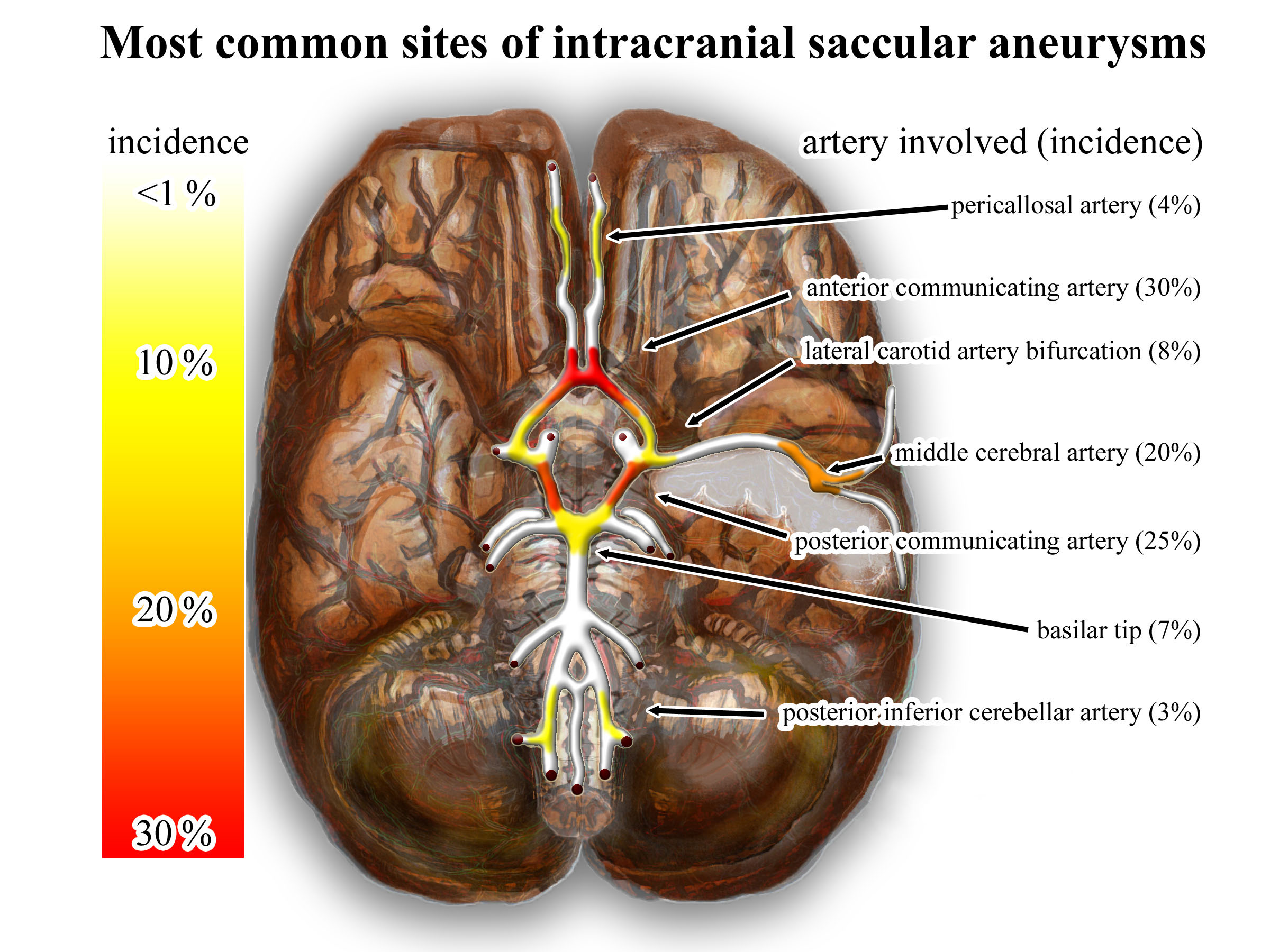
An intracranial aneurysm, also known as a cerebral aneurysm, is a Cerebrovascular disease, cerebrovascular disorder characterized by a localized dilation or ballooning of a blood vessel in the brain due to a weakness in the vessel wall. These aneurysms can occur in any part of the brain but are most commonly found in the arteries of the cerebral arterial circle. The risk of rupture varies with the size and location of the aneurysm, with those in the posterior circulation being more prone to rupture. Cerebral aneurysms are classified by size into small, large, giant, and super-giant, and by shape into saccular (berry), Fusiform gyrus, fusiform, and microaneurysms. Saccular aneurysms are the most common type and can result from various risk factors, including genetic conditions, hypertension, smoking, and Substance abuse, drug abuse. Symptoms of an unruptured aneurysm are often minimal, but a ruptured aneurysm can cause severe headaches, nausea, Visual impairment, vision impairme ... [...More Info...] [...Related Items...] OR: [Wikipedia] [Google] [Baidu] |
Oculomotor Nerve
The oculomotor nerve, also known as the third cranial nerve, cranial nerve III, or simply CN III, is a cranial nerve that enters the orbit through the superior orbital fissure and innervates extraocular muscles that enable most movements of the eye and that raise the eyelid. The nerve also contains fibers that innervate the intrinsic eye muscles that enable pupillary constriction and accommodation (ability to focus on near objects as in reading). The oculomotor nerve is derived from the basal plate of the embryonic midbrain. Cranial nerves IV and VI also participate in control of eye movement. Structure The oculomotor nerve originates from the third nerve nucleus at the level of the superior colliculus in the midbrain. The third nerve nucleus is located ventral to the cerebral aqueduct, on the pre-aqueductal grey matter. The fibers from the two third nerve nuclei located laterally on either side of the cerebral aqueduct then pass through the red nucleus. From the ... [...More Info...] [...Related Items...] OR: [Wikipedia] [Google] [Baidu] |
Anterior Perforated Substance
The anterior perforated substance is a part of the brain. It is bilateral. It is irregular and quadrilateral. It lies in front of the optic tract and behind the olfactory trigone. Structure The anterior perforated substance is bilateral. It lies in front of the optic tract. It lies behind the olfactory trigone, separated by the fissure prima. Medially and in front, it is continuous with the subcallosal gyrus. Laterally, it is bounded by the lateral stria of the olfactory tract, and is continued into the uncus. Its gray substance is confluent above with that of the corpus striatum, and is perforated anteriorly by numerous small blood vessels that supply such areas as the internal capsule. The anterior cerebral artery arises just below the anterior perforated substance. The middle cerebral artery passes through its lateral two thirds. Blood supply The anterior perforated substance is supplied by lenticulostriate arteries, which branch from the middle cerebral arte ... [...More Info...] [...Related Items...] OR: [Wikipedia] [Google] [Baidu] |
Cerebral Peduncle
The cerebral peduncles (In Latin, ''ped-'' means 'foot'.) are the two stalks that attach the cerebrum to the brainstem. They are structures at the front of the midbrain which arise from the ventral pons and contain the large ascending (sensory) and descending (motor) tracts that run to and from the cerebrum from the pons. Mainly, the three common areas that give rise to the cerebral peduncles are the cerebral cortex, the spinal cord and the cerebellum. The region includes the tegmentum, crus cerebri and pretectum. By this definition, the cerebral peduncles are also known as the basis pedunculi, while the large ventral bundle of efferent fibers is referred to as the cerebral crus (''crus'' means ‘leg’ in Latin.) or the pes pedunculi (''pes'' means 'foot' in Latin.). The cerebral peduncles are located on either side of the midbrain and are the frontmost part of the midbrain, and act as the connectors between the rest of the midbrain and the thalamic nuclei and thus the cer ... [...More Info...] [...Related Items...] OR: [Wikipedia] [Google] [Baidu] |
Interpeduncular Cistern
The interpeduncular cistern (or basal cistern) is the subarachnoid cistern situated between the dorsum sellae (anteriorly) and the two cerebral peduncles at the front of the midbrain. Its roof is represented by the floor of the third ventricle (i.e. posterior perforated substance, and the two mammillary bodies). Its floor is formed by the arachnoid membrane extending between the temporal lobes of either side. Anteriorly, it extends to the optic chiasm. The cistern communicates superiorly with the chiasmatic cistern, and inferiorly with the pontine cistern. The chiasmatic cistern, cistern of lamina terminalis, and supracallosal cistern are all extensions of the interpeduncular cistern. Anatomy Contents The cistern contains: * the posterior portion of the circle of Willis: ** basilar artery (including its bifurcation), ** (origins of the) posterior cerebral arteries, ** posterior communicating arteries, * (the origin of the) posterior thalamo-perforating arteries ... [...More Info...] [...Related Items...] OR: [Wikipedia] [Google] [Baidu] |
Optic Tract
In neuroanatomy, the optic tract () is a part of the visual system in the brain. It is a continuation of the optic nerve that relays information from the optic chiasm to the ipsilateral lateral geniculate nucleus (LGN), pretectal nuclei, and superior colliculus. It is composed of two individual tracts, the left optic tract and the right optic tract, each of which conveys visual information exclusive to its respective contralateral half of the visual field. Each of these tracts is derived from a combination of temporal and nasal retinal fibers from each eye that corresponds to one half of the visual field. In more specific terms, the optic tract contains fibers from the ipsilateral temporal hemiretina and contralateral nasal hemiretina. Anatomy Arterial supply The optic tract receives arterial supply from the anterior choroidal artery, and posterior communicating artery. Function Visual function The optic tract carries retinal information relating to the whole visua ... [...More Info...] [...Related Items...] OR: [Wikipedia] [Google] [Baidu] |
Cerebral Circulation
Cerebral circulation is the movement of blood through a network of cerebral arteries and veins supplying the brain. The rate of cerebral blood flow in an adult human is typically 750 milliliters per minute, or about 15% of cardiac output. Arteries deliver oxygenated blood, glucose and other nutrients to the brain. Veins carry "used or spent" blood back to the heart, to remove carbon dioxide, lactic acid, and other metabolic products. The neurovascular unit regulates cerebral blood flow so that activated neurons can be supplied with energy in the right amount and at the right time. Because the brain would quickly suffer damage from any stoppage in blood supply, the cerebral circulatory system has safeguards including autoregulation of the blood vessels. The failure of these safeguards may result in a stroke. The volume of blood in circulation is called the cerebral blood flow. Sudden intense accelerations change the gravitational forces perceived by bodies and can severely ... [...More Info...] [...Related Items...] OR: [Wikipedia] [Google] [Baidu] |
Anterior Communicating Artery
In human anatomy, the anterior communicating artery is a blood vessel of the brain that connects the left and right anterior cerebral arteries. Anatomy The anterior communicating artery connects the two anterior cerebral arteries across the commencement of the longitudinal fissure. Sometimes this vessel is wanting, the two arteries joining to form a single trunk, which afterward divides; or it may be wholly, or partially, divided into two. Its length averages about 4 mm, but varies greatly. It gives off some of the anteromedial ganglionic vessels, but these are principally derived from the anterior cerebral artery. It is part of the cerebral arterial circle, also known as the circle of Willis. Physiology Anatomical variations of the anterior communicating artery are relatively common. The artery is sometimes duplicated, multiplicated, fenestrated ("net-like") or very short, giving the impression that two anterior cerebral arteries are fused at the point where the anteri ... [...More Info...] [...Related Items...] OR: [Wikipedia] [Google] [Baidu] |


