|
Postcentral Sulcus
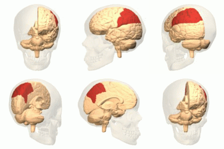
The postcentral sulcus of the parietal lobe lies parallel to, and behind, the central sulcus in the human brain. (A ''sulcus'' is one of the prominent grooves on the surface of the brain.) The postcentral sulcus divides the postcentral gyrus from the remainder of the parietal lobe. Additional images File:Gray725 postcentral sulcus.png, Lateral surface of left cerebral hemisphere, viewed from above. File:ParietCapts lateral.png, Gyri and sulci of right cerebral hemisphere. Postcentral sulcus labeled in red at top center. File:Cerebral Hemisphere Demonstration - Sanjoy Sanyal - Neuroscience Lab Fall 2013 (Cropped from 27m17s to 27m54s) Postcentral sulcus.webm, Human brain dissection video (36 sec). Demonstrating position of the postcentral sulcus of the left cerebral hemisphere The vertebrate cerebrum (brain) is formed by two cerebral hemispheres that are separated by a groove, the longitudinal fissure. The brain can thus be described as being divided into left and right cer ... [...More Info...] [...Related Items...] OR: [Wikipedia] [Google] [Baidu] |
Parietal Lobe
The parietal lobe is one of the four Lobes of the brain, major lobes of the cerebral cortex in the brain of mammals. The parietal lobe is positioned above the temporal lobe and behind the frontal lobe and central sulcus. The parietal lobe integrates sensory information among various sensory modality, modalities, including spatial sense and navigation (proprioception), the main sensory receptive area for the sense of touch in the somatosensory cortex which is just posterior to the central sulcus in the postcentral gyrus, and the two-streams hypothesis#Dorsal stream, dorsal stream of the visual system. The major sensory inputs from the skin (mechanoreceptor, touch, thermoreceptor, temperature, and nociceptor, pain receptors), relay through the thalamus to the parietal lobe. Several areas of the parietal lobe are important in language processing in the brain, language processing. The somatosensory cortex can be illustrated as a distorted figure – the cortical homunculus (Latin: "li ... [...More Info...] [...Related Items...] OR: [Wikipedia] [Google] [Baidu] |
Central Sulcus
In neuroanatomy, the central sulcus (also central fissure, fissure of Rolando, or Rolandic fissure, after Luigi Rolando) is a sulcus, or groove, in the cerebral cortex in the brains of vertebrates. It is sometimes confused with the longitudinal fissure. The central sulcus is a prominent landmark of the brain, separating the parietal lobe from the frontal lobe and the primary motor cortex from the primary somatosensory cortex. Evolution of the central sulcus The evolution of the central sulcus is theorized to have occurred in mammals when the complete dissociation of the original somatosensory cortex from its mirror duplicate developed in placental mammals such as primates, though the development did not stop there as time progressed the distinction between the two cortices grew. Evolution in primates The central sulcus is more prominent in apes as a result of fine-tuning of the motor system in apes. Hominins (bipedal apes) continued this trend through increased use of the ... [...More Info...] [...Related Items...] OR: [Wikipedia] [Google] [Baidu] |
Brain
The brain is an organ (biology), organ that serves as the center of the nervous system in all vertebrate and most invertebrate animals. It consists of nervous tissue and is typically located in the head (cephalization), usually near organs for special senses such as visual perception, vision, hearing, and olfaction. Being the most specialized organ, it is responsible for receiving information from the sensory nervous system, processing that information (thought, cognition, and intelligence) and the coordination of motor control (muscle activity and endocrine system). While invertebrate brains arise from paired segmental ganglia (each of which is only responsible for the respective segmentation (biology), body segment) of the ventral nerve cord, vertebrate brains develop axially from the midline dorsal nerve cord as a brain vesicle, vesicular enlargement at the rostral (anatomical term), rostral end of the neural tube, with centralized control over all body segments. All vertebr ... [...More Info...] [...Related Items...] OR: [Wikipedia] [Google] [Baidu] |
Postcentral Gyrus
In neuroanatomy, the postcentral gyrus is a prominent gyrus in the lateral parietal lobe of the human brain. It is the location of the primary somatosensory cortex, the main sensory receptive area for the sense of touch. Like other sensory areas, there is a map of sensory space in this location, called the '' sensory homunculus''. The primary somatosensory cortex was initially defined from surface stimulation studies of Wilder Penfield, and parallel surface potential studies of Bard, Woolsey, and Marshall. Although initially defined to be roughly the same as Brodmann areas 3, 1, and 2, more recent work by Kaas has suggested that for homogeny with other sensory fields only area 3 should be referred to as "primary somatosensory cortex", as it receives the bulk of the thalamocortical projections from the sensory input fields. Structure The lateral postcentral gyrus is bounded by: * medial longitudinal fissure medially (to the middle) * central sulcus rostrally (in front) ... [...More Info...] [...Related Items...] OR: [Wikipedia] [Google] [Baidu] |
Cerebral Hemisphere
The vertebrate cerebrum (brain) is formed by two cerebral hemispheres that are separated by a groove, the longitudinal fissure. The brain can thus be described as being divided into left and right cerebral hemispheres. Each of these hemispheres has an outer layer of grey matter, the cerebral cortex, that is supported by an inner layer of white matter. In eutherian (placental) mammals, the hemispheres are linked by the corpus callosum, a very large bundle of axon, nerve fibers. Smaller commissures, including the anterior commissure, the posterior commissure and the fornix (neuroanatomy), fornix, also join the hemispheres and these are also present in other vertebrates. These commissures transfer information between the two hemispheres to coordinate localized functions. There are three known poles of the cerebral hemispheres: the ''occipital lobe, occipital pole'', the ''frontal lobe, frontal pole'', and the ''temporal lobe, temporal pole''. The central sulcus is a prominent fissu ... [...More Info...] [...Related Items...] OR: [Wikipedia] [Google] [Baidu] |
Cerebrum
The cerebrum (: cerebra), telencephalon or endbrain is the largest part of the brain, containing the cerebral cortex (of the two cerebral hemispheres) as well as several subcortical structures, including the hippocampus, basal ganglia, and olfactory bulb. In the human brain, the cerebrum is the uppermost region of the central nervous system. The cerebrum prenatal development, develops prenatally from the forebrain (prosencephalon). In mammals, the Dorsum (biology), dorsal telencephalon, or Pallium (neuroanatomy), pallium, develops into the cerebral cortex, and the ventral telencephalon, or Pallium (neuroanatomy), subpallium, becomes the basal ganglia. The cerebrum is also divided into approximately symmetric Lateralization of brain function, left and right cerebral hemispheres. With the assistance of the cerebellum, the cerebrum controls all voluntary actions in the human body. Structure The cerebrum is the largest part of the brain. Depending upon the position of the animal, ... [...More Info...] [...Related Items...] OR: [Wikipedia] [Google] [Baidu] |
Sulci (neuroanatomy)
In neuroanatomy, a sulcus (Latin: "furrow"; : sulci) is a shallow depression or groove in the cerebral cortex. One or more sulci surround a gyrus (pl. gyri), a ridge on the surface of the cortex, creating the characteristic folded appearance of the brain in humans and most other mammals. The larger sulci are also called fissures. The cortex develops in the fetal stage of corticogenesis, preceding the cortical folding stage known as gyrification. The large fissures and main sulci are the first to develop. Mammals that have a folded cortex are known as ''gyrencephalic'', and the small-brained mammals that have a smooth cortex, such as rats and mice are termed lissencephalic. Structure Sulci, the grooves, and gyri, the folds or ridges, make up the folded surface of the cerebral cortex. Larger or deeper sulci are also often termed fissures. The folded cortex creates a larger surface area for the brain in humans and other larger mammals, without the need of increasing the size o ... [...More Info...] [...Related Items...] OR: [Wikipedia] [Google] [Baidu] |


