|
Podosome
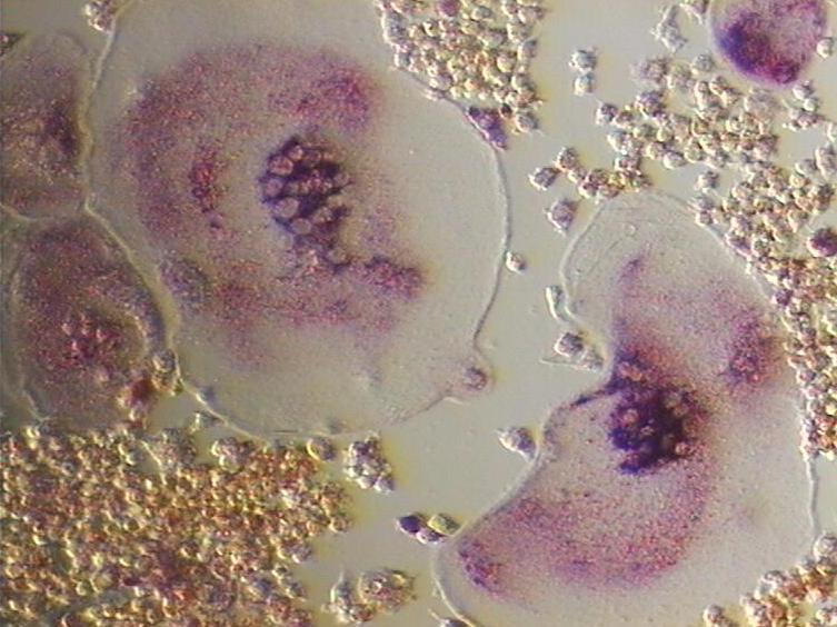
Podosomes are conical, actin-rich structures found as appendages on the outer surface of the plasma membrane of animal cells. Their size ranges from approximately 0.5 μm to 2.0 μm in diameter. While usually situated on the periphery of the cellular membrane, these unique structures display a polarized pattern of distribution in migrating cells, situating at the front border between the lamellipodium and lamellum. Their primary purpose is connected to cellular motility and invasion; therefore, they serve as both sites of attachment and degradation along the extracellular matrix. Many different specialized cells exhibit these dynamic structures such as invadopodia, (invasive cancer cells), osteoclasts, vascular smooth muscle cells, endothelial cells, and certain immune cells such as macrophages and dendritic cells. Characteristics A podosome consists of a core rich in actin surrounded by adhesion and scaffolding proteins. The actin filaments within these structures are ... [...More Info...] [...Related Items...] OR: [Wikipedia] [Google] [Baidu] |
Invadopodia
Invadopodia are actin-rich protrusions of the plasma membrane that are associated with degradation of the extracellular matrix in cancer invasiveness and metastasis. Very similar to podosomes, invadopodia are found in invasive cancer cells and are important for their ability to invade through the extracellular matrix, especially in cancer cell extravasation. Invadopodia are generally visualized by the holes they create in ECM (fibronectin, collagen etc.)-coated plates, in combination with immunohistochemistry for the invadopodia localizing proteins such as cortactin, actin, Tks5 etc. Invadopodia can also be used as a marker to quantify the invasiveness of cancer cell lines ''in vitro'' using a hyaluronic acid hydrogel assay. History and controversy In the early 1980s, researchers noticed protrusions coming from the ventral membrane of cultured chicken embryo fibroblasts that had been transformed by the Rous Sarcoma Virus and that they were at the sites of cell-to-extracellular mat ... [...More Info...] [...Related Items...] OR: [Wikipedia] [Google] [Baidu] |
Melanoma
Melanoma is the most dangerous type of skin cancer; it develops from the melanin-producing cells known as melanocytes. It typically occurs in the skin, but may rarely occur in the mouth, intestines, or eye (uveal melanoma). In very rare cases melanoma can also happen in the lung which is known as primary pulmonary melanoma and only happens in 0.01% of primary lung tumors. In women, melanomas most commonly occur on the legs; while in men, on the back. Melanoma is frequently referred to as malignant melanoma. However, the medical community stresses that there is no such thing as a 'benign melanoma' and recommends that the term 'malignant melanoma' should be avoided as redundant. About 25% of melanomas develop from nevus, moles. Changes in a mole that can indicate melanoma include increaseespecially rapid increasein size, irregular edges, change in color, itchiness, or nevus#Classification, skin breakdown. The primary cause of melanoma is ultraviolet light (UV) exposure in th ... [...More Info...] [...Related Items...] OR: [Wikipedia] [Google] [Baidu] |
Wiskott–Aldrich Syndrome Protein
The Wiskott–Aldrich syndrome protein (WASp) is a 502-amino acid protein expressed in cells of the hematopoietic system that in humans is encoded by the ''WAS'' gene. In the inactive state, WASp exists in an autoinhibited conformation with sequences near its C-terminus binding to a region near its N-terminus. Its activation is dependent upon CDC42 and PIP2 acting to disrupt this interaction, causing the WASp protein to 'open'. This exposes a domain near the WASp C-terminus that binds to and activates the Arp2/3 complex. Activated Arp2/3 nucleates new F-actin. WASp is the founding member of a gene family which also includes the broadly expressed N-WASP (neuronal Wiskott–Aldrich syndrome protein), SCAR/ WAVE1, WASH, WHAMM, and JMY. WAML (WASP and MIM like), WAWH (WASP without WH1 domain), and WHIMP (WAVE Homology in Membrane Protrusions) have more recently been discovered. Structure and function The Wiskott–Aldrich syndrome (WAS) family of proteins share similar ... [...More Info...] [...Related Items...] OR: [Wikipedia] [Google] [Baidu] |
Cortactin
Cortactin (from "''cortical actin'' binding protein") is a monomeric protein located in the cytoplasm of cells that can be activated by external stimuli to promote polymerization and rearrangement of the actin cytoskeleton, especially the actin cortex around the cellular periphery. It is present in all cell types. When activated, it will recruit Arp2/3 complex proteins to existing actin microfilaments, facilitating and stabilizing nucleation sites for actin branching. Cortactin is important in promoting lamellipodia formation, invadopodia formation, cell migration, and endocytosis. Gene In humans, cortactin is encoded by the ''CTTN'' gene on chromosome 11. Structure Cortactin is a thin, elongated monomer that consists of an amino-terminal acidic (NTA) region; 37-residue-long segments that are highly conserved among cortactin proteins of all species and repeated up to 6.5 times in tandem (“cortactin repeats”); a proline-rich region; and an SH3 domain. This basic struct ... [...More Info...] [...Related Items...] OR: [Wikipedia] [Google] [Baidu] |
Plasma Membrane
The cell membrane (also known as the plasma membrane or cytoplasmic membrane, and historically referred to as the plasmalemma) is a biological membrane that separates and protects the interior of a cell from the outside environment (the extracellular space). The cell membrane consists of a lipid bilayer, made up of two layers of phospholipids with cholesterols (a lipid component) interspersed between them, maintaining appropriate membrane fluidity at various temperatures. The membrane also contains membrane proteins, including integral proteins that span the membrane and serve as membrane transporters, and peripheral proteins that loosely attach to the outer (peripheral) side of the cell membrane, acting as enzymes to facilitate interaction with the cell's environment. Glycolipids embedded in the outer lipid layer serve a similar purpose. The cell membrane controls the movement of substances in and out of a cell, being selectively permeable to ions and organic molecu ... [...More Info...] [...Related Items...] OR: [Wikipedia] [Google] [Baidu] |
Osteoclast
An osteoclast () is a type of bone cell that breaks down bone tissue. This function is critical in the maintenance, repair, and bone remodeling, remodeling of bones of the vertebrate, vertebral skeleton. The osteoclast disassembles and digests the composite of hydrated protein and mineral at a molecular level by secreting acid and a collagenase, a process known as ''bone resorption''. This process also helps regulate the level of blood calcium. Osteoclasts are found on those surfaces of bone that are undergoing resorption. On such surfaces, the osteoclasts are seen to be located in shallow depressions called ''resorption bays (Howship's lacunae)''. The resorption bays are created by the erosive action of osteoclasts on the underlying bone. The border of the lower part of an osteoclast exhibits finger-like processes due to the presence of deep infoldings of the cell membrane; this border is called ''ruffled border''. The ruffled border lies in contact with the bone surface within a ... [...More Info...] [...Related Items...] OR: [Wikipedia] [Google] [Baidu] |
Actin Filament
Microfilaments, also called actin filaments, are protein filaments in the cytoplasm of eukaryotic cells that form part of the cytoskeleton. They are primarily composed of polymers of actin, but are modified by and interact with numerous other proteins in the cell. Microfilaments are usually about 7 nm in diameter and made up of two strands of actin. Microfilament functions include cytokinesis, amoeboid movement, cell motility, changes in cell shape, endocytosis and exocytosis, cell contractility, and mechanical stability. Microfilaments are flexible and relatively strong, resisting buckling by multi-piconewton compressive forces and filament fracture by nanonewton tensile forces. In inducing cell motility, one end of the actin filament elongates while the other end contracts, presumably by myosin II molecular motors. Additionally, they function as part of actomyosin-driven contractile molecular motors, wherein the thin filaments serve as tensile platforms for myosin's ... [...More Info...] [...Related Items...] OR: [Wikipedia] [Google] [Baidu] |
Arp2/3 Complex
Arp2/3 complex (Actin Related Protein 2/3 complex) is a seven-subunit protein complex that plays a major role in the regulation of the actin cytoskeleton. It is a major component of the actin cytoskeleton and is found in most actin cytoskeleton-containing eukaryotic cells. Two of its subunits, the ''A''ctin-''R''elated ''P''roteins ARP2 and ARP3, closely resemble the structure of monomeric actin and serve as nucleation sites for new actin filaments. The complex binds to the sides of existing ("mother") filaments and initiates growth of a new ("daughter") filament at a distinctive 70-degree angle from the mother. Branched actin networks are created as a result of this nucleation of new filaments. The regulation of rearrangements of the actin cytoskeleton is important for processes like cell locomotion, phagocytosis, and intracellular motility of lipid vesicles. The Arp2/3 complex was named after it was identified in 1994 by affinity chromatography from ''Acanthamoeba castellani ... [...More Info...] [...Related Items...] OR: [Wikipedia] [Google] [Baidu] |
Actin Nucleation Core
An actin nucleation core is a protein trimer with three actin monomers. It is called a nucleation core because it leads to the energetically favorable elongation reaction once a tetramer is formed from a trimer. Actin protein dimers and trimers are energetically unfavorable. Actin nucleators like the Arp2/3 complex of proteins from the formin family are most frequently involved in this process. Actin nucleation factors start the polymerization of actin within cells. Many distinct proteins that can mediate the de novo nucleation of filaments directly interact with actin and promote it. This gives protrusive membrane formations their initial impetus. These entities may take the form of pseudopodia, invadopodia, or non-apoptotic membrane blebs. Mechanism The unfavorable kinetics of actin oligomer production prevent spontaneous actin polymerization.Once an actin nucleus has been created, the connection of the monomers happens swiftly, with the plus end developing considerably mor ... [...More Info...] [...Related Items...] OR: [Wikipedia] [Google] [Baidu] |
Microscopy
Microscopy is the technical field of using microscopes to view subjects too small to be seen with the naked eye (objects that are not within the resolution range of the normal eye). There are three well-known branches of microscopy: optical microscope, optical, electron microscope, electron, and scanning probe microscopy, along with the emerging field of X-ray microscopy. Optical microscopy and electron microscopy involve the diffraction, reflection (physics), reflection, or refraction of electromagnetic radiation/electron beams interacting with the Laboratory specimen, specimen, and the collection of the scattered radiation or another signal in order to create an image. This process may be carried out by wide-field irradiation of the sample (for example standard light microscopy and transmission electron microscope, transmission electron microscopy) or by scanning a fine beam over the sample (for example confocal laser scanning microscopy and scanning electron microscopy). Scan ... [...More Info...] [...Related Items...] OR: [Wikipedia] [Google] [Baidu] |
Integrin
Integrins are transmembrane receptors that help cell–cell and cell–extracellular matrix (ECM) adhesion. Upon ligand binding, integrins activate signal transduction pathways that mediate cellular signals such as regulation of the cell cycle, organization of the intracellular cytoskeleton, and movement of new receptors to the cell membrane. The presence of integrins allows rapid and flexible responses to events at the cell surface (''e.g''. signal platelets to initiate an interaction with coagulation factors). Several types of integrins exist, and one cell generally has multiple different types on its surface. Integrins are found in all animals while integrin-like receptors are found in plant cells. Integrins work alongside other proteins such as cadherins, the immunoglobulin superfamily cell adhesion molecules, selectins and syndecans, to mediate cell–cell and cell–matrix interaction. Ligands for integrins include fibronectin, vitronectin, collagen and laminin. Stru ... [...More Info...] [...Related Items...] OR: [Wikipedia] [Google] [Baidu] |
Polygonal
In geometry, a polygon () is a plane (mathematics), plane Shape, figure made up of line segments connected to form a closed polygonal chain. The segments of a closed polygonal chain are called its ''edge (geometry), edges'' or ''sides''. The points where two edges meet are the polygon's ''Vertex (geometry), vertices'' or ''corners''. An ''n''-gon is a polygon with ''n'' sides; for example, a triangle is a 3-gon. A simple polygon is one which does not intersect itself. More precisely, the only allowed intersections among the line segments that make up the polygon are the shared endpoints of consecutive segments in the polygonal chain. A simple polygon is the boundary of a region of the plane that is called a ''solid polygon''. The interior of a solid polygon is its ''body'', also known as a ''polygonal region'' or ''polygonal area''. In contexts where one is concerned only with simple and solid polygons, a ''polygon'' may refer only to a simple polygon or to a solid polygon. A ... [...More Info...] [...Related Items...] OR: [Wikipedia] [Google] [Baidu] |


