|
Pituitary Tumor
Pituitary adenomas are tumors that occur in the pituitary gland. Most pituitary tumors are benign, approximately 35% are invasive and just 0.1% to 0.2% are carcinomas.Pituitary Tumors Treatment (PDQ®)–Health Professional Version NIH National Cancer Institute Pituitary adenomas represent from 10% to 25% of all intracranial , with an estimated prevalence rate in the general population of approximately 17%. Non-invasive and non-secreting pituitary adenomas are considered to be |
Visual Field
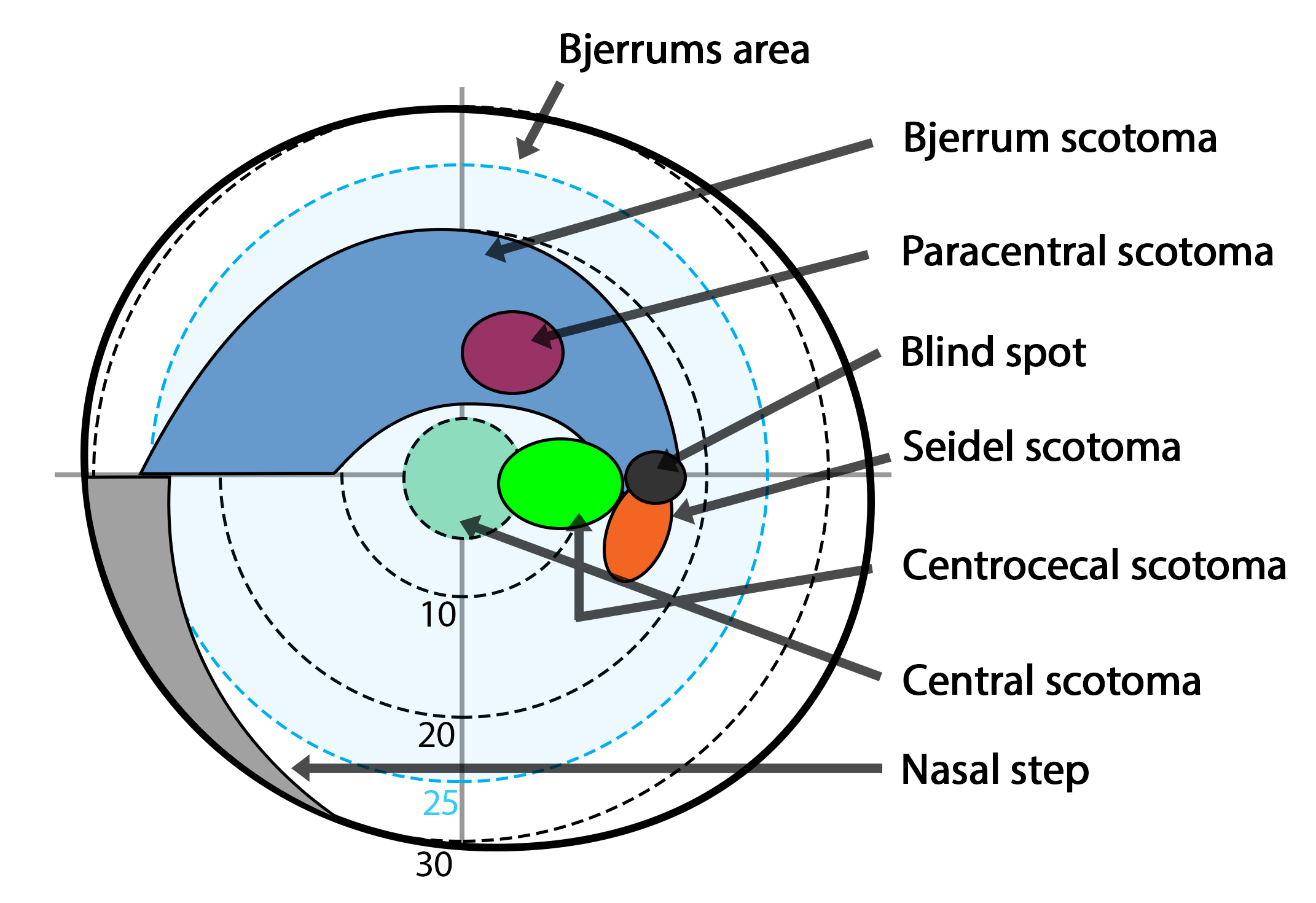
The visual field is "that portion of space in which objects are visible at the same moment during steady fixation of the gaze in one direction"; in ophthalmology and neurology the emphasis is mostly on the structure inside the visual field and it is then considered “the field of functional capacity obtained and recorded by means of perimetry”.Strasburger, Hans; Pöppel, Ernst (2002). Visual Field. In G. Adelman & B.H. Smith (Eds): ''Encyclopedia of Neuroscience''; 3rd edition, on CD-ROM. Elsevier Science B.V., Amsterdam, New York. However, the visual field can also be understood as a predominantly ''perceptual'' concept and its definition then becomes that of the "spatial array of visual sensations available to observation in introspectionist psychological experiments" (for example in van Doorn et al., 2013). The corresponding concept for optical instruments and image sensors is the field of view (FOV). In humans and animals, the FOV refers to the area visible when eye mov ... [...More Info...] [...Related Items...] OR: [Wikipedia] [Google] [Baidu] |
Hyperpituitarism
Hyperpituitarism is a condition due to the primary hypersecretion of pituitary hormones; it typically results from a pituitary adenoma. In children with hyperpituitarism, disruption of growth regulation is rare, either because of hormone hypersecretion or because of manifestations caused by local compression of the adenoma. Symptoms and signs Symptoms caused by hormone excess and associated mass effects include: Cause The cause of hyperpituitarism in most cases is due to pituitary adenomas. They usually come from the anterior lobe, are functional and secrete the hormone, GH and prolactin. Mechanism Evidence indicates that the mechanism of hyperpituitarism can originate from genetic disruption causing pituitary tumorigenesis, most pituitary adenomas are monoclonal, which in turn indicates their origin from an event in a single cell. There are three hormones that are oversecreted resulting in the pituitary adenoma: prolactin, adrenocorticotropic hormone (ACTH), and growth hormone ... [...More Info...] [...Related Items...] OR: [Wikipedia] [Google] [Baidu] |
Intracranial Pressure
Intracranial pressure (ICP) is the pressure exerted by fluids such as cerebrospinal fluid (CSF) inside the skull and on the brain tissue. ICP is measured in millimeters of mercury ( mmHg) and at rest, is normally 7–15 mmHg for a supine adult. This equals to 9–20 cmH2O, which is a common scale used in lumbar punctures. The body has various mechanisms by which it keeps the ICP stable, with CSF pressures varying by about 1 mmHg in normal adults through shifts in production and absorption of CSF. Changes in ICP are attributed to volume changes in one or more of the constituents contained in the cranium. CSF pressure has been shown to be influenced by abrupt changes in intrathoracic pressure during coughing (which is induced by contraction of the diaphragm and abdominal wall muscles, the latter of which also increases intra-abdominal pressure), the valsalva maneuver, and communication with the vasculature ( venous and arterial systems). Intracranial hypertension (IH), ... [...More Info...] [...Related Items...] OR: [Wikipedia] [Google] [Baidu] |
Lateral Rectus
The lateral rectus muscle is a muscle on the lateral side of the eye in the orbit. It is one of six extraocular muscles that control the movements of the eye. The lateral rectus muscle is responsible for lateral movement of the eyeball, specifically abduction. Abduction describes the movement of the eye away from the midline (i.a. nose), allowing the eyeball to move horizontally in the lateral direction, bringing the pupil away from the midline of the body. Structure The lateral rectus muscle originates at the lateral part of the common tendinous ring, also known as the annular tendon. The common tendinous ring is a tendinous ring that surrounds the optic nerve and serves as the origin for five of the seven extraocular muscles, excluding the inferior oblique muscle. The lateral rectus muscle inserts into the temporal side of the eyeball. This insertion is around 7 mm from the corneal limbus. It has a width of around 10 mm. Nerve supply The lateral rectus is the only muscle ... [...More Info...] [...Related Items...] OR: [Wikipedia] [Google] [Baidu] |
Abducens Nerve
The abducens nerve or abducent nerve, also known as the sixth cranial nerve, cranial nerve VI, or simply CN VI, is a cranial nerve in humans and various other animals that controls the movement of the lateral rectus muscle, one of the extraocular muscles responsible for outward Gaze (physiology), gaze. It is a Somatic (biology), somatic efferent nerve fiber, efferent nerve. Structure Nucleus The abducens nucleus is located in the pons, on the floor of the fourth ventricle, at the level of the facial colliculus. Axons from the facial nerve loop around the abducens nucleus, creating a slight bulge (the facial colliculus) that is visible on the dorsal surface of the floor of the fourth ventricle. The abducens nucleus is close to the midline, like the other motor nuclei that control eye movements (the Oculomotor nucleus, oculomotor and Trochlear nucleus, trochlear nuclei). Motor axons leaving the abducens nucleus run ventrally and caudally through the pons. They pass lateral to th ... [...More Info...] [...Related Items...] OR: [Wikipedia] [Google] [Baidu] |
Quadrantanopia
Quadrantanopia, quadrantanopsia, refers to an anopia (loss of vision) affecting a quarter of the visual field. It can be associated with a lesion of an optic radiation. While quadrantanopia can be caused by lesions in the Temporal lobe, temporal and parietal lobes of the Human brain, brain, it is most commonly associated with lesions in the occipital lobe.Kolb, B & Whishaw, I.Q. Human Neuropsychology, Sixth Edition, p.361; Worth Publishers (2008) Presentation An interesting aspect of quadrantanopia is that there exists a distinct and sharp border between the intact and damaged visual fields, due to an anatomical separation of the quadrants of the visual field. For example, information in the left half of visual field is processed in the right occipital lobe and information in the right half of the visual field is processed in the left occipital lobe. In a quadrantanopia that is partial, there also exists a distinct and sharp border between the intact and damaged field within ... [...More Info...] [...Related Items...] OR: [Wikipedia] [Google] [Baidu] |
Craniopharyngioma
A craniopharyngioma is a rare type of brain tumor derived from pituitary gland embryonic tissue that occurs most commonly in children, but also affects adults. It may present at any age, even in the prenatal and neonatal periods, but peak incidence rates are childhood-onset at 5–14 years and adult-onset at 50–74 years. People may present with Quadrantanopia, bitemporal inferior quadrantanopia leading to bitemporal hemianopsia, as the tumor may compress the optic chiasm. It has a point prevalence around two per 1,000,000. Craniopharyngiomas are distinct from Rathke's cleft cyst, Rathke's cleft tumours and intrasellar arachnoid cysts. Symptoms and signs Craniopharyngiomas are almost always Benign tumor, benign. However, as with many brain tumors, their treatment can be difficult, and significant morbidities are associated with both the tumor and treatment. * Headache (obstructive hydrocephalus) * Hypersomnia * Myxedema * Postsurgical weight gain (hypothalamic obesity) * Polydip ... [...More Info...] [...Related Items...] OR: [Wikipedia] [Google] [Baidu] |
Optic Nerve
In neuroanatomy, the optic nerve, also known as the second cranial nerve, cranial nerve II, or simply CN II, is a paired cranial nerve that transmits visual system, visual information from the retina to the brain. In humans, the optic nerve is derived from optic stalks during the seventh week of development and is composed of retinal ganglion cell axons and glial cells; it extends from the optic disc to the optic chiasma and continues as the optic tract to the lateral geniculate nucleus, Pretectal area, pretectal nuclei, and superior colliculus. Structure The optic nerve has been classified as the second of twelve paired cranial nerves, but it is technically a myelinated tract of the central nervous system, rather than a classical nerve of the peripheral nervous system because it is derived from an out-pouching of the diencephalon (optic stalks) during embryonic development. As a consequence, the fibers of the optic nerve are covered with myelin produced by oligodendrocytes, r ... [...More Info...] [...Related Items...] OR: [Wikipedia] [Google] [Baidu] |
Visual Field
The visual field is "that portion of space in which objects are visible at the same moment during steady fixation of the gaze in one direction"; in ophthalmology and neurology the emphasis is mostly on the structure inside the visual field and it is then considered “the field of functional capacity obtained and recorded by means of perimetry”.Strasburger, Hans; Pöppel, Ernst (2002). Visual Field. In G. Adelman & B.H. Smith (Eds): ''Encyclopedia of Neuroscience''; 3rd edition, on CD-ROM. Elsevier Science B.V., Amsterdam, New York. However, the visual field can also be understood as a predominantly ''perceptual'' concept and its definition then becomes that of the "spatial array of visual sensations available to observation in introspectionist psychological experiments" (for example in van Doorn et al., 2013). The corresponding concept for optical instruments and image sensors is the field of view (FOV). In humans and animals, the FOV refers to the area visible when eye mov ... [...More Info...] [...Related Items...] OR: [Wikipedia] [Google] [Baidu] |
Galactorrhea
Galactorrhea ( also spelled galactorrhoea) ( galacto- + -rrhea) or lactorrhea ( lacto- + -rrhea) is the spontaneous flow of milk from the breast, unassociated with childbirth or nursing. Galactorrhea is reported to occur in 5–32% of females. Much of the difference in reported incidence can be attributed to different definitions of galactorrhea. Although frequently benign, it may be caused by serious underlying conditions and should be properly investigated. Galactorrhea also occurs in males, newborn infants and adolescents of both sexes. Causes Galactorrhea can take place as a result of dysregulation of certain hormones. Hormonal causes most frequently associated with galactorrhea are hyperprolactinemia and thyroid conditions with elevated levels of thyroid-stimulating hormone (TSH) or thyrotropin-releasing hormone (TRH). No obvious cause is found in about 50% of cases. Lactation requires the presence of prolactin, and the evaluation of galactorrhea includes eliciting a h ... [...More Info...] [...Related Items...] OR: [Wikipedia] [Google] [Baidu] |
Acromegaly
Acromegaly is a disorder that results in excess growth of certain parts of the human body. It is caused by excess growth hormone (GH) after the growth plates have closed. The initial symptom is typically enlargement of the hands and feet. There may also be an enlargement of the forehead, jaw, and nose. Other symptoms may include joint pain, thicker skin, deepening of the voice, headaches, and Visual impairment, problems with vision. Complications of the disease may include type 2 diabetes, sleep apnea, and high blood pressure. Signs and symptoms Features that may result from a high level of GH or expanding tumor include: * Headaches * Enlargement of the hands, feet, nose, lips, and ears, and a general thickening of the skin * Soft tissue swelling of internal organs, notably the heart with the attendant weakening of its muscularity, and the kidneys, also the vocal cords resulting in a characteristic thick, deep voice and slowing of speech * Generalized expansion of the skull at ... [...More Info...] [...Related Items...] OR: [Wikipedia] [Google] [Baidu] |


