|
Perimetry
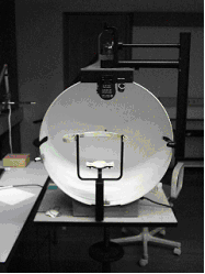
A visual field test is an eye examination that can detect dysfunction in central and peripheral vision which may be caused by various medical conditions such as glaucoma, stroke, pituitary disease, brain tumours or other neurological deficits. Visual field testing can be performed clinically by keeping the subject's gaze fixed while presenting objects at various places within their visual field. Simple manual equipment can be used such as in the tangent screen test or the Amsler grid. When dedicated machinery is used it is called a perimeter. The exam may be performed by a technician in one of several ways. The test may be performed by a technician directly, with the assistance of a machine, or completely by an automated machine. Machine-based tests aid diagnostics by allowing a detailed printout of the patient's visual field. Other names for this test may include perimetry, Tangent screen exam, Automated perimetry exam, Goldmann visual field exam, or brand names such as the H ... [...More Info...] [...Related Items...] OR: [Wikipedia] [Google] [Baidu] |
Visual Field
The visual field is "that portion of space in which objects are visible at the same moment during steady fixation of the gaze in one direction"; in ophthalmology and neurology the emphasis is mostly on the structure inside the visual field and it is then considered “the field of functional capacity obtained and recorded by means of perimetry”.Strasburger, Hans; Pöppel, Ernst (2002). Visual Field. In G. Adelman & B.H. Smith (Eds): ''Encyclopedia of Neuroscience''; 3rd edition, on CD-ROM. Elsevier Science B.V., Amsterdam, New York. However, the visual field can also be understood as a predominantly ''perceptual'' concept and its definition then becomes that of the "spatial array of visual sensations available to observation in introspectionist psychological experiments" (for example in van Doorn et al., 2013). The corresponding concept for optical instruments and image sensors is the field of view (FOV). In humans and animals, the FOV refers to the area visible when eye mov ... [...More Info...] [...Related Items...] OR: [Wikipedia] [Google] [Baidu] |
Peripheral Vision
Peripheral vision, or ''indirect vision'', is vision as it occurs outside the point of fixation, i.e. away from the center of gaze or, when viewed at large angles, in (or out of) the "corner of one's eye". The vast majority of the area in the visual field is included in the notion of peripheral vision. "Far peripheral" vision refers to the area at the edges of the visual field, "mid-peripheral" vision refers to medium eccentricities, and "near-peripheral", sometimes referred to as "para-central" vision, exists adjacent to the center of gaze. Boundaries Inner boundaries The inner boundaries of peripheral vision can be defined in any of several ways depending on the context. In everyday language the term "peripheral vision" is often used to refer to what in technical usage would be called "far peripheral vision." This is vision outside of the range of stereoscopic vision. It can be conceived as bounded at the center by a circle 60° in radius or 120° in diameter, centered aro ... [...More Info...] [...Related Items...] OR: [Wikipedia] [Google] [Baidu] |
Glaucoma
Glaucoma is a group of eye diseases that can lead to damage of the optic nerve. The optic nerve transmits visual information from the eye to the brain. Glaucoma may cause vision loss if left untreated. It has been called the "silent thief of sight" because the loss of vision usually occurs slowly over a long period of time. A major risk factor for glaucoma is increased pressure within the eye, known as Intraocular pressure, intraocular pressure (IOP). It is associated with old age, a family history of glaucoma, and certain medical conditions or the use of some medications. The word ''glaucoma'' comes from the Ancient Greek word (), meaning 'gleaming, blue-green, gray'. Of the different types of glaucoma, the most common are called open-angle glaucoma and closed-angle glaucoma. Inside the eye, a liquid called Aqueous humour, aqueous humor helps to maintain shape and provides nutrients. The aqueous humor normally drains through the trabecular meshwork. In open-angle glaucoma, ... [...More Info...] [...Related Items...] OR: [Wikipedia] [Google] [Baidu] |
Microperimetry
Microperimetry, sometimes called fundus-controlled perimetry, is a type of visual field test which uses one of several technologies to create a "retinal sensitivity map" of the quantity of light perceived in specific parts of the retina in people who have lost the ability to fixate on an object or light source. The main difference with traditional perimetry instruments is that, microperimetry includes a system to image the retina and an eye tracker to compensate eye movements during visual field testing. Usage Visual field testing is widely used to monitor pathologies affecting the periphery of vision such as glaucoma. During a conventional test, patients are asked to look steady (fixate) at a visual target, while light stimuli are projected at varying intensities in different retinal locations. This process is not, however, considered accurate in the evaluation of pathologies affecting the central part of the retina (macula and fovea centralis) as patients with these pathologies ar ... [...More Info...] [...Related Items...] OR: [Wikipedia] [Google] [Baidu] |
Human Brain
The human brain is the central organ (anatomy), organ of the nervous system, and with the spinal cord, comprises the central nervous system. It consists of the cerebrum, the brainstem and the cerebellum. The brain controls most of the activities of the human body, body, processing, integrating, and coordinating the information it receives from the sensory nervous system. The brain integrates sensory information and coordinates instructions sent to the rest of the body. The cerebrum, the largest part of the human brain, consists of two cerebral hemispheres. Each hemisphere has an inner core composed of white matter, and an outer surface – the cerebral cortex – composed of grey matter. The cortex has an outer layer, the neocortex, and an inner allocortex. The neocortex is made up of six Cerebral cortex#Layers of neocortex, neuronal layers, while the allocortex has three or four. Each hemisphere is divided into four lobes of the brain, lobes – the frontal lobe, frontal, pa ... [...More Info...] [...Related Items...] OR: [Wikipedia] [Google] [Baidu] |
Optic Nerve
In neuroanatomy, the optic nerve, also known as the second cranial nerve, cranial nerve II, or simply CN II, is a paired cranial nerve that transmits visual system, visual information from the retina to the brain. In humans, the optic nerve is derived from optic stalks during the seventh week of development and is composed of retinal ganglion cell axons and glial cells; it extends from the optic disc to the optic chiasma and continues as the optic tract to the lateral geniculate nucleus, Pretectal area, pretectal nuclei, and superior colliculus. Structure The optic nerve has been classified as the second of twelve paired cranial nerves, but it is technically a myelinated tract of the central nervous system, rather than a classical nerve of the peripheral nervous system because it is derived from an out-pouching of the diencephalon (optic stalks) during embryonic development. As a consequence, the fibers of the optic nerve are covered with myelin produced by oligodendrocytes, r ... [...More Info...] [...Related Items...] OR: [Wikipedia] [Google] [Baidu] |
Retina
The retina (; or retinas) is the innermost, photosensitivity, light-sensitive layer of tissue (biology), tissue of the eye of most vertebrates and some Mollusca, molluscs. The optics of the eye create a focus (optics), focused two-dimensional image of the visual world on the retina, which then processes that image within the retina and sends nerve impulses along the optic nerve to the visual cortex to create visual perception. The retina serves a function which is in many ways analogous to that of the photographic film, film or image sensor in a camera. The neural retina consists of several layers of neurons interconnected by Chemical synapse, synapses and is supported by an outer layer of pigmented epithelial cells. The primary light-sensing cells in the retina are the photoreceptor cells, which are of two types: rod cell, rods and cone cell, cones. Rods function mainly in dim light and provide monochromatic vision. Cones function in well-lit conditions and are responsible fo ... [...More Info...] [...Related Items...] OR: [Wikipedia] [Google] [Baidu] |
Human Eye
The human eye is a sensory organ in the visual system that reacts to light, visible light allowing eyesight. Other functions include maintaining the circadian rhythm, and Balance (ability), keeping balance. The eye can be considered as a living optics, optical device. It is approximately spherical in shape, with its outer layers, such as the outermost, white part of the eye (the sclera) and one of its inner layers (the pigmented choroid) keeping the eye essentially stray light, light tight except on the eye's optic axis. In order, along the optic axis, the optical components consist of a first lens (the cornea, cornea—the clear part of the eye) that accounts for most of the optical power of the eye and accomplishes most of the Focus (optics), focusing of light from the outside world; then an aperture (the pupil) in a Diaphragm (optics), diaphragm (the Iris (anatomy), iris—the coloured part of the eye) that controls the amount of light entering the interior of the eye; then an ... [...More Info...] [...Related Items...] OR: [Wikipedia] [Google] [Baidu] |
Scotoma
A scotoma is an area of partial alteration in the field of vision consisting of a partially diminished or entirely degenerated visual acuity that is surrounded by a field of normal – or relatively well-preserved – vision. Every normal mammalian eye has a scotoma in its field of vision, usually termed its blind spot. This is a location with no photoreceptor cells, where the retinal ganglion cell axons that compose the optic nerve exit the retina. This location is called the optic disc. There is no direct conscious awareness of visual scotomas. They are simply regions of reduced information within the visual field. Rather than recognizing an incomplete image, patients with scotomas report that things "disappear" on them. The presence of the blind spot scotoma can be demonstrated subjectively by covering one eye, carefully holding fixation with the open eye, and placing an object (such as one's thumb) in the lateral and horizontal visual field, about 15 degrees from fixati ... [...More Info...] [...Related Items...] OR: [Wikipedia] [Google] [Baidu] |
Macula Of Retina
The macula (/ˈmakjʊlə/) or macula lutea is an oval-shaped pigmented area in the center of the retina of the human eye and in other animals. The macula in humans has a diameter of around and is subdivided into the umbo, foveola, foveal avascular zone, fovea, parafovea, and perifovea areas. The anatomical macula at a size of is much larger than the clinical macula which, at a size of , corresponds to the anatomical fovea. The macula is responsible for the central, high-resolution, color vision that is possible in good light. This kind of vision is impaired if the macula is damaged, as in macular degeneration. The clinical macula is seen when viewed from the pupil, as in ophthalmoscopy or retinal photography. The term macula lutea comes from Latin ''macula'', "spot", and ''lutea'', "yellow". Structure The macula is an oval-shaped pigmented area in the center of the retina of the human eye and other animal eyes. Its center is shifted slightly away from the optical a ... [...More Info...] [...Related Items...] OR: [Wikipedia] [Google] [Baidu] |
Goldmann Perimeter
Goldmann is the surname of several people: * Erich Goldmann, German ice hockey player * Friedrich Goldmann (1941–2009), German composer and conductor * Hans Goldmann (1899–1991), Swiss ophthalmologist * Lucien Goldmann, French philosopher and sociologist * Maximilian Goldmann, real name of Max Reinhardt, Austrian theatre director * Nahum Goldmann Nahum Goldmann (; July 10, 1895 – August 29, 1982) was a leading Zionist. He was a founder of the World Jewish Congress and its president from 1951 to 1978 and was also president of the World Zionist Organization from 1956 to 1968. Biography ..., former president of the World Jewish Congress * Ulrike Goldmann, singer for German band Blutengel * Stefan Goldmann (born 1978), German-Bulgarian DJ and composer of electronic music It can also refer to: * Goldmann (publisher), large publishing house in Germany See also * Goldman (other) {{surname German-language surnames Surnames of Jewish origin Yiddish-langua ... [...More Info...] [...Related Items...] OR: [Wikipedia] [Google] [Baidu] |
Sensitivity And Specificity
In medicine and statistics, sensitivity and specificity mathematically describe the accuracy of a test that reports the presence or absence of a medical condition. If individuals who have the condition are considered "positive" and those who do not are considered "negative", then sensitivity is a measure of how well a test can identify true positives and specificity is a measure of how well a test can identify true negatives: * Sensitivity (true positive rate) is the probability of a positive test result, conditioned on the individual truly being positive. * Specificity (true negative rate) is the probability of a negative test result, conditioned on the individual truly being negative. If the true status of the condition cannot be known, sensitivity and specificity can be defined relative to a " gold standard test" which is assumed correct. For all testing, both diagnoses and screening, there is usually a trade-off between sensitivity and specificity, such that higher sensiti ... [...More Info...] [...Related Items...] OR: [Wikipedia] [Google] [Baidu] |


