|
Orbital Canal
The infraorbital canal is a canal found at the base of the orbit that opens on to the maxilla. It is continuous with the infraorbital groove and opens onto the maxilla at the infraorbital foramen. The infraorbital nerve and infraorbital artery travel through the canal. Structure One of the canals of the orbital surface of the maxilla, the infraorbital canal, opens just below the margin of the orbit, the area of the skull containing the eye and related structures. It should not be confused with the infraorbital foramen, with which it is continuous. Function It transmits the infraorbital nerve as well as infraorbital artery, both of which enter this canal at the infraorbital groove and after coursing through the maxillary sinus exit via the infraorbital foramen. Before exiting, the anterior superior alveolar nerve, middle superior alveolar nerve The middle superior alveolar nerve is a nerve that drops from the infraorbital portion of the maxillary nerve to supply the sinus mu ... [...More Info...] [...Related Items...] OR: [Wikipedia] [Google] [Baidu] |
Infraorbital Groove
The infraorbital groove (or sulcus) is located in the middle of the posterior part of the orbital surface of the maxilla. Its function is to act as the passage of the infraorbital artery, the infraorbital vein, and the infraorbital nerve. Structure The infraorbital groove begins at the middle of the posterior border of the maxilla (with which it is continuous). This is near the upper edge of the infratemporal surface of the maxilla. It passes forward, and ends in a canal which subdivides into two branches. The infraorbital groove has an average length of 16.7 mm, with a small amount of variation between people. It is similar in men and women. Function The infraorbital groove creates space that allows for passage of the infraorbital artery, the infraorbital vein, and the infraorbital nerve. Clinical significance The infraorbital groove is an important surgical landmark for local anaesthesia of the infraorbital nerve. See also * Infraorbital foramen In human a ... [...More Info...] [...Related Items...] OR: [Wikipedia] [Google] [Baidu] |
Infraorbital Foramen
In human anatomy, the infraorbital foramen is one of two small holes in the skull's upper jawbone (maxillary bone), located below the eye socket and to the left and right of the nose. Both holes are used for blood vessels and nerves. In anatomical terms, it is located below the infraorbital margin of the orbit. It transmits the infraorbital artery and vein, and the infraorbital nerve, a branch of the maxillary nerve. It is typically from the infraorbital margin. Structure Forming the exterior end of the infraorbital canal, the infraorbital foramen communicates with the infraorbital groove, the canal's opening on the interior side. The ramifications of the three principal branches of the trigeminal nerve—at the supraorbital, infraorbital, and mental foramen—are distributed on a vertical line (in anterior view) passing through the middle of the pupil. The infraorbital foramen is used as a pressure point to test the sensitivity of the infraorbital nerve. Palpation of the ... [...More Info...] [...Related Items...] OR: [Wikipedia] [Google] [Baidu] |
Canal (anatomy)
* In anatomy, a canal (or canalis in Latin) is a tubular passage or channel which connects different regions of the body. Examples include: * Cranial Region ** Alveolar canals ** Carotid canal ** Facial canal ** Greater palatine canal ** Incisive canals ** Infraorbital canal ** Mandibular canal ** Optic canal ** Palatovaginal canal ** Pterygoid canal * Abdominal Region ** Inguinal canal * Pelvic Region ** Anal canal ** Pudendal canal * Upper Extremities ** Suprascapular canal ** Carpal canal ** Ulnar canal ** Radial canal * Lower Extremities ** Adductor canal ** Femoral canal ** Obturator canal See also * Foramen In anatomy and osteology, a foramen (; in [...More Info...] [...Related Items...] OR: [Wikipedia] [Google] [Baidu] |
Orbit (anatomy)
In anatomy, the orbit is the cavity or socket of the skull in which the eye and its appendages are situated. "Orbit" can refer to the bony socket, or it can also be used to imply the contents. In the adult human, the volume of the orbit is , of which the eye occupies . The orbital contents comprise the eye, the orbital and retrobulbar fascia, extraocular muscles, cranial nerves II, III, IV, V, and VI, blood vessels, fat, the lacrimal gland with its sac and duct, the eyelids, medial and lateral palpebral ligaments, cheek ligaments, the suspensory ligament, septum, ciliary ganglion and short ciliary nerves. Structure The orbits are conical or four-sided pyramidal cavities, which open into the midline of the face and point back into the head. Each consists of a base, an apex and four walls."eye, human."Encyclopædia Britannica from Encyclopædia Britannica 2006 Ultimate Reference Suite DVD 2009 Openings There are two important foramina, or windows, two important ... [...More Info...] [...Related Items...] OR: [Wikipedia] [Google] [Baidu] |
Maxilla
The maxilla (plural: ''maxillae'' ) in vertebrates is the upper fixed (not fixed in Neopterygii) bone of the jaw formed from the fusion of two maxillary bones. In humans, the upper jaw includes the hard palate in the front of the mouth. The two maxillary bones are fused at the intermaxillary suture, forming the anterior nasal spine. This is similar to the mandible (lower jaw), which is also a fusion of two mandibular bones at the mandibular symphysis. The mandible is the movable part of the jaw. Structure In humans, the maxilla consists of: * The body of the maxilla * Four processes ** the zygomatic process ** the frontal process of maxilla ** the alveolar process ** the palatine process * three surfaces – anterior, posterior, medial * the Infraorbital foramen * the maxillary sinus * the incisive foramen Articulations Each maxilla articulates with nine bones: * two of the cranium: the frontal and ethmoid * seven of the face: the nasal, zygomatic, lacrimal ... [...More Info...] [...Related Items...] OR: [Wikipedia] [Google] [Baidu] |
Infraorbital Nerve
The infraorbital nerve is a branch of the maxillary nerve, itself a branch of the trigeminal nerve (CN V). It travels through the orbit and enters the infraorbital canal to exit onto the face through the infraorbital foramen. It provides sensory innervation to the skin and mucous membranes around the middle of the face. Structure The infraorbital nerve is a branch of the maxillary nerve (CN V2), itself a branch of the trigeminal nerve (CN V). It travels with the infraorbital artery and vein. It branches from the maxillary nerve in the pterygopalatine fossa and travels through the inferior orbital fissure to enter the orbit. It runs anteriorly along the floor of the orbit in the infraorbital groove to the infraorbital canal of the maxilla. Within the infraorbital canal it has three branches, the posterior superior alveolar nerve, middle superior alveolar nerve and anterior superior alveolar nerve. After traversing the canal it emerges onto the anterior surface of the maxill ... [...More Info...] [...Related Items...] OR: [Wikipedia] [Google] [Baidu] |
Infraorbital Artery
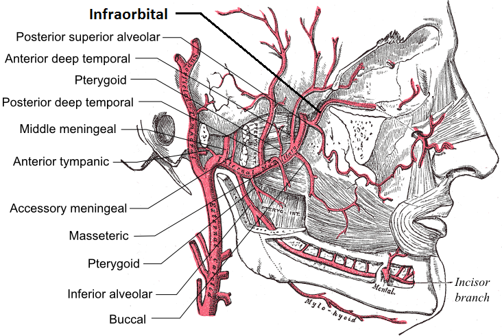
The infraorbital artery is an artery in the head that branches off the maxillary artery, emerging through the infraorbital foramen, just under the orbit of the eye. Course The infraorbital artery appears, from its direction, to be the continuation of the trunk of the maxillary artery, but often arises in conjunction with the posterior superior alveolar artery. It runs along the infraorbital groove and canal with the infraorbital nerve, and emerges on the face through the infraorbital foramen, beneath the infraorbital head of the levator labii superioris muscle. Branches While in the canal, it gives off * (a) orbital branches which assist in supplying the inferior rectus and inferior oblique and the lacrimal sac, and * (b) anterior superior alveolar arteries - branches which descend through the anterior alveolar canals to supply the upper incisor and canine teeth and the mucous membrane of the maxillary sinus. On the face, some branches pass upward to the medial angle of the o ... [...More Info...] [...Related Items...] OR: [Wikipedia] [Google] [Baidu] |
Canal (other)
A canal is a human-made channel for water. Canal may also refer to: People (Alphabetical by surname) * David Canal (born 1978), Spanish sprinter * Esteban Canal (1896-1981), Peruvian chess player * Giovanni Antonio Canal (1697–1768), Venetian painter, better known as Canaletto * Richard Canal (born 1953), French science fiction writer * B. de Canals, was a 14th-century Spanish author of a Latin chronicle * María Antònia Canals (1930–2022), Spanish mathematician * Agustín de la Canal (born 1980), Argentine football player * Ramón Alva de la Canal (1892–1985), Mexican painter Places * Canal Flats, British Columbia, a village in British Columbia, Canada * Canal Fulton, Ohio, United States * Canal Street (other), name of numerous roads and neighborhoods * Canal Township, Venango County, Pennsylvania, United States * Canal Winchester, Ohio, United States * Canals, Tarn-et-Garonne, a commune in the Tarn-et-Garonne department, France * Canals, Valencia, a munic ... [...More Info...] [...Related Items...] OR: [Wikipedia] [Google] [Baidu] |
Skull
The skull is a bone protective cavity for the brain. The skull is composed of four types of bone i.e., cranial bones, facial bones, ear ossicles and hyoid bone. However two parts are more prominent: the cranium and the mandible. In humans, these two parts are the neurocranium and the viscerocranium ( facial skeleton) that includes the mandible as its largest bone. The skull forms the anterior-most portion of the skeleton and is a product of cephalisation—housing the brain, and several sensory structures such as the eyes, ears, nose, and mouth. In humans these sensory structures are part of the facial skeleton. Functions of the skull include protection of the brain, fixing the distance between the eyes to allow stereoscopic vision, and fixing the position of the ears to enable sound localisation of the direction and distance of sounds. In some animals, such as horned ungulates (mammals with hooves), the skull also has a defensive function by providing the mount (on the ... [...More Info...] [...Related Items...] OR: [Wikipedia] [Google] [Baidu] |
Anterior Superior Alveolar Nerve
The anterior superior alveolar nerve (or anterior superior dental nerve), is a branch of the infraorbital nerve, itself a branch of the maxillary nerve (V2). It branches from the infraorbital nerve within the infraorbital canal before the infraorbital nerve exits through the infraorbital foramen. It descends in a canal in the anterior wall of the maxillary sinus, and divides into branches which supply the incisor and canine teeth. It communicates with the middle superior alveolar nerve, and gives off a nasal branch, which passes through a minute canal in the lateral wall of the inferior meatus, and supplies the mucous membrane of the anterior part of the inferior meatus and the floor of the nasal cavity, communicating with the nasal branches from the sphenopalatine ganglion. Dental considerations for this nerve are important. The anterior superior alveolar usually innervates all anterior teeth, loops backwards to join the middle superior alveolar nerve to form the superior dental ... [...More Info...] [...Related Items...] OR: [Wikipedia] [Google] [Baidu] |
Middle Superior Alveolar Nerve
The middle superior alveolar nerve is a nerve that drops from the infraorbital portion of the maxillary nerve to supply the sinus mucosa, the roots of the maxillary premolars, and the mesiobuccal root of the first maxillary molar. It is not always present; in 72% of cases it is non existent with the anterior superior alveolar nerve innervating the premolars and the posterior superior alveolar nerve The posterior superior alveolar branches (posterior superior dental branches) arise from the trunk of the maxillary nerve just before it enters the infraorbital groove; they are generally two in number, but sometimes arise by a single trunk. They ... innervating the molars, including the mesiobuccal root of the first molar. External links * Maxillary nerve {{Neuroanatomy-stub ... [...More Info...] [...Related Items...] OR: [Wikipedia] [Google] [Baidu] |


