|
Occipital Gyri
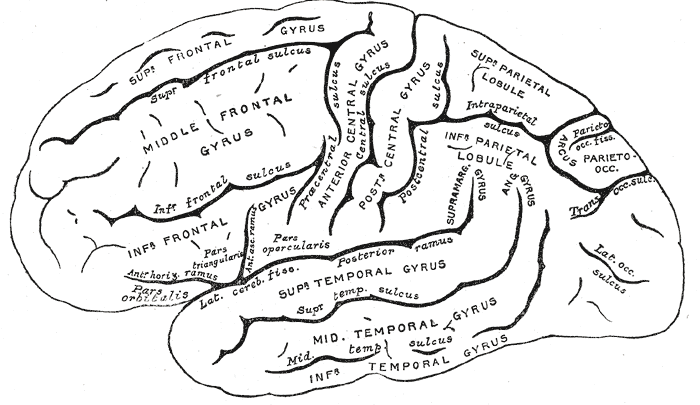
The occipital gyri (OcG) are three gyri in parallel, along the lateral portion of the occipital lobe, also referred to as a composite structure in the brain. The gyri are the superior occipital gyrus, the middle occipital gyrus, and the inferior occipital gyrus, and these are also known as the occipital face area. The superior and inferior occipital sulci separates the three occipital gyri. The intraoccipital sulcus, also known as the superior occipital sulcus, stems from the intraparietal sulcus and continues until the sulcus reaches the transverse occipital sulcus, separating the superior occipital gyrus from the middle occipital gyrus. The transverse occipital sulcus comes down along the lateral occipital surface or the inferior occipital sulcus. Structural anatomy The border between the occipital lobe and the parietal and temporal lobes is characterized by different gyri: the superior occipital gyrus (also known as gyrus occipitalis superior), middle occipital gyrus (or ... [...More Info...] [...Related Items...] OR: [Wikipedia] [Google] [Baidu] |
Gyrus
In neuroanatomy, a gyrus (pl. gyri) is a ridge on the cerebral cortex. It is generally surrounded by one or more sulci (depressions or furrows; sg. ''sulcus''). Gyri and sulci create the folded appearance of the brain in humans and other mammals. Structure The gyri are part of a system of folds and ridges that create a larger surface area for the human brain and other mammalian brains. Because the brain is confined to the skull, brain size is limited. Ridges and depressions create folds allowing a larger cortical surface area, and greater cognitive function, to exist in the confines of a smaller cranium. Development The human brain undergoes gyrification during fetal and neonatal development. In embryonic development, all mammalian brains begin as smooth structures derived from the neural tube. A cerebral cortex without surface convolutions is lissencephalic, meaning 'smooth-brained'. As development continues, gyri and sulci begin to take shape on the fetal brain, w ... [...More Info...] [...Related Items...] OR: [Wikipedia] [Google] [Baidu] |
Occipital Lobe
The occipital lobe is one of the four major lobes of the cerebral cortex in the brain of mammals. The name derives from its position at the back of the head, from the Latin ''ob'', "behind", and ''caput'', "head". The occipital lobe is the visual processing center of the mammalian brain containing most of the anatomical region of the visual cortex. The primary visual cortex is Brodmann area 17, commonly called V1 (visual one). Human V1 is located on the medial side of the occipital lobe within the calcarine sulcus; the full extent of V1 often continues onto the occipital pole. V1 is often also called striate cortex because it can be identified by a large stripe of myelin, the Stria of Gennari. Visually driven regions outside V1 are called extrastriate cortex. There are many extrastriate regions, and these are specialized for different visual tasks, such as visuospatial processing, color differentiation, and motion perception. Bilateral lesions of the occipital lobe can l ... [...More Info...] [...Related Items...] OR: [Wikipedia] [Google] [Baidu] |
Brain
The brain is an organ that serves as the center of the nervous system in all vertebrate and most invertebrate animals. It consists of nervous tissue and is typically located in the head ( cephalization), usually near organs for special senses such as vision, hearing and olfaction. Being the most specialized organ, it is responsible for receiving information from the sensory nervous system, processing those information (thought, cognition, and intelligence) and the coordination of motor control (muscle activity and endocrine system). While invertebrate brains arise from paired segmental ganglia (each of which is only responsible for the respective body segment) of the ventral nerve cord, vertebrate brains develop axially from the midline dorsal nerve cord as a vesicular enlargement at the rostral end of the neural tube, with centralized control over all body segments. All vertebrate brains can be embryonically divided into three parts: the forebrain (prosencep ... [...More Info...] [...Related Items...] OR: [Wikipedia] [Google] [Baidu] |
Occipital Face Area
The occipital face area (OFA) is a region of the human cerebral cortex which is specialised for face perception. The OFA is located on the lateral surface of the occipital lobe adjacent to the inferior occipital gyrus. The OFA comprises a network of brain regions including the fusiform face area (FFA) and posterior superior temporal sulcus (STS) which support facial processing. Structure Like other regions of cerebral cortex, the OFA is functionally defined by using neuroimaging techniques to localise changes in neural activity in response to different face stimuli. Typically, participants will view different kinds of face stimuli which can be contrasted with scrambled images, letter strings or non-face objects to localise the OFA. While the exact location of the OFA varies between individuals and according to the specific paradigm used, it usually corresponds to Brodmann areas 18 or 19. Function The OFA is believed to be functionally necessary for some face computations ... [...More Info...] [...Related Items...] OR: [Wikipedia] [Google] [Baidu] |
Transverse Occipital Sulcus
The transverse occipital sulcus is a structure in the occipital lobe. The transverse occipital sulcus is continuous with the posterior end of the occipital ramus of the intraparietal sulcus, and runs across the upper part of the lobe, a short distance behind the parietooccipital fissure. References Sulci (neuroanatomy) Occipital lobe {{neuroanatomy-stub ... [...More Info...] [...Related Items...] OR: [Wikipedia] [Google] [Baidu] |
Gyri Of Lateral Cortex
In neuroanatomy, a gyrus (pl. gyri) is a ridge on the cerebral cortex. It is generally surrounded by one or more sulci (depressions or furrows; sg. ''sulcus''). Gyri and sulci create the folded appearance of the brain in humans and other mammals. Structure The gyri are part of a system of folds and ridges that create a larger surface area for the human brain and other mammalian brains. Because the brain is confined to the skull, brain size is limited. Ridges and depressions create folds allowing a larger cortical surface area, and greater cognitive function, to exist in the confines of a smaller cranium. Development The human brain undergoes gyrification during fetal and neonatal development. In embryonic development, all mammalian brains begin as smooth structures derived from the neural tube. A cerebral cortex without surface convolutions is lissencephalic, meaning 'smooth-brained'. As development continues, gyri and sulci begin to take shape on the fetal brain, wit ... [...More Info...] [...Related Items...] OR: [Wikipedia] [Google] [Baidu] |
Parietal Lobe
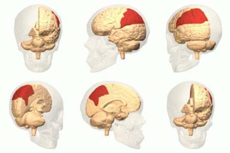
The parietal lobe is one of the four major lobes of the cerebral cortex in the brain of mammals. The parietal lobe is positioned above the temporal lobe and behind the frontal lobe and central sulcus. The parietal lobe integrates sensory information among various modalities, including spatial sense and navigation (proprioception), the main sensory receptive area for the sense of touch in the somatosensory cortex which is just posterior to the central sulcus in the postcentral gyrus, and the dorsal stream of the visual system. The major sensory inputs from the skin (touch, temperature, and pain receptors), relay through the thalamus to the parietal lobe. Several areas of the parietal lobe are important in language processing. The somatosensory cortex can be illustrated as a distorted figure – the cortical homunculus (Latin: "little man") in which the body parts are rendered according to how much of the somatosensory cortex is devoted to them. The superior parietal lobule an ... [...More Info...] [...Related Items...] OR: [Wikipedia] [Google] [Baidu] |
Temporal Lobe
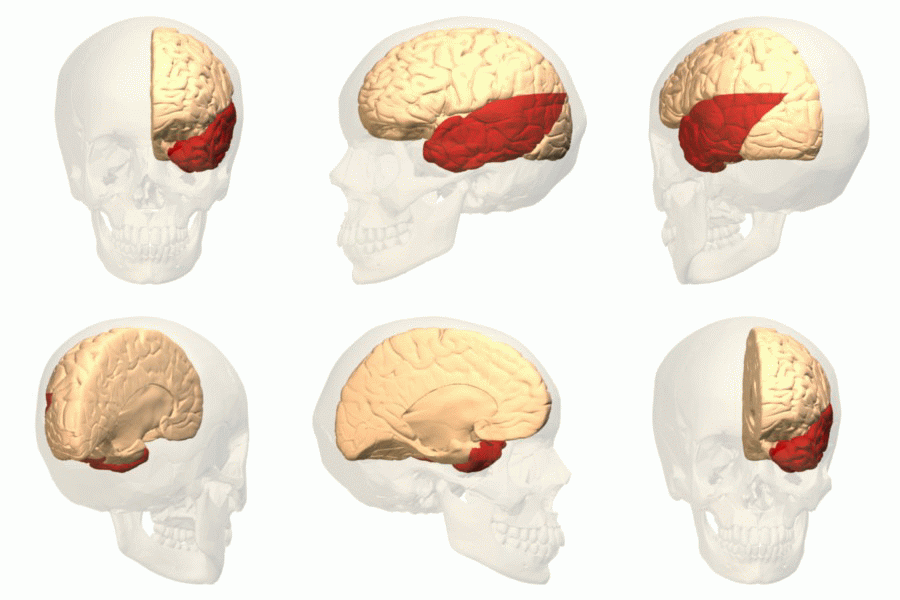
The temporal lobe is one of the four major lobes of the cerebral cortex in the brain of mammals. The temporal lobe is located beneath the lateral fissure on both cerebral hemispheres of the mammalian brain. The temporal lobe is involved in processing sensory input into derived meanings for the appropriate retention of visual memory, language comprehension, and emotion association. ''Temporal'' refers to the head's temples. Structure The temporal lobe consists of structures that are vital for declarative or long-term memory. Declarative (denotative) or explicit memory is conscious memory divided into semantic memory (facts) and episodic memory (events). Medial temporal lobe structures that are critical for long-term memory include the hippocampus, along with the surrounding hippocampal region consisting of the perirhinal, parahippocampal, and entorhinal neocortical regions. The hippocampus is critical for memory formation, and the surrounding medial temporal cortex is curre ... [...More Info...] [...Related Items...] OR: [Wikipedia] [Google] [Baidu] |
Lingual Gyrus
The lingual gyrus, also known as the ''medial'' occipitotemporal gyrus, is a brain structure that is linked to processing vision, especially related to letters. It is thought to also play a role in analysis of logical conditions (i.e., logical order of events) and encoding visual memories. It is named after its shape, which is somewhat similar to a tongue. Contrary to the name, the region has little to do with speech. It is believed that a hypermetabolism of the lingual gyrus is associated with visual snow. Location The lingual gyrus of the occipital lobe lies between the calcarine sulcus and the posterior part of the collateral sulcus; behind, it reaches the occipital pole; in front, it is continued on to the tentorial surface of the temporal lobe, and joins the parahippocampal gyrus. Function Role in vision This region is believed to play an important role in vision and dreaming. Visual memory dysfunction and visuo- limbic disconnection have been shown in cases where the l ... [...More Info...] [...Related Items...] OR: [Wikipedia] [Google] [Baidu] |
Cerebrum
The cerebrum, telencephalon or endbrain is the largest part of the brain containing the cerebral cortex (of the two cerebral hemispheres), as well as several subcortical structures, including the hippocampus, basal ganglia, and olfactory bulb. In the human brain, the cerebrum is the uppermost region of the central nervous system. The cerebrum develops prenatally from the forebrain (prosencephalon). In mammals, the dorsal telencephalon, or pallium, develops into the cerebral cortex, and the ventral telencephalon, or subpallium, becomes the basal ganglia. The cerebrum is also divided into approximately symmetric left and right cerebral hemispheres. With the assistance of the cerebellum, the cerebrum controls all voluntary actions in the human body. Structure The cerebrum is the largest part of the brain. Depending upon the position of the animal it lies either in front or on top of the brainstem. In humans, the cerebrum is the largest and best-developed of the five majo ... [...More Info...] [...Related Items...] OR: [Wikipedia] [Google] [Baidu] |


