|
Nitrogen Washout
Nitrogen washout (or Fowler's method) is a test for measuring anatomic dead space in the lung during a respiratory cycle, as well as some parameters related to the closure of airways. Procedure A nitrogen washout can be performed with a single nitrogen breath, or multiple ones. Both tests use similar tools, both can estimate functional residual capacity and the degree of nonuniformity of gas distribution in the lungs, but the multiple-breath test more accurately measures absolute lung volumes. The following describes a single-breath nitrogen test: A subject takes a breath of 100% oxygen and exhales through a one-way valve measuring nitrogen content and volume. A plot of the nitrogen concentration (as a % of total gas) vs. expired volume is obtained by increasing the nitrogen concentration from zero to the percentage of nitrogen in the alveoli Alveolus (; pl. alveoli, adj. alveolar) is a general anatomical term for a concave cavity or pit. Uses in anatomy and zoology * Pulmonar ... [...More Info...] [...Related Items...] OR: [Wikipedia] [Google] [Baidu] |
Dead Space (physiology)
Dead space is the volume of air that is inhaled that does not take part in the gas exchange, because it either remains in the conducting airways or reaches alveoli that are not perfused or poorly perfused. It means that not all the air in each breath Breathing (or ventilation) is the process of moving air into and from the lungs to facilitate gas exchange with the internal environment, mostly to flush out carbon dioxide and bring in oxygen. All aerobic creatures need oxygen for cell ... is available for the exchange of oxygen and carbon dioxide. Mammals breathe in and out of their lungs, wasting that part of the inhalation which remains in the conducting airways where no gas exchange can occur. Components ''Total dead space'' (also known as physiological dead space) is the sum of the anatomical dead space and the alveolar dead space. Benefits do accrue to a seemingly wasteful design for ventilation that includes dead space. #Carbon dioxide is retained, making a Aci ... [...More Info...] [...Related Items...] OR: [Wikipedia] [Google] [Baidu] |
Respiratory System
The respiratory system (also respiratory apparatus, ventilatory system) is a biological system consisting of specific organs and structures used for gas exchange in animals and plants. The anatomy and physiology that make this happen varies greatly, depending on the size of the organism, the environment in which it lives and its evolutionary history. In land animals the respiratory surface is internalized as linings of the lungs. Gas exchange in the lungs occurs in millions of small air sacs; in mammals and reptiles these are called alveoli, and in birds they are known as atria. These microscopic air sacs have a very rich blood supply, thus bringing the air into close contact with the blood. These air sacs communicate with the external environment via a system of airways, or hollow tubes, of which the largest is the trachea, which branches in the middle of the chest into the two main bronchi. These enter the lungs where they branch into progressively narrower secondary ... [...More Info...] [...Related Items...] OR: [Wikipedia] [Google] [Baidu] |
Functional Residual Capacity
Functional residual capacity (FRC) is the volume of air present in the lungs at the end of passive expiration. At FRC, the opposing elastic recoil forces of the lungs and chest wall are in equilibrium and there is no exertion by the diaphragm or other respiratory muscles. FRC is the sum of expiratory reserve volume (ERV) and residual volume (RV) and measures approximately 2500 mL in a 70 kg, average-sized male (or approximately 30ml/kg). It cannot be estimated through spirometry, since it includes the residual volume. In order to measure RV precisely, one would need to perform a test such as nitrogen washout, helium dilution or body plethysmography. A lowered or elevated FRC is often an indication of some form of respiratory disease. For instance, in emphysema, FRC is increased, because the lungs are more compliant and the equilibrium between the inward recoil of the lungs and outward recoil of the chest wall is disturbed. As such, patients with emphysema often have not ... [...More Info...] [...Related Items...] OR: [Wikipedia] [Google] [Baidu] |
Lung Volumes
Lung volumes and lung capacities refer to the volume of air in the lungs at different phases of the respiratory cycle. The average total lung capacity of an adult human male is about 6 litres of air. Tidal breathing is normal, resting breathing; the tidal volume is the volume of air that is inhaled or exhaled in only a single such breath. The average human respiratory rate is 30–60 breaths per minute at birth, decreasing to 12–20 breaths per minute in adults. Factors affecting volumes Several factors affect lung volumes; some can be controlled, and some cannot be controlled. Lung volumes vary with different people as follows: A person who is born and lives at sea level will develop a slightly smaller lung capacity than a person who spends their life at a high altitude. This is because the partial pressure of oxygen is lower at higher altitude which, as a result means that oxygen less readily diffuses into the bloodstream. In response to higher altitude, the body's di ... [...More Info...] [...Related Items...] OR: [Wikipedia] [Google] [Baidu] |
Pulmonary Alveolus
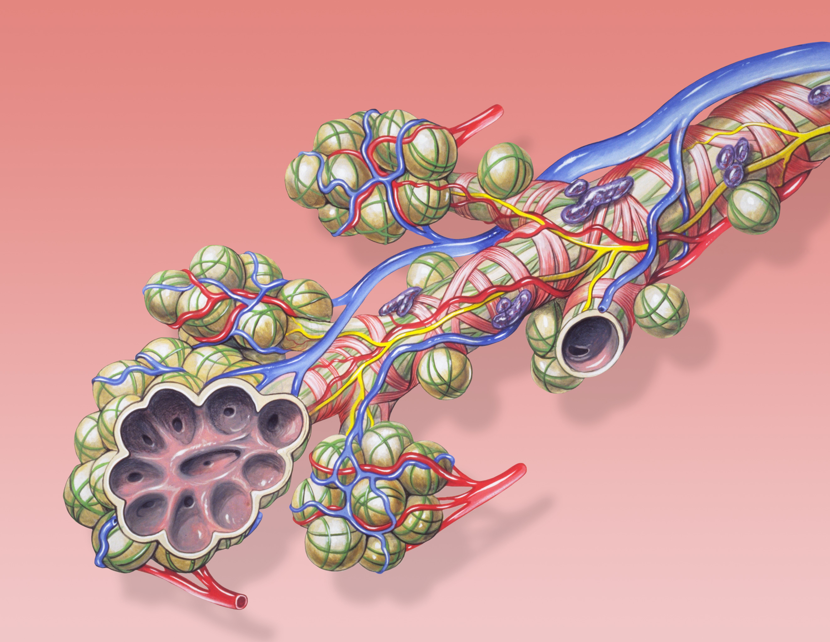
A pulmonary alveolus (plural: alveoli, from Latin ''alveolus'', "little cavity"), also known as an air sac or air space, is one of millions of hollow, distensible cup-shaped cavities in the lungs where oxygen is exchanged for carbon dioxide. Alveoli make up the functional tissue of the mammalian lungs known as the lung parenchyma, which takes up 90 percent of the total lung volume. Alveoli are first located in the respiratory bronchioles that mark the beginning of the respiratory zone. They are located sparsely in these bronchioles, line the walls of the alveolar ducts, and are more numerous in the blind-ended alveolar sacs. The acini are the basic units of respiration, with gas exchange taking place in all the alveoli present. The alveolar membrane is the gas exchange surface, surrounded by a network of capillaries. Across the membrane oxygen is diffused into the capillaries and carbon dioxide released from the capillaries into the alveoli to be breathed out. Alveoli are ... [...More Info...] [...Related Items...] OR: [Wikipedia] [Google] [Baidu] |
Total Lung Capacity
Lung volumes and lung capacities refer to the volume of air in the lungs at different phases of the respiratory cycle. The average total lung capacity of an adult human male is about 6 litres of air. Tidal breathing is normal, resting breathing; the tidal volume is the volume of air that is inhaled or exhaled in only a single such breath. The average human respiratory rate is 30–60 breaths per minute at birth, decreasing to 12–20 breaths per minute in adults. Factors affecting volumes Several factors affect lung volumes; some can be controlled, and some cannot be controlled. Lung volumes vary with different people as follows: A person who is born and lives at sea level will develop a slightly smaller lung capacity than a person who spends their life at a high altitude. This is because the partial pressure of oxygen is lower at higher altitude which, as a result means that oxygen less readily diffuses into the bloodstream. In response to higher altitude, the body's diff ... [...More Info...] [...Related Items...] OR: [Wikipedia] [Google] [Baidu] |
Vital Capacity
Vital capacity (VC) is the maximum amount of air a person can inhale after a maximum exhalation. It is equal to the sum of inspiratory reserve volume, tidal volume, and expiratory reserve volume. It is approximately equal to Forced Vital Capacity (FVC). A person's vital capacity can be measured by a wet or regular spirometer. In combination with other physiological measurements, the vital capacity can help make a diagnosis of underlying lung disease. Furthermore, the vital capacity is used to determine the severity of respiratory muscle involvement in neuromuscular disease, and can guide treatment decisions in Guillain–Barré syndrome and myasthenic crisis. A normal adult has a vital capacity between 3 and 5 litres. A human's vital capacity depends on age, sex, height, mass, and possibly ethnicity. However, the dependence on ethnicity is poorly understood or defined, as it was first established by studying black slaves in the 19th century and may be the result of conflation ... [...More Info...] [...Related Items...] OR: [Wikipedia] [Google] [Baidu] |
Medical Tests
A medical test is a medical procedure performed to detect, diagnose, or monitor diseases, disease processes, susceptibility, or to determine a course of treatment. Medical tests such as, physical and visual exams, diagnostic imaging, genetic testing, chemical and cellular analysis, relating to clinical chemistry and molecular diagnostics, are typically performed in a medical setting. Types of tests By purpose Medical tests can be classified by their purposes, the most common of which are diagnosis, screening and evaluation. Diagnostic A diagnostic test is a procedure performed to confirm or determine the presence of disease in an individual suspected of having a disease, usually following the report of symptoms, or based on other medical test results. This includes posthumous diagnosis. Examples of such tests are: * Using nuclear medicine to examine a patient suspected of having a lymphoma. * Measuring the blood sugar in a person suspected of having diabetes mellitus afte ... [...More Info...] [...Related Items...] OR: [Wikipedia] [Google] [Baidu] |
Pulmonary Function Testing
Pulmonary function testing (PFT) is a complete evaluation of the respiratory system including patient history, physical examinations, and tests of pulmonary function. The primary purpose of pulmonary function testing is to identify the severity of pulmonary impairment. Pulmonary function testing has diagnostic and therapeutic roles and helps clinicians answer some general questions about patients with lung disease. PFTs are normally performed by a pulmonary function technician, respiratory therapist, respiratory physiologist, physiotherapist, pulmonologist, or general practitioner. Indications Pulmonary function testing is a diagnostic and management tool used for a variety of reasons, such as: * Diagnose lung disease. * Monitor the effect of chronic diseases like asthma, chronic obstructive lung disease, or cystic fibrosis. * Detect early changes in lung function. * Identify narrowing in the airways. * Evaluate airway bronchodilator reactivity. * Show if environmental fac ... [...More Info...] [...Related Items...] OR: [Wikipedia] [Google] [Baidu] |


