|
Nerve Plexus
A nerve plexus is a plexus (branching network) of intersecting nerves. A nerve plexus is composed of afferent and efferent fibers that arise from the merging of the anterior rami of spinal nerves and blood vessels. There are five spinal nerve plexuses, except in the thoracic region, as well as other forms of autonomic plexuses, many of which are a part of the enteric nervous system. The nerves that arise from the plexuses have both sensory and motor functions. These functions include muscle contraction, the maintenance of body coordination and control, and the reaction to sensations such as heat, cold, pain, and pressure. There are several plexuses in the body, including: *Spinal Plexuses **Cervical plexus - serves the head, neck and shoulders **Brachial plexus - serves the chest, shoulders, arms and hands ** Lumbosacral plexus *** Lumbar plexus - serves the back, abdomen, groin, thighs, knees, and calves ****Subsartorial plexus - below the sartorius muscle of thigh *** Sacral ple ... [...More Info...] [...Related Items...] OR: [Wikipedia] [Google] [Baidu] |
Plexus
In neuroanatomy, a plexus (from the Latin term for "braid") is a branching network of vessels or nerves. The vessels may be blood vessels (veins, capillaries) or lymphatic vessels. The nerves are typically axons outside the central nervous system. The standard plural form in English is plexuses. Alternatively, the Latin plural plexūs may be used. Types Nerve plexuses The four primary nerve plexuses are the cervical plexus, brachial plexus, lumbar plexus, and the sacral plexus. Cardiac plexus Celiac plexus Renal plexus Venous plexus Choroid plexus The choroid plexus is a part of the central nervous system in the brain and consists of capillaries, brain ventricles, and ependymal cells. Invertebrates The plexus is the characteristic form of nervous system in the coelenterates and persists with modifications in the flatworms. The nerves of the radially symmetric echinoderm An echinoderm () is any member of the phylum Echinodermata (). The adults are recognis ... [...More Info...] [...Related Items...] OR: [Wikipedia] [Google] [Baidu] |
Cardiac Plexus
The cardiac plexus is a plexus of nerves situated at the base of the heart that innervates the heart. Structure The cardiac plexus is divided into a superficial part, which lies in the concavity of the aortic arch, and a deep part, between the aortic arch and the trachea. The two parts are, however, closely connected. The sympathetic component of the cardiac plexus comes from cardiac nerves, which originate from the sympathetic trunk. The parasympathetic component of the cardiac plexus originates from the cardiac branches of the vagus nerve. Superficial part The superficial part of the cardiac plexus lies beneath the arch of the aorta, in front of the right pulmonary artery. It is formed by the superior cervical cardiac branch of the left sympathetic trunk and the inferior cardiac branch of the left vagus nerve. A small ganglion, the ''cardiac ganglion of Wrisberg'', is occasionally found connected with these nerves at their point of junction. This ganglion, when present, is si ... [...More Info...] [...Related Items...] OR: [Wikipedia] [Google] [Baidu] |
Iliohypogastric Nerve
The iliohypogastric nerve is a nerve that originates from the lumbar plexus that supplies sensation to skin over the lateral gluteal and hypogastric regions and motor to the internal oblique muscles and transverse abdominal muscles. Structure The iliohypogastric nerve originates from the superior branch of the anterior ramus of spinal nerve L1. It also receives fibers from T12 via the subcostal nerve. The branch below it is the ilioinguinal nerve. It emerges from the upper lateral border of the psoas major. It then crosses in front of the quadratus lumborum muscle to an area superior to the iliac crest. It runs behind the kidneys. Just superior to the iliac crest, it pierces the posterior part of the transversus abdominis muscle and continues anteriorly in the abdominal wall between the transversus abdominis and internal oblique muscles. It divides into a lateral cutaneous branch and an anterior cutaneous branch between the transversus abdominis muscle and the internal obli ... [...More Info...] [...Related Items...] OR: [Wikipedia] [Google] [Baidu] |
Ulnar Nerve
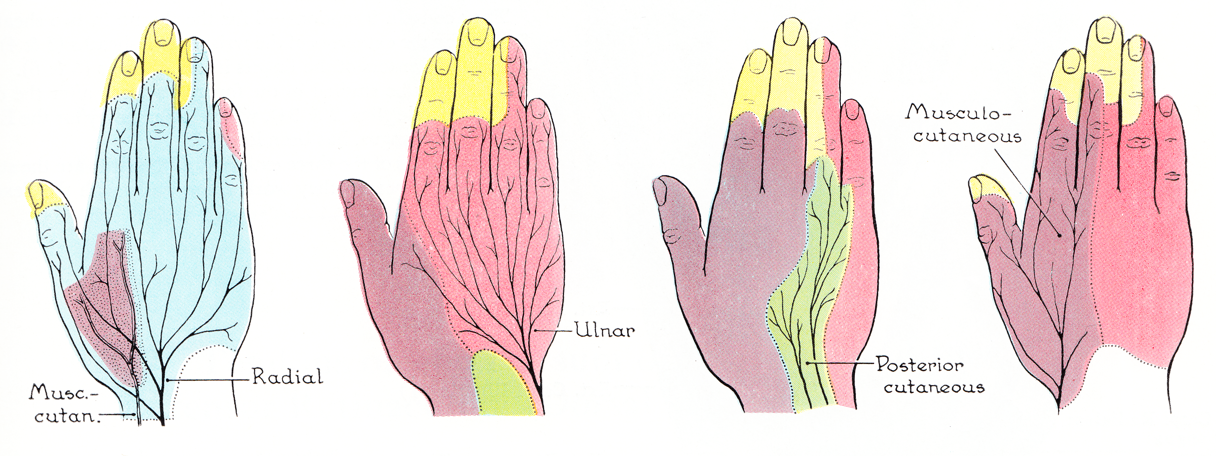
In human anatomy, the ulnar nerve is a nerve that runs near the ulna bone. The ulnar collateral ligament of elbow joint is in relation with the ulnar nerve. The nerve is the largest in the human body unprotected by muscle or bone, so injury is common. This nerve is directly connected to the little finger, and the adjacent half of the ring finger, innervating the palmar aspect of these fingers, including both front and back of the tips, perhaps as far back as the fingernail beds. This nerve can cause an electric shock-like sensation by striking the medial epicondyle of the humerus posteriorly, or inferiorly with the elbow flexed. The ulnar nerve is trapped between the bone and the overlying skin at this point. This is commonly referred to as bumping one's "funny bone". This name is thought to be a pun, based on the sound resemblance between the name of the bone of the upper arm, the humerus, and the word " humorous". Alternatively, according to the Oxford English Dictiona ... [...More Info...] [...Related Items...] OR: [Wikipedia] [Google] [Baidu] |
Median Nerve
The median nerve is a nerve in humans and other animals in the upper limb. It is one of the five main nerves originating from the brachial plexus. The median nerve originates from the lateral and medial cords of the brachial plexus, and has contributions from ventral roots of C5-C7 (lateral cord) and C8 and T1 (medial cord). The median nerve is the only nerve that passes through the carpal tunnel. Carpal tunnel syndrome is the disability that results from the median nerve being pressed in the carpal tunnel. Structure The median nerve arises from the branches from lateral and medial cords of the brachial plexus, courses through the anterior part of arm, forearm, and hand, and terminates by supplying the muscles of the hand. Arm After receiving inputs from both the lateral and medial cords of the brachial plexus, the median nerve enters the arm from the axilla at the inferior margin of the teres major muscle. It then passes vertically down and courses lateral to the brachial a ... [...More Info...] [...Related Items...] OR: [Wikipedia] [Google] [Baidu] |
Radial Nerve
The radial nerve is a nerve in the human body that supplies the posterior portion of the upper limb. It innervates the medial and lateral heads of the triceps brachii muscle of the arm, as well as all 12 muscles in the posterior osteofascial compartment of the forearm and the associated joints and overlying skin. It originates from the brachial plexus, carrying fibers from the ventral roots of spinal nerves C5, C6, C7, C8 & T1. The radial nerve and its branches provide motor innervation to the dorsal arm muscles (the triceps brachii and the anconeus) and the extrinsic extensors of the wrists and hands; it also provides cutaneous sensory innervation to most of the back of the hand, except for the back of the little finger and adjacent half of the ring finger (which are innervated by the ulnar nerve). The radial nerve divides into a deep branch, which becomes the posterior interosseous nerve, and a superficial branch, which goes on to innervate the dorsum (back) of the hand ... [...More Info...] [...Related Items...] OR: [Wikipedia] [Google] [Baidu] |
Axillary Nerve
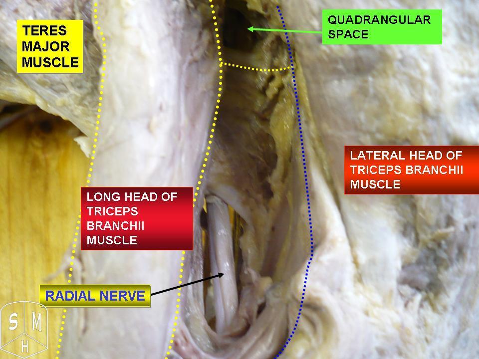
The axillary nerve or the circumflex nerve is a nerve of the human body, that originates from the brachial plexus ( upper trunk, posterior division, posterior cord) at the level of the axilla (armpit) and carries nerve fibers from C5 and C6. The axillary nerve travels through the quadrangular space with the posterior circumflex humeral artery and vein to innervate the deltoid and teres minor. Structure The nerve lies at first behind the axillary artery, and in front of the subscapularis, and passes downward to the lower border of that muscle. It then winds from anterior to posterior around the neck of the humerus, in company with the posterior humeral circumflex artery, through the quadrangular space (bounded above by the teres minor, below by the teres major, medially by the long head of the triceps brachii, and laterally by the surgical neck of the humerus), and divides into an anterior, a posterior, and a collateral branch to the long head of the triceps brachii bra ... [...More Info...] [...Related Items...] OR: [Wikipedia] [Google] [Baidu] |
Musculocutaneous Nerve
The musculocutaneous nerve arises from the lateral cord of the brachial plexus, opposite the lower border of the pectoralis major, its fibers being derived from C5, C6 and C7. Structure The musculocutaneous nerve arises from the lateral cord of the brachial plexus, courses through the anterior part of the arm, and terminates at 2 cm above elbow as the lateral cutaneous nerve of the forearm. Musculocutaneous nerve arises from the lateral cord of the brachial plexus with root value of C5 to C7 of the spinal cord. It follows the course of the third part of the axillary artery (part of the axillary artery distal to the pectoralis minor) laterally and enters the frontal aspect of the arm where it penetrates the coracobrachialis muscle. It then passes downwards and laterally between the biceps brachii (above) and the brachialis muscles (below), to the lateral side of the arm; at 2 cm above the elbow it pierces the deep fascia lateral to the tendon of the biceps brachii ... [...More Info...] [...Related Items...] OR: [Wikipedia] [Google] [Baidu] |
Phrenic Nerve
The phrenic nerve is a mixed motor/sensory nerve which originates from the C3-C5 spinal nerves in the neck. The nerve is important for breathing because it provides exclusive motor control of the diaphragm, the primary muscle of respiration. In humans, the right and left phrenic nerves are primarily supplied by the C4 spinal nerve, but there is also contribution from the C3 and C5 spinal nerves. From its origin in the neck, the nerve travels downward into the chest to pass between the heart and lungs towards the diaphragm. In addition to motor fibers, the phrenic nerve contains sensory fibers, which receive input from the central tendon of the diaphragm and the mediastinal pleura, as well as some sympathetic nerve fibers. Although the nerve receives contributions from nerves roots of the cervical plexus and the brachial plexus, it is usually considered separate from either plexus. The name of the nerve comes from Ancient Greek ''phren'' 'diaphragm'. Structure The phreni ... [...More Info...] [...Related Items...] OR: [Wikipedia] [Google] [Baidu] |
Supraclavicular Nerves
The supraclavicular nerves (descending branches) arise from the third and fourth cervical nerves. They emerge beneath the posterior border of the sternocleidomastoideus (sternocleidomastoid muscle), and descend in the posterior triangle of the neck beneath the platysma muscle and the deep cervical fascia. Together, they innervate skin over the shoulder. The supraclavicular nerve can be blocked during shoulder surgery. Branches The supraclavicular nerves arise from C3 and C4 spinal nerve roots. Near the clavicle, the supraclavicular nerves perforate the fascia and the platysma muscle to become cutaneous. They are arranged, according to their position, into three groups—anterior, middle, and posterior. Medial supraclavicular nerve The medial supraclavicular nerves or ''anterior supraclavicular nerves'' (nn. supraclaviculares anteriores; suprasternal nerves) cross obliquely over the external jugular vein and the clavicular and sternal heads of the sternocleidomastoideus, and supp ... [...More Info...] [...Related Items...] OR: [Wikipedia] [Google] [Baidu] |
Ansa Cervicalis
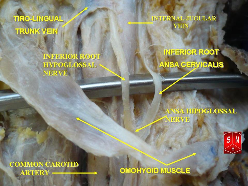
The ansa cervicalis (or ansa hypoglossi in older literature) is a loop of nerves that are part of the cervical plexus. It lies superficial to the internal jugular vein in the carotid triangle. Its name means "handle of the neck" in Latin. Branches from the ansa cervicalis innervate most of the infrahyoid muscles, including the sternothyroid muscle, sternohyoid muscle and the omohyoid muscle. Note that the thyrohyoid muscle, which is also an infrahyoid muscle and the geniohyoid muscle which is a suprahyoid muscle are innervated by cervical spinal nerve 1 via the hypoglossal nerve. Roots Two roots make up the ansa cervicalis, a superior root, and an inferior root. The superior root of the ansa cervicalis is formed from cervical spinal nerve 1 of the cervical plexus. These nerve fibers travel in the hypoglossal nerve before separating in the carotid triangle to form the superior root. The superior root goes around the occipital artery and then descends on the carotid s ... [...More Info...] [...Related Items...] OR: [Wikipedia] [Google] [Baidu] |
Transverse Cervical Nerve
The transverse cervical nerve (superficial cervical or cutaneous cervical) arises from the second and third spinal nerves, turns around the posterior border of the sternocleidomastoideus about its middle, and, passing obliquely forward beneath the external jugular vein to the anterior border of the muscle, it perforates the deep cervical fascia, and divides beneath the Platysma into ascending and descending branches, which are distributed to the antero-lateral parts of the neck The neck is the part of the body on many vertebrates that connects the head with the torso. The neck supports the weight of the head and protects the nerves that carry sensory and motor information from the brain down to the rest of the body. In .... It provides cutaneous innervation to this area. During dissection, the sternocleidomastoid (SCM) is the landmark. The transverse cervical nerves will pass horizontally directly over the SCM from Erb's point. Additional images File:Gray784.png, Dermato ... [...More Info...] [...Related Items...] OR: [Wikipedia] [Google] [Baidu] |


