|
Nociceptin
Nociceptin/orphanin FQ (N/OFQ), a 17-amino acid neuropeptide, is the endogenous ligand for the nociceptin receptor (NOP, ORL-1). Nociceptin acts as a potent anti-analgesic, effectively counteracting the effect of pain-relievers; its activation is associated with brain functions such as pain sensation and fear learning. The gene coding for prepronociceptin is located on Ch8p21 in humans. Nociceptin is derived from the prepronociceptin protein, as are a further two peptides, nocistatin and NocII, both of which inhibit N/OFQ receptor function. Nociceptin is the first example of reverse pharmacology; the NOP receptor was discovered before the endogenous ligand which was discovered by two separate groups in 1995. Roles of nociceptin Since its discovery, nociceptin has been of great interest to researchers. Nociceptin is a peptide related to the kappa opioid receptor ligand dynorphin A, but it does not act at the classic opioid receptors (namely, mu, kappa, and delta opioid re ... [...More Info...] [...Related Items...] OR: [Wikipedia] [Google] [Baidu] [Amazon] |
Nociceptin Receptor
The nociceptin opioid peptide receptor (NOP), also known as the nociceptin/orphanin FQ (N/OFQ) receptor or kappa-type 3 opioid receptor, is a protein that in humans is encoded by the ''OPRL1'' (opioid receptor-like 1) gene. The nociceptin receptor is a member of the opioid subfamily of G protein-coupled receptors whose natural Ligand (biochemistry), ligand is the 17 amino acid neuropeptide known as nociceptin, nociceptin (N/OFQ). This receptor is involved in the regulation of numerous brain activities, particularly instinctive and emotional behaviors. Antagonists targeting NOP are under investigation for their role as treatments for depression and Parkinson's disease, whereas NOP agonists have been shown to act as powerful, non-addictive painkillers in non-human primates. Although NOP shares high sequence identity (~60%) with the ‘classical’ opioid receptors Μ-opioid receptor, μ-OP (MOP), Κ-opioid receptor, κ-OP (KOP), and Δ-opioid receptor, δ-OP (DOP), it possesses litt ... [...More Info...] [...Related Items...] OR: [Wikipedia] [Google] [Baidu] [Amazon] |
Κ-opioid Receptor
The κ-opioid receptor or kappa opioid receptor, abbreviated KOR or KOP for its ligand ketazocine, is a G protein-coupled receptor that in humans is encoded by the ''OPRK1'' gene. The KOR is coupled to the G protein Gi/G0 and is one of four related receptors that bind opioid-like compounds in the brain and are responsible for mediating the effects of these compounds. These effects include altering nociception, consciousness, motor control, and mood. Dysregulation of this receptor system has been implicated in alcohol and drug addiction. The KOR is a type of opioid receptor that binds the opioid peptide dynorphin as the primary endogenous ligand (substrate naturally occurring in the body). In addition to dynorphin, a variety of natural alkaloids, terpenes and synthetic ligands bind to the receptor. The KOR may provide a natural addiction control mechanism, and therefore, drugs that target this receptor may have therapeutic potential in the treatment of addiction . There is ... [...More Info...] [...Related Items...] OR: [Wikipedia] [Google] [Baidu] [Amazon] |
Amino Acid
Amino acids are organic compounds that contain both amino and carboxylic acid functional groups. Although over 500 amino acids exist in nature, by far the most important are the 22 α-amino acids incorporated into proteins. Only these 22 appear in the genetic code of life. Amino acids can be classified according to the locations of the core structural functional groups ( alpha- , beta- , gamma- amino acids, etc.); other categories relate to polarity, ionization, and side-chain group type ( aliphatic, acyclic, aromatic, polar, etc.). In the form of proteins, amino-acid '' residues'' form the second-largest component (water being the largest) of human muscles and other tissues. Beyond their role as residues in proteins, amino acids participate in a number of processes such as neurotransmitter transport and biosynthesis. It is thought that they played a key role in enabling life on Earth and its emergence. Amino acids are formally named by the IUPAC- IUBMB Joint Commi ... [...More Info...] [...Related Items...] OR: [Wikipedia] [Google] [Baidu] [Amazon] |
Spinal Cord
The spinal cord is a long, thin, tubular structure made up of nervous tissue that extends from the medulla oblongata in the lower brainstem to the lumbar region of the vertebral column (backbone) of vertebrate animals. The center of the spinal cord is hollow and contains a structure called the central canal, which contains cerebrospinal fluid. The spinal cord is also covered by meninges and enclosed by the neural arches. Together, the brain and spinal cord make up the central nervous system. In humans, the spinal cord is a continuation of the brainstem and anatomically begins at the occipital bone, passing out of the foramen magnum and then enters the spinal canal at the beginning of the cervical vertebrae. The spinal cord extends down to between the first and second lumbar vertebrae, where it tapers to become the cauda equina. The enclosing bony vertebral column protects the relatively shorter spinal cord. It is around long in adult men and around long in adult women. The diam ... [...More Info...] [...Related Items...] OR: [Wikipedia] [Google] [Baidu] [Amazon] |
Nociception
In physiology, nociception , also nocioception; ) is the Somatosensory system, sensory nervous system's process of encoding Noxious stimulus, noxious stimuli. It deals with a series of events and processes required for an organism to receive a painful stimulus, convert it to a molecular signal, and recognize and characterize the signal to trigger an appropriate defensive response. In nociception, intense chemical (e.g., capsaicin present in chili pepper or cayenne pepper), mechanical (e.g., cutting, crushing), or thermal (heat and cold) stimulation of sensory neurons called nociceptors produces a signal that travels along a chain of nerve fibers to the brain. Nociception triggers a variety of physiological and behavioral responses to protect the organism against an aggression, and usually results in a subjective experience, or perception, of pain in Sentience, sentient beings. Detection of noxious stimuli Potentially damaging mechanical, thermal, and chemical stimuli are detecte ... [...More Info...] [...Related Items...] OR: [Wikipedia] [Google] [Baidu] [Amazon] |
Locus Coeruleus
The locus coeruleus () (LC), also spelled locus caeruleus or locus ceruleus, is a nucleus in the pons of the brainstem involved with physiological responses to stress and panic. It is a part of the reticular activating system in the reticular formation. The locus coeruleus, which in Latin means "blue spot", is the principal site for brain synthesis of norepinephrine (noradrenaline). The locus coeruleus and the areas of the body affected by the norepinephrine it produces are described collectively as the locus coeruleus-noradrenergic system or LC-NA system. Norepinephrine may also be released directly into the blood from the adrenal medulla. Anatomy The locus coeruleus (LC) is located in the posterior area of the rostral pons in the lateral floor of the fourth ventricle. It is composed of mostly medium-size neurons. Melanin granules inside the neurons contribute to its blue colour. Thus, it is also known as the ''blue nucleus'', or the ''nucleus pigmentosus pontis'' (hea ... [...More Info...] [...Related Items...] OR: [Wikipedia] [Google] [Baidu] [Amazon] |
Raphe Nuclei
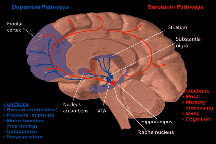
The raphe nuclei (, "seam") are a moderate-size cluster of nuclei found in the brain stem. They have 5-HT1 receptors which are coupled with Gi/Go-protein-inhibiting adenyl cyclase. They function as autoreceptors in the brain and decrease the release of serotonin. The anxiolytic drug Buspirone acts as partial agonist against these receptors. Selective serotonin reuptake inhibitor (SSRI) antidepressants are believed to act in these nuclei, as well as at their targets. Anatomy The raphe nuclei are traditionally considered to be the medial portion of the reticular formation, and appear as a ridge of cells in the center and most medial portion of the brain stem. In order from caudal to rostral, the raphe nuclei are known as the '' nucleus raphe obscurus'', the '' nucleus raphe pallidus'', the '' nucleus raphe magnus'', the '' nucleus raphe pontis'', the '' median raphe nucleus'', ''dorsal raphe nucleus'', ''caudal linear nucleus''. In the first systematic examination of the raph ... [...More Info...] [...Related Items...] OR: [Wikipedia] [Google] [Baidu] [Amazon] |
Substantia Nigra
The substantia nigra (SN) is a basal ganglia structure located in the midbrain that plays an important role in reward and movement. ''Substantia nigra'' is Latin for "black substance", reflecting the fact that parts of the substantia nigra appear darker than neighboring areas due to high levels of neuromelanin in dopaminergic neurons. Parkinson's disease is characterized by the loss of dopaminergic neurons in the substantia nigra pars compacta. Although the substantia nigra appears as a continuous band in brain sections, anatomical studies have found that it actually consists of two parts with very different connections and functions: the pars compacta (SNpc) and the pars reticulata (SNpr). The pars compacta serves mainly as a projection to the basal ganglia circuit, supplying the striatum with dopamine. The pars reticulata conveys signals from the basal ganglia to numerous other brain structures. Structure The substantia nigra, along with four other nuclei, is ... [...More Info...] [...Related Items...] OR: [Wikipedia] [Google] [Baidu] [Amazon] |
Interpeduncular Fossa
The interpeduncular fossa is a deep depression of the ventral surface of the midbrain between the two cerebral crus, cerebal crura. It has been found in humans and macaques, but not in rats or mice, showing that this is a relatively new evolutionary region. Structure The interpeduncular fossa is a somewhat rhomboid-shaped area of the base of the brain. Features The lateral wall of the interpeduncular fossa bears a groove - the oculomotor sulcus - from which rootlets of the oculomotor nerve emerge from the substance of the brainstem and aggregate into a single fascicle. Anatomical relations The ventral tegmental area lies at the depth of the interpeduncular fossa. Boundaries The interpeduncular fossa is in front by the optic chiasma, behind by the antero-superior surface of the pons, antero-laterally by the converging optic tracts, and postero-laterally by the diverging cerebral peduncles. The floor of interpeduncular fossa, from behind forward, are the posterior perfor ... [...More Info...] [...Related Items...] OR: [Wikipedia] [Google] [Baidu] [Amazon] |
Pons
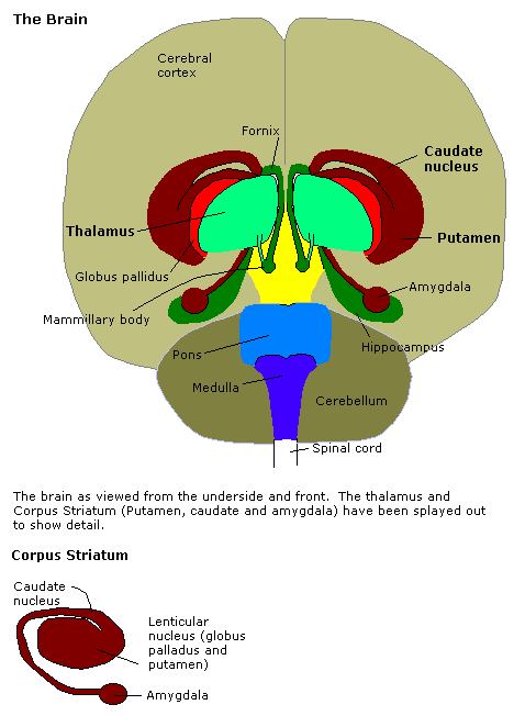
The pons (from Latin , "bridge") is part of the brainstem that in humans and other mammals, lies inferior to the midbrain, superior to the medulla oblongata and anterior to the cerebellum. The pons is also called the pons Varolii ("bridge of Varolius"), after the Italian anatomist and surgeon Costanzo Varolio (1543–75). This region of the brainstem includes neural pathways and tracts that conduct signals from the brain down to the cerebellum and medulla, and tracts that carry the sensory signals up into the thalamus. Structure The pons in humans measures about in length. It is the part of the brainstem situated between the midbrain and the medulla oblongata. The horizontal ''medullopontine sulcus'' demarcates the boundary between the pons and medulla oblongata on the ventral aspect of the brainstem, and the roots of cranial nerves VI/VII/VIII emerge from the brainstem along this groove. The junction of pons, medulla oblongata, and cerebellum forms the cerebellopontine ... [...More Info...] [...Related Items...] OR: [Wikipedia] [Google] [Baidu] [Amazon] |
Periaqueductal Gray
The periaqueductal gray (PAG), also known as the central gray, is a brain region that plays a critical role in autonomic function, motivated behavior and behavioural responses to threatening stimuli. PAG is also the primary control center for descending pain modulation. It has enkephalin-producing cells that suppress pain. The periaqueductal gray is the gray matter located around the cerebral aqueduct within the tegmentum of the midbrain. It projects to the nucleus raphe magnus, and also contains descending autonomic tracts. The ascending pain and temperature fibers of the spinothalamic tract send information to the PAG via the spinomesencephalic pathway (so-named because the fibers originate in the spine and terminate in the PAG, in the mesencephalon or midbrain). This region has been used as the target for brain-stimulating implants in patients with chronic pain. Role in analgesia Stimulation of the periaqueductal gray matter of the midbrain activates enkephalin-r ... [...More Info...] [...Related Items...] OR: [Wikipedia] [Google] [Baidu] [Amazon] |
Amygdala
The amygdala (; : amygdalae or amygdalas; also '; Latin from Greek language, Greek, , ', 'almond', 'tonsil') is a paired nucleus (neuroanatomy), nuclear complex present in the Cerebral hemisphere, cerebral hemispheres of vertebrates. It is considered part of the limbic system. In Primate, primates, it is located lateral and medial, medially within the temporal lobes. It consists of many nuclei, each made up of further subnuclei. The subdivision most commonly made is into the Basolateral amygdala, basolateral, Central nucleus of the amygdala, central, cortical, and medial nuclei together with the intercalated cells of the amygdala, intercalated cell clusters. The amygdala has a primary role in the processing of memory, decision making, decision-making, and emotions, emotional responses (including fear, anxiety, and aggression). The amygdala was first identified and named by Karl Friedrich Burdach in 1822. Structure Thirteen Nucleus (neuroanatomy), nuclei have been identif ... [...More Info...] [...Related Items...] OR: [Wikipedia] [Google] [Baidu] [Amazon] |


