|
Levator Anguli Oris
The levator anguli oris (caninus) is a facial muscle of the mouth arising from the canine fossa, immediately below the infraorbital foramen. It elevates angle of mouth medially. Its fibers are inserted into the angle of the mouth, intermingling with those of the zygomaticus, triangularis, and orbicularis oris. Specifically, the levator anguli oris is innervated by the buccal branches of the facial nerve The buccal branches of the facial nerve (infraorbital branches), are of larger size than the rest of the branches, pass horizontally forward to be distributed below the orbit and around the mouth. Branches The ''superficial branches'' run beneath .... Additional images File:Sobo 1909 264.png File:Sobo 1909 263.png, Seen from the inside. References External links PTCentral Muscles of the head and neck {{muscle-stub ... [...More Info...] [...Related Items...] OR: [Wikipedia] [Google] [Baidu] |
Orbicularis Oris
In human anatomy, the orbicularis oris muscle is a complex of muscles in the lips that encircles the mouth. It is a sphincter, or circular muscle, but it is actually composed of four independent quadrants that interlace and give only an appearance of circularity.Saladin, "Anatomy & Physiology: The Unity of Form and Function". 5th edition. McGraw Hill. Page 330 It is also one of the muscles used in the playing of all brass instruments and some woodwind instruments. This muscle closes the mouth and puckers the lips when it contracts. Structure The orbicularis oris is not a simple sphincter muscle like the orbicularis oculi; it consists of numerous strata of muscular fibers surrounding the orifice of the mouth, but having different direction. It consists partly of fibers derived from the other facial muscles which are inserted into the lips, and partly of fibers proper to the lips. Of the former, a considerable number are derived from the buccinator and form the deeper stratum of ... [...More Info...] [...Related Items...] OR: [Wikipedia] [Google] [Baidu] |
Maxilla
The maxilla (plural: ''maxillae'' ) in vertebrates is the upper fixed (not fixed in Neopterygii) bone of the jaw formed from the fusion of two maxillary bones. In humans, the upper jaw includes the hard palate in the front of the mouth. The two maxillary bones are fused at the intermaxillary suture, forming the anterior nasal spine. This is similar to the mandible (lower jaw), which is also a fusion of two mandibular bones at the mandibular symphysis. The mandible is the movable part of the jaw. Structure In humans, the maxilla consists of: * The body of the maxilla * Four processes ** the zygomatic process ** the frontal process of maxilla ** the alveolar process ** the palatine process * three surfaces – anterior, posterior, medial * the Infraorbital foramen * the maxillary sinus * the incisive foramen Articulations Each maxilla articulates with nine bones: * two of the cranium: the frontal and ethmoid * seven of the face: the nasal, zygomatic, lacrimal ... [...More Info...] [...Related Items...] OR: [Wikipedia] [Google] [Baidu] |
Modiolus (face)
In facial anatomy, the modiolus is a chiasma of facial muscles held together by fibrous tissue, located lateral and slightly superior to each angle of the mouth. It is important in moving the mouth, facial expression and in dentistry. It is extremely important in relation to stability of lower denture, because of the strength and variability of movement of the area. It derives its motor nerve supply from the facial nerve, and its blood supply from labial branches of the facial artery. It is contributed to by the following muscles: orbicularis oris, buccinator, levator anguli oris, depressor anguli oris, zygomaticus major, risorius The risorius muscle is a muscle of facial expression. It arises from the fascia over the parotid gland, and inserts into the angle of the mouth. It is supplied by the facial nerve (CN VII). It may be absent or asymmetrical in some people. It retr ..., quadratus labii superioris, quadratus labii inferioris.Drake, Vogl, Mitchell. Gray's Anatomy ... [...More Info...] [...Related Items...] OR: [Wikipedia] [Google] [Baidu] |
Facial Artery
The facial artery (external maxillary artery in older texts) is a branch of the external carotid artery that supplies structures of the superficial face. Structure The facial artery arises in the carotid triangle from the external carotid artery, a little above the lingual artery and, sheltered by the ramus of the mandible. It passes obliquely up beneath the digastric and stylohyoid muscles, over which it arches to enter a groove on the posterior surface of the submandibular gland. It then curves upward over the body of the mandible at the antero-inferior angle of the masseter; passes forward and upward across the cheek to the angle of the mouth, then ascends along the side of the nose, and ends at the medial commissure of the eye, under the name of the angular artery. The facial artery is remarkably tortuous. This is to accommodate itself to neck movements such as those of the pharynx in deglutition; and facial movements such as those of the mandible, lips, and cheeks. ... [...More Info...] [...Related Items...] OR: [Wikipedia] [Google] [Baidu] |
Buccal Branches Of The Facial Nerve
The buccal branches of the facial nerve (infraorbital branches), are of larger size than the rest of the branches, pass horizontally forward to be distributed below the orbit and around the mouth. Branches The ''superficial branches'' run beneath the skin and above the superficial muscles of the face, which they supply: some are distributed to the procerus, joining at the medial angle of the orbit with the infratrochlear and nasociliary branches of the ophthalmic. The ''deep branches'' pass beneath the zygomaticus and the quadratus labii superioris, supplying them and forming an infraorbital plexus with the infraorbital branch of the maxillary nerve. These branches also supply the small muscles of the nose. The ''lower deep branches'' supply the buccinator and orbicularis oris, and join with filaments of the buccinator branch of the mandibular nerve. Muscles of facial expression The facial nerve innervates the muscles of facial expression. The buccal branch supplies these ... [...More Info...] [...Related Items...] OR: [Wikipedia] [Google] [Baidu] |
Smile
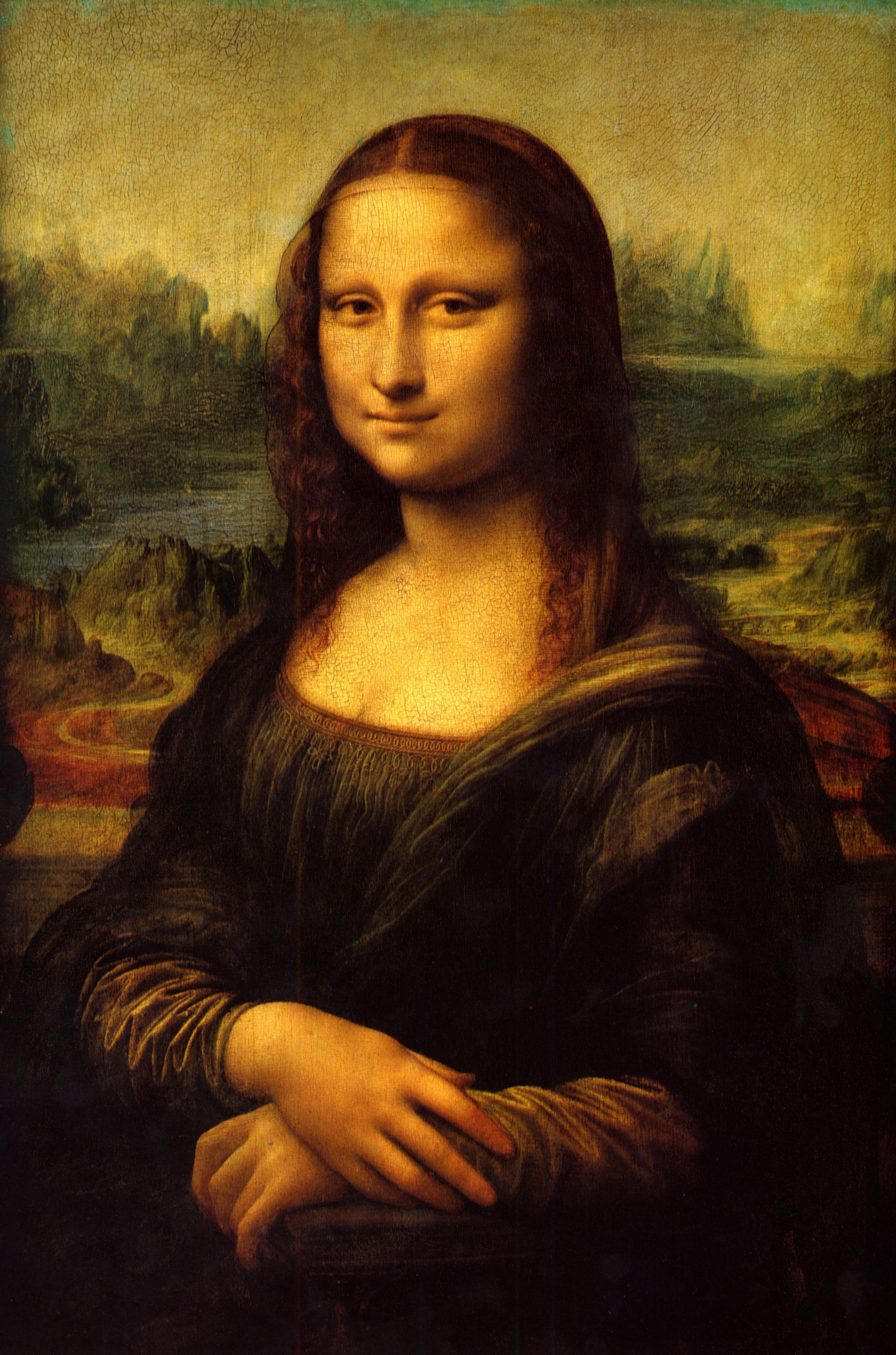
A smile is a facial expression formed primarily by flexing the muscles at the sides of the mouth. Some smiles include a contraction of the muscles at the corner of the eyes, an action known as a Duchenne smile. Among humans, a smile expresses delight, sociability, happiness, joy, or amusement. It is distinct from a similar but usually involuntary expression of anxiety known as a grimace. Although cross-cultural studies have shown that smiling is a means of communication throughout the world, there are large differences among different cultures, religions, and societies, with some using smiles to convey confusion or embarrassment. Evolutionary background Primatologist Signe Preuschoft traces the smile back over 30 million years of evolution to a "fear grin" stemming from monkeys and apes, who often used barely clenched teeth to portray to predators that they were harmless or to signal submission to more dominant group members. The smile may have evolved differently among s ... [...More Info...] [...Related Items...] OR: [Wikipedia] [Google] [Baidu] |
Elevation (kinesiology)
Motion, the process of movement, is described using specific anatomical terms. Motion includes movement of organs, joints, limbs, and specific sections of the body. The terminology used describes this motion according to its direction relative to the anatomical position of the body parts involved. Anatomists and others use a unified set of terms to describe most of the movements, although other, more specialized terms are necessary for describing unique movements such as those of the hands, feet, and eyes. In general, motion is classified according to the anatomical plane it occurs in. ''Flexion'' and ''extension'' are examples of ''angular'' motions, in which two axes of a joint are brought closer together or moved further apart. ''Rotational'' motion may occur at other joints, for example the shoulder, and are described as ''internal'' or ''external''. Other terms, such as ''elevation'' and ''depression'', describe movement above or below the horizontal plane. Many anatomic ... [...More Info...] [...Related Items...] OR: [Wikipedia] [Google] [Baidu] |
Mouth
In animal anatomy, the mouth, also known as the oral cavity, or in Latin cavum oris, is the opening through which many animals take in food and issue vocal sounds. It is also the cavity lying at the upper end of the alimentary canal, bounded on the outside by the lips and inside by the pharynx. In tetrapods, it contains the tongue and, except for some like birds, teeth. This cavity is also known as the buccal cavity, from the Latin ''bucca'' ("cheek"). Some animal phyla, including arthropods, molluscs and chordates, have a complete digestive system, with a mouth at one end and an anus at the other. Which end forms first in ontogeny is a criterion used to classify bilaterian animals into protostomes and deuterostomes. Development In the first multicellular animals, there was probably no mouth or gut and food particles were engulfed by the cells on the exterior surface by a process known as endocytosis. The particles became enclosed in vacuoles into which enzymes were secre ... [...More Info...] [...Related Items...] OR: [Wikipedia] [Google] [Baidu] |
Facial Muscles
The facial muscles are a group of striated skeletal muscles supplied by the facial nerve (cranial nerve VII) that, among other things, control facial expression. These muscles are also called mimetic muscles. They are only found in mammals, although they derive from neural crest cells found in all vertebrates. They are the only muscles that attach to the dermis. Structure The facial muscles are just under the skin ( subcutaneous) muscles that control facial expression. They generally originate from the surface of the skull bone (rarely the fascia), and insert on the skin of the face. When they contract, the skin moves. These muscles also cause wrinkles at right angles to the muscles’ action line. Nerve supply The facial muscles are supplied by the facial nerve (cranial nerve VII), with each nerve serving one side of the face. In contrast, the nearby masticatory muscles are supplied by the mandibular nerve, a branch of the trigeminal nerve (cranial nerve V). List of m ... [...More Info...] [...Related Items...] OR: [Wikipedia] [Google] [Baidu] |
Muscle
Skeletal muscles (commonly referred to as muscles) are organs of the vertebrate muscular system and typically are attached by tendons to bones of a skeleton. The muscle cells of skeletal muscles are much longer than in the other types of muscle tissue, and are often known as muscle fibers. The muscle tissue of a skeletal muscle is striated – having a striped appearance due to the arrangement of the sarcomeres. Skeletal muscles are voluntary muscles under the control of the somatic nervous system. The other types of muscle are cardiac muscle which is also striated and smooth muscle which is non-striated; both of these types of muscle tissue are classified as involuntary, or, under the control of the autonomic nervous system. A skeletal muscle contains multiple fascicles – bundles of muscle fibers. Each individual fiber, and each muscle is surrounded by a type of connective tissue layer of fascia. Muscle fibers are formed from the fusion of developmental myoblasts ... [...More Info...] [...Related Items...] OR: [Wikipedia] [Google] [Baidu] |
Canine Fossa
In the musculoskeletal anatomy of the human head, lateral to the incisive fossa of the maxilla is a depression called the canine fossa. It is larger and deeper than the comparable incisive fossa, and is separated from it by a vertical ridge, the canine eminence, corresponding to the socket of the canine tooth In mammalian oral anatomy, the canine teeth, also called cuspids, dog teeth, or (in the context of the upper jaw) fangs, eye teeth, vampire teeth, or vampire fangs, are the relatively long, pointed teeth. They can appear more flattened howeve ...; See also * Fossa References External links UNC Bones of the head and neck Facial features Biological anthropology {{musculoskeletal-stub ... [...More Info...] [...Related Items...] OR: [Wikipedia] [Google] [Baidu] |
Infraorbital Foramen
In human anatomy, the infraorbital foramen is one of two small holes in the skull's upper jawbone (maxillary bone), located below the eye socket and to the left and right of the nose. Both holes are used for blood vessels and nerves. In anatomical terms, it is located below the infraorbital margin of the orbit. It transmits the infraorbital artery and vein, and the infraorbital nerve, a branch of the maxillary nerve. It is typically from the infraorbital margin. Structure Forming the exterior end of the infraorbital canal, the infraorbital foramen communicates with the infraorbital groove, the canal's opening on the interior side. The ramifications of the three principal branches of the trigeminal nerve—at the supraorbital, infraorbital, and mental foramen—are distributed on a vertical line (in anterior view) passing through the middle of the pupil. The infraorbital foramen is used as a pressure point to test the sensitivity of the infraorbital nerve. Palpation of the ... [...More Info...] [...Related Items...] OR: [Wikipedia] [Google] [Baidu] |


