|
Lusitropic
Lusitropy or lucitropy is the rate of myocardial relaxation. The increase in cytosolic calcium of cardiomyocytes via increased uptake leads to increased myocardial contractility (positive inotropic effect), but the myocardial relaxation, or lusitropy, decreases. This should not be confused, however, with catecholamine-induced calcium uptake into the sarcoplasmic reticulum, which increases lusitropy. __TOC__ Positive lusitropy Increased catecholamine levels promote positive lusitropy, enabling the heart to relax more rapidly. This effect is mediated by the phosphorylation of phospholamban and troponin I via a cAMP-dependent pathway. Catecholamine-induced calcium influx into the sarcoplasmic reticulum increases both inotropy and lusitropy. In other words, a quicker reduction in cytosolic calcium levels (because the calcium enters the sarcoplasmic reticulum) causes an increased rate of relaxation (a positive lusitropy), however, this also enables a greater degree of calcium efflux, ... [...More Info...] [...Related Items...] OR: [Wikipedia] [Google] [Baidu] [Amazon] |
Phospholamban
Phospholamban, also known as PLN or PLB, is a micropeptide protein that in humans is encoded by the ''PLN'' gene. Phospholamban is a 52-amino acid integral membrane protein that regulates the calcium (Ca2+) pump in cardiac muscle cells. Function This protein is found as a pentamer and is a major substrate for the cAMP-dependent protein kinase (PKA) in cardiac muscle. In the unphosphorylated state, phospholamban is an inhibitor of cardiac muscle sarcoplasmic reticulum Ca2+-ATPase ( SERCA2) which transports calcium from cytosol into the sarcoplasmic reticulum. When phosphorylated (by PKA) - disinhibition of Ca2+-ATPase of SR leads to faster Ca2+ uptake into the sarcoplasmic reticulum, thereby contributing to the lusitropic response elicited in heart by beta-agonists. The protein is a key regulator of cardiac diastolic function. Mutations in this gene are a cause of inherited human dilated cardiomyopathy with refractory congestive heart failure. When phospholamban is phosp ... [...More Info...] [...Related Items...] OR: [Wikipedia] [Google] [Baidu] [Amazon] |
Myocardial
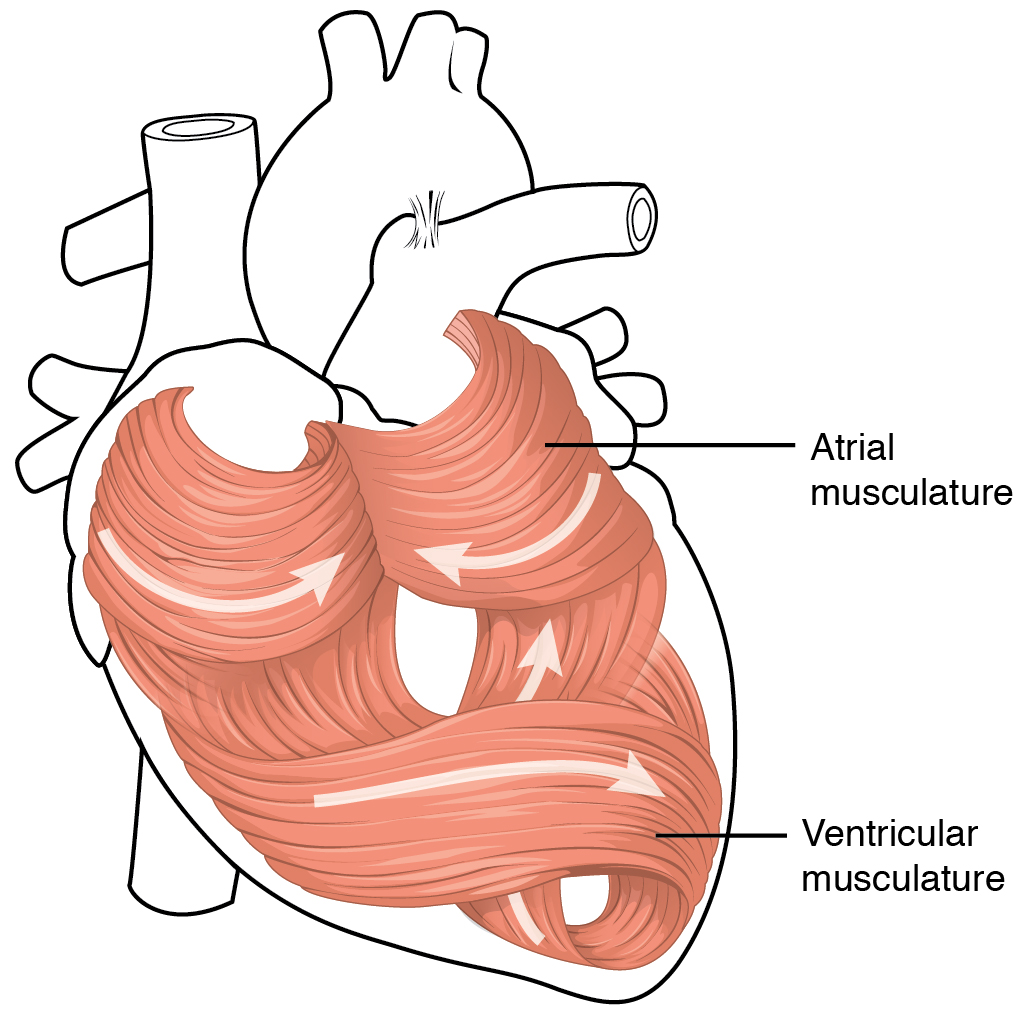
Cardiac muscle (also called heart muscle or myocardium) is one of three types of vertebrate muscle tissues, the others being skeletal muscle and smooth muscle. It is an involuntary, striated muscle that constitutes the main tissue of the wall of the heart. The cardiac muscle (myocardium) forms a thick middle layer between the outer layer of the heart wall (the pericardium) and the inner layer (the endocardium), with blood supplied via the coronary circulation. It is composed of individual cardiac muscle cells joined by intercalated discs, and encased by collagen fibers and other substances that form the extracellular matrix. Cardiac muscle contracts in a similar manner to skeletal muscle, although with some important differences. Electrical stimulation in the form of a cardiac action potential triggers the release of calcium from the cell's internal calcium store, the sarcoplasmic reticulum. The rise in calcium causes the cell's myofilaments to slide past each other in a pro ... [...More Info...] [...Related Items...] OR: [Wikipedia] [Google] [Baidu] [Amazon] |
Cytosol
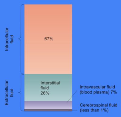
The cytosol, also known as cytoplasmic matrix or groundplasm, is one of the liquids found inside cells ( intracellular fluid (ICF)). It is separated into compartments by membranes. For example, the mitochondrial matrix separates the mitochondrion into many compartments. In the eukaryotic cell, the cytosol is surrounded by the cell membrane and is part of the cytoplasm, which also comprises the mitochondria, plastids, and other organelles (but not their internal fluids and structures); the cell nucleus is separate. The cytosol is thus a liquid matrix around the organelles. In prokaryotes, most of the chemical reactions of metabolism take place in the cytosol, while a few take place in membranes or in the periplasmic space. In eukaryotes, while many metabolic pathways still occur in the cytosol, others take place within organelles. The cytosol is a complex mixture of substances dissolved in water. Although water forms the large majority of the cytosol, its structure and proper ... [...More Info...] [...Related Items...] OR: [Wikipedia] [Google] [Baidu] [Amazon] |
Calcium
Calcium is a chemical element; it has symbol Ca and atomic number 20. As an alkaline earth metal, calcium is a reactive metal that forms a dark oxide-nitride layer when exposed to air. Its physical and chemical properties are most similar to its heavier homologues strontium and barium. It is the fifth most abundant element in Earth's crust, and the third most abundant metal, after iron and aluminium. The most common calcium compound on Earth is calcium carbonate, found in limestone and the fossils of early sea life; gypsum, anhydrite, fluorite, and apatite are also sources of calcium. The name comes from Latin ''calx'' " lime", which was obtained from heating limestone. Some calcium compounds were known to the ancients, though their chemistry was unknown until the seventeenth century. Pure calcium was isolated in 1808 via electrolysis of its oxide by Humphry Davy, who named the element. Calcium compounds are widely used in many industries: in foods and pharmaceuticals for ... [...More Info...] [...Related Items...] OR: [Wikipedia] [Google] [Baidu] [Amazon] |
Cardiomyocyte
Cardiac muscle (also called heart muscle or myocardium) is one of three types of vertebrate muscle tissues, the others being skeletal muscle and smooth muscle. It is an involuntary, striated muscle that constitutes the main tissue of the wall of the heart. The cardiac muscle (myocardium) forms a thick middle layer between the outer layer of the heart wall (the pericardium) and the inner layer (the endocardium), with blood supplied via the coronary circulation. It is composed of individual cardiac muscle cells joined by intercalated discs, and encased by collagen fibers and other substances that form the extracellular matrix. Cardiac muscle contracts in a similar manner to skeletal muscle, although with some important differences. Electrical stimulation in the form of a cardiac action potential triggers the release of calcium from the cell's internal calcium store, the sarcoplasmic reticulum. The rise in calcium causes the cell's myofilaments to slide past each other in a p ... [...More Info...] [...Related Items...] OR: [Wikipedia] [Google] [Baidu] [Amazon] |
Inotropic
An inotrope or inotropic is a drug or any substance that alters the force or energy of muscular contractions. Negatively inotropic agents weaken the force of muscular contractions. Positively inotropic agents increase the strength of muscular contraction. The term ''inotropic state'' is most commonly used in reference to various drugs that affect the strength of contraction of heart muscle. However, it can also refer to pathological conditions. For example, enlarged heart muscle can increase inotropic state, whereas dead heart muscle can decrease it. Medical uses Both positive and negative inotropes are used in the management of various cardiovascular conditions. The choice of agent depends largely on specific pharmacological effects of individual agents with respect to the condition. One of the most important factors affecting inotropic state is the level of calcium in the cytoplasm of the muscle cell. Positive inotropes usually increase this level, while negative inotropes ... [...More Info...] [...Related Items...] OR: [Wikipedia] [Google] [Baidu] [Amazon] |
Catecholamine
A catecholamine (; abbreviated CA), most typically a 3,4-dihydroxyphenethylamine, is a monoamine neurotransmitter, an organic compound that has a catechol (benzene with two hydroxyl side groups next to each other) and a side-chain amine. Catechol can be either a free molecule or a substituent of a larger molecule, where it represents a 1,2-dihydroxybenzene group. Catecholamines are derived from the amino acid tyrosine, which is derived from dietary sources as well as synthesis from phenylalanine. Catecholamines are water-soluble and are 50% bound to plasma proteins in circulation. Included among catecholamines are epinephrine (adrenaline), norepinephrine (noradrenaline), and dopamine. Release of the hormones epinephrine and norepinephrine from the adrenal medulla of the adrenal glands is part of the fight-or-flight response. Tyrosine is created from phenylalanine by hydroxylation by the enzyme phenylalanine hydroxylase. Tyrosine is also ingested directly from dietary prote ... [...More Info...] [...Related Items...] OR: [Wikipedia] [Google] [Baidu] [Amazon] |
Sarcoplasmic Reticulum
The sarcoplasmic reticulum (SR) is a membrane-bound structure found within muscle cells that is similar to the smooth endoplasmic reticulum in other cells. The main function of the SR is to store calcium ions (Ca2+). Calcium ion levels are kept relatively constant, with the concentration of calcium ions within a cell being 10,000 times smaller than the concentration of calcium ions outside the cell. This means that small increases in calcium ions within the cell are easily detected and can bring about important cellular changes (the calcium is said to be a second messenger). Calcium is used to make calcium carbonate (found in chalk) and calcium phosphate, two compounds that the body uses to make teeth and bones. This means that too much calcium within the cells can lead to hardening (calcification) of certain intracellular structures, including the mitochondria, leading to cell death. Therefore, it is vital that calcium ion levels are controlled tightly, and can be released int ... [...More Info...] [...Related Items...] OR: [Wikipedia] [Google] [Baidu] [Amazon] |
Troponin I
Troponin I is a cardiac and skeletal muscle protein family. It is a part of the troponin protein complex, where it binds to actin in thin myofilaments to hold the actin-tropomyosin complex in place. Troponin I prevents myosin from binding to actin in relaxed muscle. When calcium binds to the troponin C, it causes conformational changes which lead to dislocation of troponin I. Afterwards, tropomyosin leaves the binding site for myosin on actin leading to contraction of muscle. The letter ''I'' is given due to its inhibitory character. It is a useful marker in the laboratory diagnosis of heart attack. It occurs in different plasma concentration but the same circumstances as troponin T - either test can be performed for confirmation of cardiac muscle damage and laboratories usually offer one test or the other. Three paralogs with unique tissue-specific expression patterns are expressed in humans, listed below with their locations and OMIM accessions: * Slow-twitch skeletal muscle i ... [...More Info...] [...Related Items...] OR: [Wikipedia] [Google] [Baidu] [Amazon] |
CAMP-dependent Pathway
In the field of molecular biology, the cAMP-dependent pathway, also known as the adenylyl cyclase pathway, is a G protein-coupled receptor-triggered signaling cascade used in cell communication. Discovery cAMP was discovered by Earl Sutherland and Ted Rall in the mid 1950s. cAMP is considered a secondary messenger along with Ca2+. Sutherland won the Nobel Prize in 1971 for his discovery of the mechanism of action of epinephrine in glycogenolysis, that requires cAMP as secondary messenger. Mechanism G protein-coupled receptors (GPCRs) are a large family of integral membrane proteins that respond to a variety of extracellular stimuli. Each GPCR binds to and is activated by a specific ligand stimulus that ranges in size from small molecule catecholamines, lipids, or neurotransmitters to large protein hormones. When a GPCR is activated by its extracellular ligand, a conformational change is induced in the receptor that is transmitted to an attached intracellular heterotrimer ... [...More Info...] [...Related Items...] OR: [Wikipedia] [Google] [Baidu] [Amazon] |
Sodium-calcium Exchanger
The sodium-calcium exchanger (often denoted Na+/Ca2+ exchanger, exchange protein, or NCX) is an antiporter membrane protein that removes calcium from cells. It uses the energy that is stored in the electrochemical gradient of sodium (Na+) by allowing Na+ to flow down its gradient across the plasma membrane in exchange for the countertransport of calcium ions (Ca2+). A single calcium ion is exported for the import of three sodium ions. The exchanger exists in many different cell types and animal species. The NCX is considered one of the most important cellular mechanisms for removing Ca2+. The exchanger is usually found in the plasma membranes and the mitochondria and endoplasmic reticulum of excitable cells. Function The sodium–calcium exchanger is only one of the systems by which the cytoplasmic concentration of calcium ions in the cell is kept low. The exchanger does not bind very tightly to Ca2+ (has a low affinity), but it can transport the ions rapidly (has a high c ... [...More Info...] [...Related Items...] OR: [Wikipedia] [Google] [Baidu] [Amazon] |


