|
Koniocellular Cell
A koniocellular cell (konio: Greek, ''dust'' or ''poison'', also known as K cell) is a neuron with a small cell body that is located in the koniocellular layer of the lateral geniculate nucleus (LGN) in primates, including humans. Koniocellular layers are located ventral to each parvocellular and magnocellular layer of the LGN. Even if the quantity of neurons is approximately equal to the number of magnocellular cells the koniocellular layers are much thinner due to their size. In comparison to the parvocellular and magnocellular system, fewer studies have been conducted to investigate the koniocellular system. Koniocellular cells are a heterogeneous population differing in many aspects, such as response properties and connectivity. Structure K cells are neurochemically and anatomically distinct from M and P cells. There are three proteins by which K cells can be clearly distinguished: * Calbindin (28kDa calcium binding protein, CALB) * The alpha subunit of type II calmodulin- ... [...More Info...] [...Related Items...] OR: [Wikipedia] [Google] [Baidu] |
Lateral Geniculate Nucleus
In neuroanatomy, the lateral geniculate nucleus (LGN; also called the lateral geniculate body or lateral geniculate complex) is a structure in the thalamus and a key component of the mammalian visual pathway. It is a small, ovoid, ventral projection of the thalamus where the thalamus connects with the optic nerve. There are two LGNs, one on the left and another on the right side of the thalamus. In humans, both LGNs have six layers of neurons (grey matter) alternating with optic fibers (white matter). The LGN receives information directly from the ascending retinal ganglion cells via the optic tract and from the reticular activating system. Neurons of the LGN send their axons through the optic radiation, a direct pathway to the primary visual cortex. In addition, the LGN receives many strong feedback connections from the primary visual cortex. In humans as well as other mammals, the two strongest pathways linking the eye to the brain are those projecting to the dorsal part of th ... [...More Info...] [...Related Items...] OR: [Wikipedia] [Google] [Baidu] |
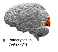
Primary Visual Cortex
The visual cortex of the brain is the area of the cerebral cortex that processes visual information. It is located in the occipital lobe. Sensory input originating from the eyes travels through the lateral geniculate nucleus in the thalamus and then reaches the visual cortex. The area of the visual cortex that receives the sensory input from the lateral geniculate nucleus is the primary visual cortex, also known as visual area 1 ( V1), Brodmann area 17, or the striate cortex. The extrastriate areas consist of visual areas 2, 3, 4, and 5 (also known as V2, V3, V4, and V5, or Brodmann area 18 and all Brodmann area 19). Both hemispheres of the brain include a visual cortex; the visual cortex in the left hemisphere receives signals from the right visual field, and the visual cortex in the right hemisphere receives signals from the left visual field. Introduction The primary visual cortex (V1) is located in and around the calcarine fissure in the occipital lobe. Each hemisphere' ... [...More Info...] [...Related Items...] OR: [Wikipedia] [Google] [Baidu] |
List Of Human Cell Types Derived From The Germ Layers
This is a list of cells in humans derived from the three embryonic germ layers – ectoderm, mesoderm, and endoderm. Cells derived from ectoderm Surface ectoderm Skin * Trichocyte * Keratinocyte Anterior pituitary * Gonadotrope * Corticotrope * Thyrotrope * Somatotrope * Lactotroph Tooth enamel * Ameloblast Neural crest Peripheral nervous system * Neuron * Glia ** Schwann cell ** Satellite glial cell Neuroendocrine system * Chromaffin cell * Glomus cell Skin * Melanocyte ** Nevus cell * Merkel cell Teeth * Odontoblast * Cementoblast Eyes * Corneal keratocyte Neural tube Central nervous system * Neuron * Glia ** Astrocyte ** Ependymocytes ** Muller glia (retina) ** Oligodendrocyte ** Oligodendrocyte progenitor cell ** Pituicyte (posterior pituitary) Pineal gland * Pinealocyte Cells derived from mesoderm Paraxial mesoderm Mesenchymal stem cell =Osteochondroprogenitor cell= * Bone (Osteoblast → Osteocyte) * Cartilage (Chondroblast → Chond ... [...More Info...] [...Related Items...] OR: [Wikipedia] [Google] [Baidu] |
Visual Cortex
The visual cortex of the brain is the area of the cerebral cortex that processes visual information. It is located in the occipital lobe. Sensory input originating from the eyes travels through the lateral geniculate nucleus in the thalamus and then reaches the visual cortex. The area of the visual cortex that receives the sensory input from the lateral geniculate nucleus is the primary visual cortex, also known as visual area 1 ( V1), Brodmann area 17, or the striate cortex. The extrastriate areas consist of visual areas 2, 3, 4, and 5 (also known as V2, V3, V4, and V5, or Brodmann area 18 and all Brodmann area 19). Both hemispheres of the brain include a visual cortex; the visual cortex in the left hemisphere receives signals from the right visual field, and the visual cortex in the right hemisphere receives signals from the left visual field. Introduction The primary visual cortex (V1) is located in and around the calcarine fissure in the occipital lobe. Each hemisphere's V ... [...More Info...] [...Related Items...] OR: [Wikipedia] [Google] [Baidu] |
Blindsight
Blindsight is the ability of people who are cortically blind to respond to visual stimuli that they do not consciously see due to lesions in the primary visual cortex, also known as the striate cortex or Brodmann Area 17. The term was coined by Lawrence Weiskrantz and his colleagues in a paper published in a 1974 issue of ''Brain''. A previous paper studying the discriminatory capacity of a cortically blind patient was published in ''Nature'' in 1973. Type classification The majority of studies on blindsight are conducted on patients who are hemianopic, i.e. blind in one half of their visual field. Following the destruction of the left or right striate cortex, patients are asked to detect, localize, and discriminate amongst visual stimuli that are presented to their blind side, often in a forced-response or guessing situation, even though they may not consciously recognize the visual stimulus. Research shows that such blind patients may achieve a higher accuracy than would be ex ... [...More Info...] [...Related Items...] OR: [Wikipedia] [Google] [Baidu] |
Visual Cortex
The visual cortex of the brain is the area of the cerebral cortex that processes visual information. It is located in the occipital lobe. Sensory input originating from the eyes travels through the lateral geniculate nucleus in the thalamus and then reaches the visual cortex. The area of the visual cortex that receives the sensory input from the lateral geniculate nucleus is the primary visual cortex, also known as visual area 1 ( V1), Brodmann area 17, or the striate cortex. The extrastriate areas consist of visual areas 2, 3, 4, and 5 (also known as V2, V3, V4, and V5, or Brodmann area 18 and all Brodmann area 19). Both hemispheres of the brain include a visual cortex; the visual cortex in the left hemisphere receives signals from the right visual field, and the visual cortex in the right hemisphere receives signals from the left visual field. Introduction The primary visual cortex (V1) is located in and around the calcarine fissure in the occipital lobe. Each hemisphere's V ... [...More Info...] [...Related Items...] OR: [Wikipedia] [Google] [Baidu] |
Inferotemporal Cortex
The inferior temporal gyrus is one of three gyri of the temporal lobe and is located below the middle temporal gyrus, connected behind with the inferior occipital gyrus; it also extends around the infero-lateral border on to the inferior surface of the temporal lobe, where it is limited by the inferior sulcus. This region is one of the higher levels of the ventral stream of visual processing, associated with the representation of objects, places, faces, and colors. It may also be involved in face perception, and in the recognition of numbers and words. The inferior temporal gyrus is the anterior region of the temporal lobe located underneath the central temporal sulcus. The primary function of the occipital temporal gyrus – otherwise referenced as IT cortex – is associated with visual stimuli processing, namely visual object recognition, and has been suggested by recent experimental results as the final location of the ventral cortical visual system. The IT cortex in humans ... [...More Info...] [...Related Items...] OR: [Wikipedia] [Google] [Baidu] |
Superficial Blobs
Superficial may refer to: * Superficial anatomy, is the study of the external features of the body *Superficiality, the discourses in philosophy regarding social relation * Superficial charm, the tendency to be smooth, engaging, charming, slick and verbally facile *Superficial sympathy, false or insincere display of emotion such as a hypocrite crying fake tears of grief In entertainment * ''Superficial'' (album), an album by Heidi Montag, or its title track * The Superficial The Superficial was a website devoted to celebrity gossip. It was founded on May 23, 2004, and quickly grew in popularity. The Superficial was a part of Anticlown Media, along with other sites such as IWatchStuff.com and Geekologie.com. The websit ..., a website devoted to celebrity gossip * "Superficial", a song by Natalia Kills from the album '' Perfectionist'' See also * Artificial (other) * Synthetic (other) * Man-made (other) {{disambiguation ... [...More Info...] [...Related Items...] OR: [Wikipedia] [Google] [Baidu] |
Superior Colliculus
In neuroanatomy, the superior colliculus () is a structure lying on the roof of the mammalian midbrain. In non-mammalian vertebrates, the homologous structure is known as the optic tectum, or optic lobe. The adjective form '' tectal'' is commonly used for both structures. In mammals, the superior colliculus forms a major component of the midbrain. It is a paired structure and together with the paired inferior colliculi forms the corpora quadrigemina. The superior colliculus is a layered structure, with a pattern that is similar to all mammals. The layers can be grouped into the superficial layers ( stratum opticum and above) and the deeper remaining layers. Neurons in the superficial layers receive direct input from the retina and respond almost exclusively to visual stimuli. Many neurons in the deeper layers also respond to other modalities, and some respond to stimuli in multiple modalities. The deeper layers also contain a population of motor-related neurons, capable of acti ... [...More Info...] [...Related Items...] OR: [Wikipedia] [Google] [Baidu] |
Extrastriate Cortex
The extrastriate cortex is the region of the occipital cortex of the mammalian brain located next to the primary visual cortex. Primary visual cortex (V1) is also named striate cortex because of its striped appearance in the microscope. The extrastriate cortex encompasses multiple functional areas, including V3, V4, V5/MT, which is sensitive to motion,Guy A. Orban. Higher Order Visual Processing in Macaque Extrastriate Cortex. ''Physiol Rev'' January 1, 2008 88:(1) 59-89; or the extrastriate body area (EBA) used in the perception of human bodies. Anatomy In terms of Brodmann areas, the extrastriate cortex comprises Brodmann area 18 and Brodmann area 19, while the striate cortex comprises Brodmann area 17. In primates, the extrastriate cortex includes visual area V3, visual area V4, and visual area MT The visual system comprises the sensory organ (the eye) and parts of the central nervous system (the retina containing photoreceptor cells, the optic nerve, the optic tra ... [...More Info...] [...Related Items...] OR: [Wikipedia] [Google] [Baidu] |
Bistratified Cell
Bistratified ganglion cell can refer to either of two kinds of retinal ganglion cells whose cell body is located in the ganglion cell layer of the retina, the small-field bistratified ganglion cell, also known as small bistratified cell (SBC), and the large-field bistratified ganglion cell or large bistratified cell (LBC). Bistratified cells receive their input from bipolar cells and amacrine cells. The bistratified cells project their axons through the optic nerve and optic tract to the koniocellular layers in the lateral geniculate nucleus (LGN), synapsing with koniocellular cells. Koniocellular means "cells as small as dust"; their small size made them hard to find. About 8 to 10% of retinal ganglion cells are bistratified cells. They receive inputs from intermediate numbers of rods and cones. They have moderate spatial resolution, moderate conduction velocity, and can respond to moderate-contrast stimuli. They may be involved in color vision. See also * Midget cell * Paras ... [...More Info...] [...Related Items...] OR: [Wikipedia] [Google] [Baidu] |
Blob (visual System)
Blobs are sections of the visual cortex where groups of neurons that are sensitive to color assemble in cylindrical shapes. They were first identified in 1979 by Margaret Wong-Riley when she used a cytochrome oxidase stain, from which they get their name. These areas receive input from parvocellular cells in layer 4Cβ of the primary visual cortex and output to the thin stripes of area V2. Interblobs are areas between blobs which receive the same input, but are sensitive to orientation Orientation may refer to: Positioning in physical space * Map orientation, the relationship between directions on a map and compass directions * Orientation (housing), the position of a building with respect to the sun, a concept in building de ... instead of color. They output to the pale and thick stripes of area V2. Blobs are on the parvocellular pathway. This pathway begins at the photoreceptors which then relay signals to the 'P' ganglion cells in the retina. The pathway then continues out ... [...More Info...] [...Related Items...] OR: [Wikipedia] [Google] [Baidu] |


