|
Interosseous Membrane Of Leg
The interosseous membrane of the leg (middle tibiofibular ligament) extends between the interosseous crests of the tibia and fibula, helps stabilize the Tib-Fib relationship and separates the muscles on the front from those on the back of the leg. It consists of a thin, aponeurotic joint lamina composed of oblique fibers, which for the most part run downward and lateralward; some few fibers, however, pass in the opposite direction. It is broader above than below. Its upper margin does not quite reach the tibiofibular joint, but presents a free concave border, above which is a large, oval aperture for the passage of the anterior tibial vessels to the front of the leg. In its lower part is an opening for the passage of the anterior peroneal vessels. It is continuous below with the interosseous ligament of the tibiofibular syndesmosis In anatomy, fibrous joints are joints connected by fibrous tissue, consisting mainly of collagen. These are fixed joints where bones are unit ... [...More Info...] [...Related Items...] OR: [Wikipedia] [Google] [Baidu] |
Tibia
The tibia (; ), also known as the shinbone or shankbone, is the larger, stronger, and anterior (frontal) of the two bones in the leg below the knee in vertebrates (the other being the fibula, behind and to the outside of the tibia); it connects the knee with the ankle. The tibia is found on the medial side of the leg next to the fibula and closer to the median plane. The tibia is connected to the fibula by the interosseous membrane of leg, forming a type of fibrous joint called a syndesmosis with very little movement. The tibia is named for the flute '' tibia''. It is the second largest bone in the human body, after the femur. The leg bones are the strongest long bones as they support the rest of the body. Structure In human anatomy, the tibia is the second largest bone next to the femur. As in other vertebrates the tibia is one of two bones in the lower leg, the other being the fibula, and is a component of the knee and ankle joints. The ossification or formation of the ... [...More Info...] [...Related Items...] OR: [Wikipedia] [Google] [Baidu] |
Fibula
The fibula or calf bone is a leg bone on the lateral side of the tibia, to which it is connected above and below. It is the smaller of the two bones and, in proportion to its length, the most slender of all the long bones. Its upper extremity is small, placed toward the back of the head of the tibia, below the knee joint and excluded from the formation of this joint. Its lower extremity inclines a little forward, so as to be on a plane anterior to that of the upper end; it projects below the tibia and forms the lateral part of the ankle joint. Structure The bone has the following components: * Lateral malleolus * Interosseous membrane connecting the fibula to the tibia, forming a syndesmosis joint * The superior tibiofibular articulation is an arthrodial joint between the lateral condyle of the tibia and the head of the fibula. * The inferior tibiofibular articulation (tibiofibular syndesmosis) is formed by the rough, convex surface of the medial side of the lower end of th ... [...More Info...] [...Related Items...] OR: [Wikipedia] [Google] [Baidu] |
Lamina
Lamina may refer to: Science and technology * Planar lamina, a two-dimensional planar closed surface with mass and density, in mathematics * Laminar flow, (or streamline flow) occurs when a fluid flows in parallel layers, with no disruption between the layers * Lamina (algae), a structure in seaweeds * Lamina (anatomy), with several meanings * Lamina (leaf), the flat part of a leaf, an organ of a plant * Lamina, the largest petal of a floret in an aster family flowerhead: see * ''Lamina'' (spider), a genus in the family Toxopidae * Lamina (neuropil), the most peripheral neuropil of the insect visual system *Nuclear lamina, another structure of a living cell *Basal lamina, a structure of a living cell *Lamina propria, the connective part of the mucous *Lamina of the vertebral arch *Lamination (geology), a layering structure in sedimentary rocks usually less than 1 cm in thickness * Laminae, a part of the horse hoof * Laminae, another name for the core lamiids, a clade in botany ... [...More Info...] [...Related Items...] OR: [Wikipedia] [Google] [Baidu] |
Superior Tibiofibular Joint
The proximal tibiofibular articulation (also called superior tibiofibular joint) is an arthrodial joint between the lateral condyle of the tibia and the head of the fibula. The contiguous surfaces of the bones present flat, oval facets covered with cartilage and connected together by an articular capsule and by anterior and posterior ligaments. When the term ''tibiofibular articulation'' is used without a modifier, it refers to the proximal, not the distal (i.e., inferior) tibiofibular articulation. Clinical significance Injuries to the proximal tibiofibular joint are uncommon and usually associated with other injuries to the lower leg. Dislocations can be classified into the following five types: * Anterolateral dislocation (most common) * Posteromedial dislocation * Superior dislocation (uncommon, associated with shortened tibia fractures or severe ankle injuries) * Inferior dislocation (rare, associated with lengthened tibia fractures or avulsion of the foot, usua ... [...More Info...] [...Related Items...] OR: [Wikipedia] [Google] [Baidu] |
Anterior Tibial Vessels
The anterior tibial artery is an artery of the leg. It carries blood to the anterior compartment of the leg and dorsal surface of the foot, from the popliteal artery. Structure Course The anterior tibial artery is a branch of the popliteal artery. It originates at the distal end of the popliteus muscle posterior to the tibia. The artery typically passes anterior to the popliteus muscle prior to passing between the tibia and fibula through an oval opening at the superior aspect of the interosseus membrane. The artery then descends between the tibialis anterior and extensor digitorum longus muscles. It is accompanied by the anterior tibial vein, and the deep peroneal nerve, along its course. It crosses the anterior aspect of the ankle joint, at which point it becomes the dorsalis pedis artery. Branches The branches of the anterior tibial artery are: *posterior tibial recurrent artery * anterior tibial recurrent artery * muscular branches * anterior medial malleolar artery *a ... [...More Info...] [...Related Items...] OR: [Wikipedia] [Google] [Baidu] |
Anterior Peroneal Vessels
In anatomy, the fibular artery, also known as the peroneal artery, supplies blood to the lateral compartment of the leg. It arises from the tibial-fibular trunk. Structure The fibular artery arises from the bifurcation of tibial-fibular trunk into the fibular and posterior tibial arteries in the upper part of the leg proper, just below the knee. It runs towards the foot in the deep posterior compartment of the leg, just medial to the fibula. It supplies a perforating branch to both the lateral and anterior compartments of the leg; it also provides a nutrient artery to the fibula. Some sources claim that the fibular artery arises directly from the posterior tibial artery, but vascular and plastic surgeons note the clinical significance of the tibial-fibular trunk. The fibular artery is accompanied by small veins (venae comitantes) known as fibular veins. Branches Communication branch to posterior tibial artery. Perforating branch to anterior lateral malleolar artery. A calcan ... [...More Info...] [...Related Items...] OR: [Wikipedia] [Google] [Baidu] |
Syndesmosis
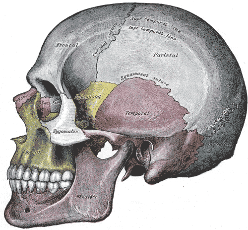
In anatomy, fibrous joints are joints connected by fibrous tissue, consisting mainly of collagen. These are fixed joints where bones are united by a layer of white fibrous tissue of varying thickness. In the skull the joints between the bones are called sutures. Such immovable joints are also referred to as synarthroses. Types Most fibrous joints are also called "fixed" or "immovable". These joints have no joint cavity and are connected via fibrous connective tissue. The skull bones are connected by fibrous joints called '' sutures''. In fetal skulls the sutures are wide to allow slight movement during birth. They later become rigid ( synarthrodial). Some of the long bones in the body such as the radius and ulna in the forearm are joined by a '' syndesmosis'' (along the interosseous membrane). Syndemoses are slightly moveable ( amphiarthrodial). The distal tibiofibular joint is another example. A '' gomphosis'' is a joint between the root of a tooth and the socket in the max ... [...More Info...] [...Related Items...] OR: [Wikipedia] [Google] [Baidu] |


