|
Internal Vertebral Venous Plexuses
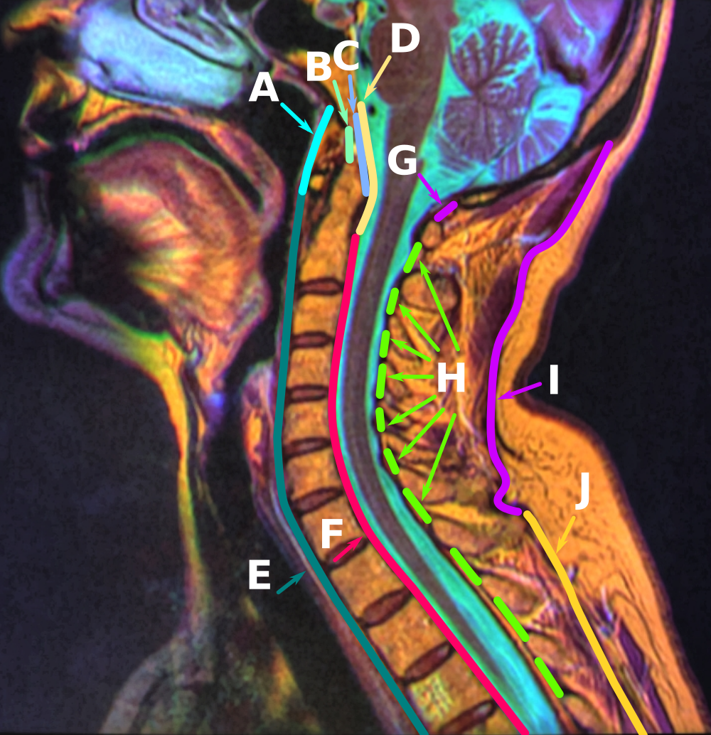
The internal vertebral venous plexuses (intraspinal veins) lie within the vertebral canal in the epidural space, and receive tributaries from the bones and from the spinal cord. They form a closer network than the external plexuses, and, running mainly in a vertical direction, form four longitudinal veins, two in front and two behind; they therefore may be divided into anterior and posterior groups. * The ''anterior internal plexuses'' consist of large veins which lie on the posterior surfaces of the vertebral bodies and intervertebral fibrocartilages on either side of the posterior longitudinal ligament; under cover of this ligament they are connected by transverse branches into which the basivertebral veins open. * The ''posterior internal plexuses'' are placed, one on either side of the middle line in front of the vertebral arches and ligamenta flava, and anastomose by veins passing through those ligaments with the posterior external plexuses. The anterior and posterior plexu ... [...More Info...] [...Related Items...] OR: [Wikipedia] [Google] [Baidu] |
Thoracic Vertebra
In vertebrates, thoracic vertebrae compose the middle segment of the vertebral column, between the cervical vertebrae and the lumbar vertebrae. In humans, there are twelve thoracic vertebrae and they are intermediate in size between the cervical and lumbar vertebrae; they increase in size going towards the lumbar vertebrae, with the lower ones being much larger than the upper. They are distinguished by the presence of facets on the sides of the bodies for articulation with the heads of the ribs, as well as facets on the transverse processes of all, except the eleventh and twelfth, for articulation with the tubercles of the ribs. By convention, the human thoracic vertebrae are numbered T1–T12, with the first one (T1) located closest to the skull and the others going down the spine toward the lumbar region. General characteristics These are the general characteristics of the second through eighth thoracic vertebrae. The first and ninth through twelfth vertebrae contain certain ... [...More Info...] [...Related Items...] OR: [Wikipedia] [Google] [Baidu] |
Posterior External Plexuses
Posterior may refer to: * Posterior (anatomy), the end of an organism opposite to its head ** Buttocks, as a euphemism * Posterior horn (other) * Posterior probability The posterior probability is a type of conditional probability that results from updating the prior probability with information summarized by the likelihood via an application of Bayes' rule. From an epistemological perspective, the posterior ..., the conditional probability that is assigned when the relevant evidence is taken into account * Posterior tense, a relative future tense {{disambiguation ... [...More Info...] [...Related Items...] OR: [Wikipedia] [Google] [Baidu] |
Rete Canalis Hypoglossi
The venous plexus of hypoglossal canal ( ''TA'') – also known as ''plexus venosus canalis nervi hypoglossi'' ( ''TA''), ''circellus venosus hypoglossi'' and ''rete canalis hypoglossi'' – is a small venous plexus around the hypoglossal nerve that connects with the occipital sinus, the inferior petrosal sinus and the internal jugular vein The internal jugular vein is a paired jugular vein that collects blood from the brain and the superficial parts of the face and neck. This vein runs in the carotid sheath with the common carotid artery and vagus nerve. It begins in the poste .... Occasionally, it may be a single vein rather than a venous plexus. Notes References Veins of the head and neck {{Circulatory-stub ... [...More Info...] [...Related Items...] OR: [Wikipedia] [Google] [Baidu] |
Condyloid Emissary Vein
A condyloid joint (also called condylar, ellipsoidal, or bicondylar) is an ovoid articular surface, or condyle that is received into an elliptical cavity. This permits movement in two planes, allowing flexion, extension, adduction, abduction, and circumduction. Examples Examples include: * the * s * |
Basilar Plexus
The basilar plexus (transverse or basilar sinus) consists of several interlacing venous channels between the layers of the dura mater In neuroanatomy, dura mater is a thick membrane made of dense irregular connective tissue that surrounds the brain and spinal cord. It is the outermost of the three layers of membrane called the meninges that protect the central nervous system. ... over the basilar part of the occipital bone (the clivus), and serves to connect the two inferior petrosal sinuses. It communicates with the anterior vertebral venous plexus. References Veins of the head and neck {{circulatory-stub ... [...More Info...] [...Related Items...] OR: [Wikipedia] [Google] [Baidu] |
Occipital Sinus
The occipital sinus is the smallest of the dural venous sinuses. It is usually unpaired, and is sometimes altogether absent. It is situated in the attached margin of the falx cerebelli. It commences near the foramen magnum, and ends by draining into the confluence of sinuses. Occipital sinuses were discovered by Guichard Joseph Duverney. Anatomy The occipital sinus is present in around 65% of individuals. It is usually single, but occasionally paired. It is situated in the attached margin of the falx cerebelli. Course The occipital sinus commences around the margin of the foramen magnum by several small venous channels (one of which joins the terminal part of the sigmoid sinus The sigmoid sinuses (sigma- or s-shaped hollow curve), also known as the , are venous sinuses within the skull that receive blood from posterior dural venous sinus veins. Structure The sigmoid sinus is a dural venous sinus situated within the ...). It terminates by draining into the confluenc ... [...More Info...] [...Related Items...] OR: [Wikipedia] [Google] [Baidu] |
Vertebral Veins
The vertebral vein is formed in the suboccipital triangle, from numerous small tributaries which spring from the internal vertebral venous plexuses and issue from the vertebral canal above the posterior arch of the atlas An atlas is a collection of maps; it is typically a bundle of maps of Earth or of a region of Earth. Atlases have traditionally been bound into book form, but today many atlases are in multimedia formats. In addition to presenting geograp .... They unite with small veins from the deep muscles at the upper part of the back of the neck, and form a vessel which enters the foramen in the transverse process of the atlas, and descends, forming a dense plexus around the vertebral artery, in the canal formed by the transverse foramina of the upper six cervical vertebrae. This plexus ends in a single trunk, which emerges from the transverse foramina of the sixth cervical vertebra, and opens at the root of the neck into the back part of the innominate vein ne ... [...More Info...] [...Related Items...] OR: [Wikipedia] [Google] [Baidu] |
Foramen Magnum
The foramen magnum ( la, great hole) is a large, oval-shaped opening in the occipital bone of the skull. It is one of the several oval or circular openings (foramina) in the base of the skull. The spinal cord, an extension of the medulla oblongata, passes through the foramen magnum as it exits the cranial cavity. Apart from the transmission of the medulla oblongata and its membranes, the foramen magnum transmits the vertebral arteries, the anterior and posterior spinal arteries, the tectorial membranes and alar ligaments. It also transmits the accessory nerve into the skull. The foramen magnum is a very important feature in bipedal mammals. One of the attributes of a biped's foramen magnum is a forward shift of the anterior border of the cerebellar tentorium; this is caused by the shortening of the cranial base. Studies on the foramen magnum position have shown a connection to the functional influences of both posture and locomotion. The forward shift of the foramen magn ... [...More Info...] [...Related Items...] OR: [Wikipedia] [Google] [Baidu] |
Ligamenta Flava
The ligamenta flava (singular, ''ligamentum flavum'', Latin for ''yellow ligament'') are a series of ligaments that connect the ventral parts of the laminae of adjacent vertebrae. They help to preserve upright posture, preventing hyperflexion, and ensuring that the vertebral column straightens after flexion. Hypertrophy can cause spinal stenosis. Structure Each ligamentum flavum connects the laminae two adjacent vertebrae. They begin with the junction of the axis and third cervical vertebra, continuing down to the junction of the fifth lumbar vertebra and the sacrum. They are best seen from the interior of the vertebral canal. when looked at from the outer surface they appear short, being overlapped by the lamina of the vertebral arch. Each ligament consists of two lateral portions which commence one on either side of the roots of the articular processes, and extend backward to the point where the laminae meet to form the spinous process; the posterior margins of the t ... [...More Info...] [...Related Items...] OR: [Wikipedia] [Google] [Baidu] |
Vertebral Canal
The spinal canal (or vertebral canal or spinal cavity) is the canal that contains the spinal cord within the vertebral column. The spinal canal is formed by the vertebrae through which the spinal cord passes. It is a process of the dorsal body cavity. This canal is enclosed within the foramen of the vertebrae. In the intervertebral spaces, the canal is protected by the ligamentum flavum posteriorly and the posterior longitudinal ligament anteriorly. Structure The outermost layer of the meninges, the dura mater, is closely associated with the arachnoid mater which in turn is loosely connected to the innermost layer, the pia mater. The meninges divide the spinal canal into the epidural space and the subarachnoid space. The pia mater is closely attached to the spinal cord. A subdural space is generally only present due to trauma and/or pathological situations. The subarachnoid space is filled with cerebrospinal fluid and contains the vessels that supply the spinal cord, nam ... [...More Info...] [...Related Items...] OR: [Wikipedia] [Google] [Baidu] |
Vertebral Arches
The vertebral column, also known as the backbone or spine, is part of the axial skeleton. The vertebral column is the defining characteristic of a vertebrate in which the notochord (a flexible rod of uniform composition) found in all chordates has been replaced by a segmented series of bone: vertebrae separated by intervertebral discs. Individual vertebrae are named according to their region and position, and can be used as anatomical landmarks in order to guide procedures such as lumbar punctures. The vertebral column houses the spinal canal, a cavity that encloses and protects the spinal cord. There are about 50,000 species of animals that have a vertebral column. The human vertebral column is one of the most-studied examples. Many different diseases in humans can affect the spine, with spina bifida and scoliosis being recognisable examples. The general structure of human vertebrae is fairly typical of that found in mammals, reptiles, and birds. The shape of the verte ... [...More Info...] [...Related Items...] OR: [Wikipedia] [Google] [Baidu] |
Basivertebral Veins
The basivertebral veins are veins within the vertebral column. They are contained in large, tortuous channels in the substance of the bones, similar in every respect to those found in the diploë of the cranial bones. They emerge from the foramina on the posterior surfaces of the vertebral bodies The spinal column, a defining synapomorphy shared by nearly all vertebrates,Hagfish are believed to have secondarily lost their spinal column is a moderately flexible series of vertebrae (singular vertebra), each constituting a characteristic .... They communicate through small openings on the front and sides of the vertebral bodies with the anterior external vertebral plexuses, and converge behind to the principal canal, which is sometimes double toward its posterior part, and open by valved orifices into the transverse branches which unite the anterior internal vertebral plexuses. The basivertebral veins become greatly enlarged in advanced age. References External links * ... [...More Info...] [...Related Items...] OR: [Wikipedia] [Google] [Baidu] |


