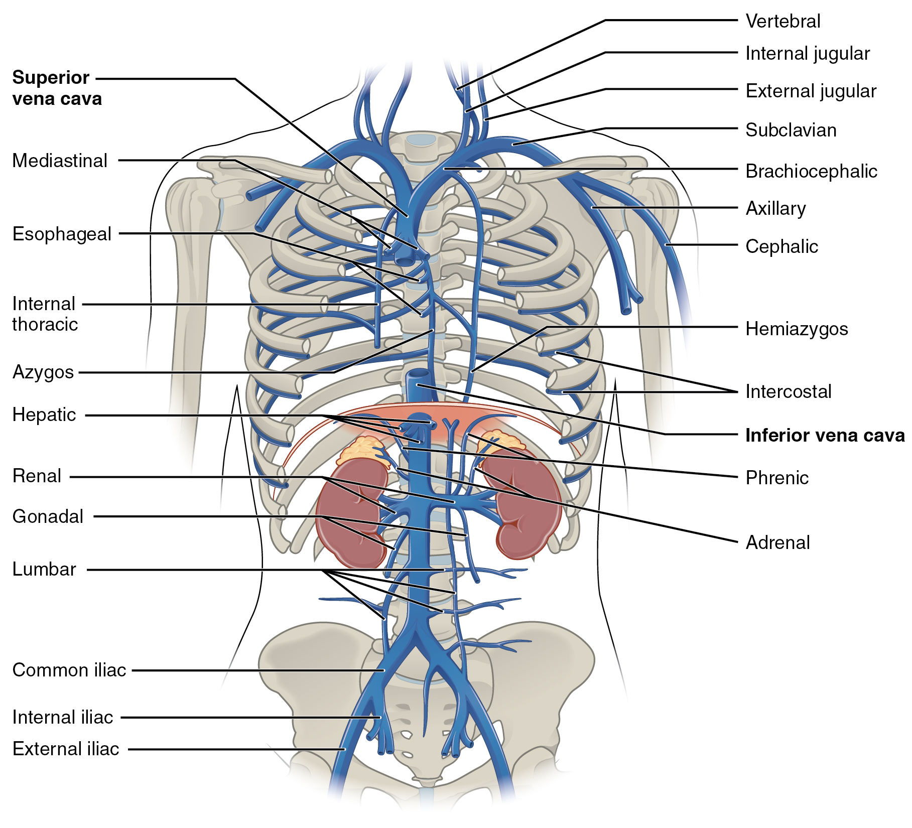
|
Innominate Vein
The left and right brachiocephalic veins (previously called innominate veins) are major veins in the upper chest, formed by the union of the ipsilateral internal jugular vein and subclavian vein (the so-called venous angle) behind the sternoclavicular joint. The left brachiocephalic vein is more than twice the length of the right brachiocephalic vein. These veins merge to form the superior vena cava, a great vessel, posterior to the junction of the first costal cartilage with the manubrium of the sternum. The brachiocephalic veins are the major veins returning blood to the superior vena cava. Left and right veins Left brachiocephalic vein The left brachiocephalic vein is about 6cm, more than twice the length of the right brachiocephalic vein. and is formed by the confluence of the left subclavian and left internal jugular veins. In addition the left vein receives drainage from the following tributaries: * The left vertebral vein, internal thoracic vein, inferior thyroid vein ... [...More Info...] [...Related Items...] OR: [Wikipedia] [Google] [Baidu] |
Internal Jugular Vein
The internal jugular vein is a paired jugular vein that collects blood from the brain and the superficial parts of the face and neck. This vein runs in the carotid sheath with the common carotid artery and vagus nerve. It begins in the posterior compartment of the jugular foramen, at the base of the skull. It is somewhat dilated at its origin, which is called the ''superior bulb''. This vein also has a common trunk into which drains the anterior branch of the retromandibular vein, the facial vein, and the lingual vein. It runs down the side of the neck in a vertical direction, being at one end lateral to the internal carotid artery, and then lateral to the common carotid artery, and at the root of the neck, it unites with the subclavian vein to form the brachiocephalic vein (innominate vein); a little above its termination is a second dilation, the ''inferior bulb''. Above, it lies upon the rectus capitis lateralis, behind the internal carotid artery and the nerves pa ... [...More Info...] [...Related Items...] OR: [Wikipedia] [Google] [Baidu] |
Great Vessel
Great vessels are the large vessels that bring blood to and from the heart. These are: * Superior vena cava * Inferior vena cava * Pulmonary arteries * Pulmonary veins * Aorta Transposition of the great vessels is a group of congenital A birth defect is an abnormal condition that is present at childbirth, birth, regardless of its cause. Birth defects may result in disability, disabilities that may be physical disability, physical, intellectual disability, intellectual, or dev ... heart defects involving an abnormal spatial arrangement of any of the great vessels. References Angiology {{Anatomy-stub ... [...More Info...] [...Related Items...] OR: [Wikipedia] [Google] [Baidu] |
Innominate Artery
The brachiocephalic artery, brachiocephalic trunk, or innominate artery is an artery of the mediastinum that supplies blood to the right arm, head, and neck. It is the first branch of the aortic arch. Soon after it emerges, the brachiocephalic artery divides into the right common carotid artery and the right subclavian artery. There is no brachiocephalic artery for the left side of the body. The left common carotid artery and the left subclavian artery come directly off the aortic arch. Despite this, there are two brachiocephalic veins. Structure The brachiocephalic artery arises on a level with the upper border of the second right costal cartilage from the start of the aortic arch on a plane anterior to the origin of the left carotid artery. It ascends obliquely upward, backward, and to the right to the level of the upper border of the right sternoclavicular articulation, where it divides into the right common carotid artery and right subclavian arteries. The artery then cros ... [...More Info...] [...Related Items...] OR: [Wikipedia] [Google] [Baidu] |
Innominate Bone
The hip bone (os coxae, innominate bone, pelvic bone or coxal bone) is a large flat bone, constricted in the center and expanded above and below. In some vertebrates (including humans before puberty) it is composed of three parts: the ilium, ischium, and the pubis. The two hip bones join at the pubic symphysis and together with the sacrum and coccyx (the pelvic part of the spine) comprise the skeletal component of the pelvis – the pelvic girdle which surrounds the pelvic cavity. They are connected to the sacrum, which is part of the axial skeleton, at the sacroiliac joint. Each hip bone is connected to the corresponding femur (thigh bone) (forming the primary connection between the bones of the lower limb and the axial skeleton) through the large ball and socket joint of the hip. Structure The hip bone is formed by three parts: the ilium, ischium, and pubis. At birth, these three components are separated by hyaline cartilage. They join each other in a Y-shaped portion of ca ... [...More Info...] [...Related Items...] OR: [Wikipedia] [Google] [Baidu] |
Pericardial Veins
The smallest cardiac veins (also known as the Thebesian veins (named for Adam Christian Thebesius) are small, valveless veins in the walls of all four heart chambers that drain venous blood from the myocardium directly into any of the heart chambers. They are most abundant in the right atrium, and least abundant in the left ventricle. Structure The smallest cardiac veins vary greatly in size and number. Those draining the right atrium have a lumen of up to 2 mm in diameter, whereas those draining the right ventricle have lumens as small as 0.5 mm in diameter. Course They run a perpendicular course to the endocardial surface, directly connecting the heart chambers to the medium-sized, and larger coronary veins. Openings The ''openings of the smallest cardiac veins'' are located in the endocardium. Here the smallest cardiac veins return blood into the heart chambers from the capillary bed in the muscular cardiac wall, enabling a form of collateral circulation unique to the ... [...More Info...] [...Related Items...] OR: [Wikipedia] [Google] [Baidu] |
Thymic Veins
Thymic veins are veins which drain the thymus. They are tributaries of the left brachiocephalic vein The left and right brachiocephalic veins (previously called innominate veins) are major veins in the Thorax, upper chest, formed by the union of the ipsilateral internal jugular vein and subclavian vein (the so-called venous angle) behind the ster .... External links * * * http://www.instantanatomy.net/thorax/vessels/vinsuperiormediastinum.html * https://radiopaedia.org/cases/thymic-vein-on-ct Veins of the head and neck {{circulatory-stub ... [...More Info...] [...Related Items...] OR: [Wikipedia] [Google] [Baidu] |
Internal Thoracic Vein
In human anatomy, the internal thoracic vein (previously known as the internal mammary vein) is the vein that drains the chest wall and breasts. Structure Bilaterally, the internal thoracic vein arises from the superior epigastric vein, and accompanies the internal thoracic artery along its course. It drains the intercostal veins, although the posterior drainage is often handled by the azygous veins. It terminates in the brachiocephalic vein. It has a width of 2-3 mm. There is either one or two internal thoracic veins accompanying the corresponding artery (internal thoracic artery). If internal thoracic vein is single, it usually runs medial to the artery. If there are double thoracic veins, they run on either side of the internal thoracic artery. Variations Bifurcation of each internal thoracic vein is common. The left internal thoracic vein may bifurcate between ribs 3-4 or remain as a single vein. The right internal thoracic vein may bifurcate between ribs 2-4 or re ... [...More Info...] [...Related Items...] OR: [Wikipedia] [Google] [Baidu] |
Vertebral Vein
The vertebral vein is formed in the suboccipital triangle, from numerous small tributaries which spring from the internal vertebral venous plexuses and issue from the vertebral canal above the posterior arch of the atlas. They unite with small veins from the deep muscles at the upper part of the back of the neck, and form a vessel which enters the foramen in the transverse process of the atlas, and descends, forming a dense plexus around the vertebral artery, in the canal formed by the transverse foramina of the upper six cervical vertebrae In tetrapods, cervical vertebrae (: vertebra) are the vertebrae of the neck, immediately below the skull. Truncal vertebrae (divided into thoracic and lumbar vertebrae in mammals) lie caudal (toward the tail) of cervical vertebrae. In saurop .... This plexus ends in a single trunk, which emerges from the transverse foramina of the sixth cervical vertebra, and opens at the root of the neck into the back part of the innominate vein ne ... [...More Info...] [...Related Items...] OR: [Wikipedia] [Google] [Baidu] |
2132 Thoracic Abdominal Veins
Thirteen or 13 may refer to: * 13 (number) * Any of the years 13 BC, AD 13, 1913, or 2013 Music Albums * ''13'' (Black Sabbath album), 2013 * ''13'' (Blur album), 1999 * ''13'' (Borgeous album), 2016 * ''13'' (Brian Setzer album), 2006 * ''13'' (Die Ärzte album), 1998 * ''13'' (The Doors album), 1970 * ''13'' (Havoc album), 2013 * ''13'' (HLAH album), 1993 * ''13'' (Indochine album), 2017 * ''13'' (Marta Savić album), 2011 * ''13'' (Norman Westberg album), 2015 * ''13'' (Ozark Mountain Daredevils album), 1997 * ''13'' (Six Feet Under album), 2005 * ''13'' (Suicidal Tendencies album), 2013 * ''13'' (Solace album), 2003 * ''13'' (Second Coming album), 2003 * 13 (Timati album), 2013 * ''13'' (Ces Cru EP), 2012 * ''13'' (Denzel Curry EP), 2017 * ''Thirteen'' (CJ & The Satellites album), 2007 * ''Thirteen'' (Emmylou Harris album), 1986 * ''Thirteen'' (Harem Scarem album), 2014 * ''Thirteen'' (James Reyne album), 2012 * ''Thirteen'' (Megadeth album), 2011 * ' ... [...More Info...] [...Related Items...] OR: [Wikipedia] [Google] [Baidu] |
Manubrium
The sternum (: sternums or sterna) or breastbone is a long flat bone located in the central part of the chest. It connects to the ribs via cartilage and forms the front of the rib cage, thus helping to protect the heart, human lung, lungs, and major blood vessels from injury. Shaped roughly like a necktie, it is one of the largest and longest flat bones of the body. Its three regions are the manubrium, the body, and the xiphoid process. The word ''sternum'' originates from Ancient Greek στέρνον (''stérnon'') 'chest'. Structure The sternum is a narrow, flat bone, forming the middle portion of the front of the chest. The top of the sternum supports the clavicles (collarbones) and its edges join with the costal cartilages of the first two pairs of ribs. The inner surface of the sternum is also the attachment of the sternopericardial ligaments. Its top is also connected to the sternocleidomastoid muscle. The sternum consists of three main parts, listed from the top: * Manubr ... [...More Info...] [...Related Items...] OR: [Wikipedia] [Google] [Baidu] |
Costal Cartilage
Costal cartilage, also known as rib cartilage, are bars of hyaline cartilage that serve to prolong the ribs forward and contribute to the elasticity of the walls of the thorax. Costal cartilage is only found at the anterior ends of the ribs, providing medial extension. Differences from ribs 1-12 The first seven pairs are connected with the sternum; the next three are each articulated with the lower border of the cartilage of the preceding rib; the last two have pointed extremities, which end in the wall of the abdomen. Like the ribs, the costal cartilages vary in their length, breadth, and direction. They increase in length from the first to the seventh, then gradually decrease to the twelfth. Their breadth, as well as that of the intervals between them, diminishes from the first to the last. They are broad at their attachments to the ribs, and taper toward their sternal extremities, excepting the first two, which are of the same breadth throughout, and the sixth, seventh, an ... [...More Info...] [...Related Items...] OR: [Wikipedia] [Google] [Baidu] |

