|
Inferior Turbinate
The inferior nasal concha (inferior turbinated bone or inferior turbinal/turbinate) is one of the three paired nasal conchae in the nose. It extends horizontally along the lateral wall of the nasal cavity and consists of a lamina of spongy bone, curled upon itself like a scroll, (''turbinate'' meaning inverted cone). The inferior nasal conchae are considered a pair of facial bones. As the air passes through the turbinates, the air is churned against these mucosa-lined bones in order to receive warmth, moisture and cleansing. Superior to inferior nasal concha are the middle nasal concha and superior nasal concha which both arise from the ethmoid bone, of the cranial portion of the skull. Hence, these two are considered as a part of the cranial bones. It has two surfaces, two borders, and two extremities. Structure Surfaces The medial surface is convex, perforated by numerous apertures, and traversed by longitudinal grooves for the lodgement of vessels. The lateral surface i ... [...More Info...] [...Related Items...] OR: [Wikipedia] [Google] [Baidu] [Amazon] |
Ethmoid
The ethmoid bone (; from ) is an unpaired bone in the skull that separates the nasal cavity from the brain. It is located at the roof of the nose, between the two orbit (anatomy), orbits. The cubical (cube-shaped) bone is lightweight due to a spongy construction. The ethmoid bone is one of the bones that make up the orbit of the eye. Structure The ethmoid bone is an anterior cranial bone located between the eyes. It contributes to the medial wall of the orbit, the nasal cavity, and the nasal septum. The ethmoid has three parts: cribriform plate, ethmoidal labyrinth, and Perpendicular plate of ethmoid bone, perpendicular plate. The cribriform plate forms the roof of the nasal cavity and also contributes to formation of the anterior cranial fossa, the ethmoidal labyrinth consists of a large mass on either side of the perpendicular plate, and the perpendicular plate forms the superior two-thirds of the nasal septum. Between the orbital lamina of ethmoid bone, orbital plate and the n ... [...More Info...] [...Related Items...] OR: [Wikipedia] [Google] [Baidu] [Amazon] |
Palatine Bone
In anatomy, the palatine bones (; derived from the Latin ''palatum'') are two irregular bones of the facial skeleton in many animal species, located above the uvula in the throat. Together with the maxilla, they comprise the hard palate. Structure The palatine bones are situated at the back of the nasal cavity between the maxilla and the pterygoid process of the sphenoid bone. They contribute to the walls of three cavities: the floor and lateral walls of the nasal cavity, the roof of the mouth, and the floor of the orbits. They help to form the pterygopalatine and pterygoid fossae, and the inferior orbital fissures. Each palatine bone somewhat resembles the letter L, and consists of a horizontal plate, a perpendicular plate, and three projecting processes—the pyramidal process, which is directed backward and lateral from the junction of the two parts, and the orbital and sphenoidal processes, which surmount the vertical part, and are separated by a deep notch, the s ... [...More Info...] [...Related Items...] OR: [Wikipedia] [Google] [Baidu] [Amazon] |
Nasal Septum
The nasal septum () separates the left and right airways of the Human nose, nasal cavity, dividing the two nostrils. It is Depression (kinesiology), depressed by the depressor septi nasi muscle. Structure The fleshy external end of the nasal septum is called the Human nose#Cartilages, columella or columella nasi, and is made up of cartilage and soft tissue. The nasal septum contains bone and hyaline cartilage. It is normally about 2 mm thick. The nasal septum is composed of four structures: * Maxillary bone (the crest) * Perpendicular plate of ethmoid bone * Septal nasal cartilage (ie, quandrangular cartilage) * Vomer bone The lowest part of the septum is a narrow strip of bone that projects from the maxilla and the palatine bones, and is the length of the septum. This strip of bone is called the maxillary crest; it articulates in front with the septal nasal cartilage, and at the back with the vomer. The maxillary crest is described in the anatomy of the nasal septum as h ... [...More Info...] [...Related Items...] OR: [Wikipedia] [Google] [Baidu] [Amazon] |
Inflammation
Inflammation (from ) is part of the biological response of body tissues to harmful stimuli, such as pathogens, damaged cells, or irritants. The five cardinal signs are heat, pain, redness, swelling, and loss of function (Latin ''calor'', ''dolor'', ''rubor'', ''tumor'', and ''functio laesa''). Inflammation is a generic response, and therefore is considered a mechanism of innate immunity, whereas adaptive immunity is specific to each pathogen. Inflammation is a protective response involving immune cells, blood vessels, and molecular mediators. The function of inflammation is to eliminate the initial cause of cell injury, clear out damaged cells and tissues, and initiate tissue repair. Too little inflammation could lead to progressive tissue destruction by the harmful stimulus (e.g. bacteria) and compromise the survival of the organism. However inflammation can also have negative effects. Too much inflammation, in the form of chronic inflammation, is associated with variou ... [...More Info...] [...Related Items...] OR: [Wikipedia] [Google] [Baidu] [Amazon] |
Irritation
Irritation, in biology and physiology, is a state of inflammation or painful reaction to allergy or cell-lining damage. A stimulus or agent which induces the state of irritation is an irritant. Irritants are typically thought of as chemical agents (for example phenol and capsaicin) but mechanical, thermal (heat), and radiative stimuli (for example ultraviolet light or ionising radiations) can also be irritants. Irritation also has non-clinical usages referring to bothersome physical or psychological pain or discomfort. Irritation can also be induced by some allergic response due to exposure of some allergens for example contact dermatitis, irritation of mucosal membranes and pruritus. Mucosal membrane is the most common site of irritation because it contains secretory glands that release mucus which attracts the allergens due to its sticky nature. Chronic irritation is a medical term signifying that afflictive health conditions have been present for a while. There are many dis ... [...More Info...] [...Related Items...] OR: [Wikipedia] [Google] [Baidu] [Amazon] |
Allergies
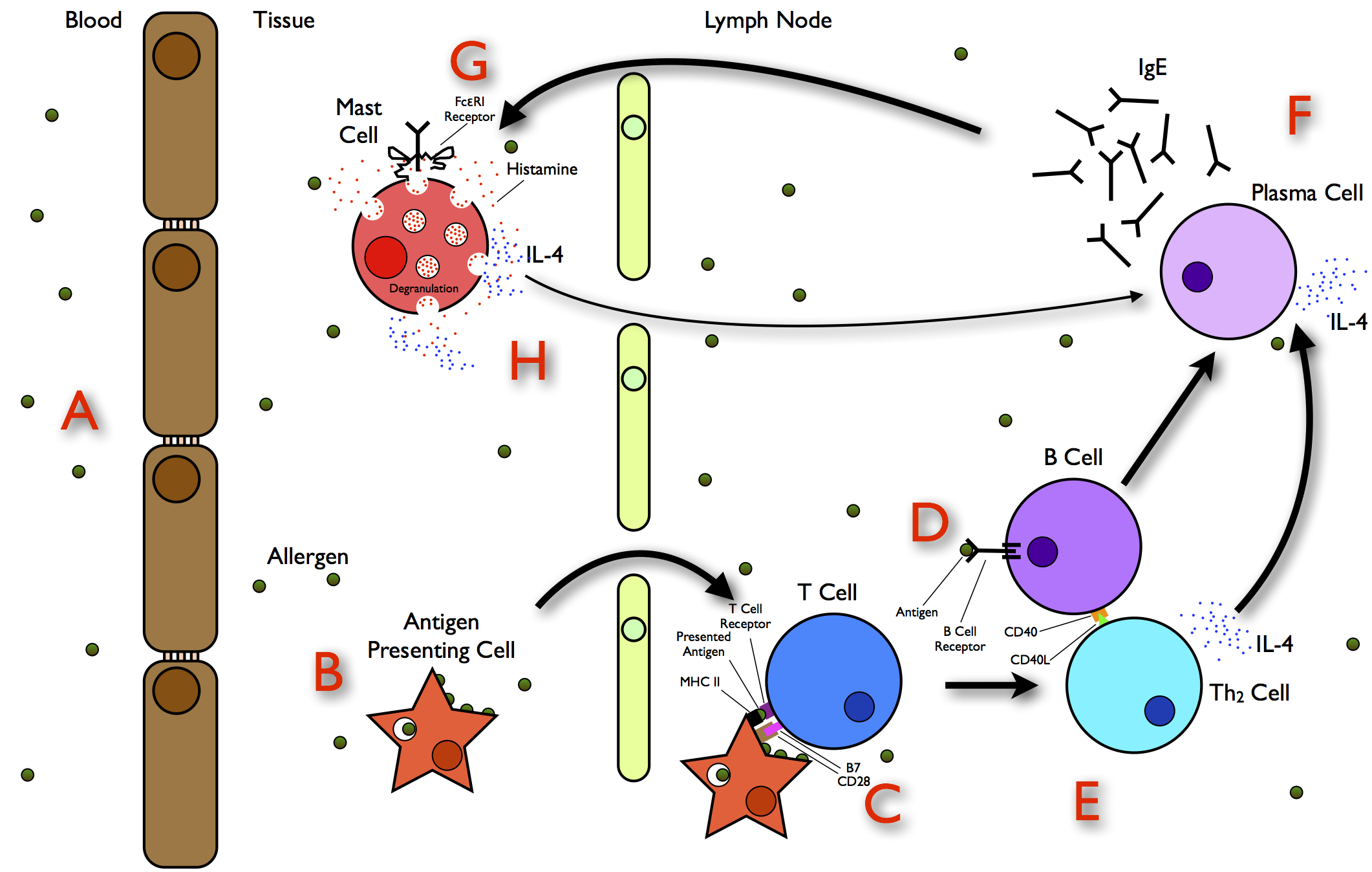
Allergies, also known as allergic diseases, are various conditions caused by hypersensitivity of the immune system to typically harmless substances in the environment. These diseases include Allergic rhinitis, hay fever, Food allergy, food allergies, atopic dermatitis, allergic asthma, and anaphylaxis. Symptoms may include allergic conjunctivitis, red eyes, an itchy rash, sneeze, sneezing, coughing, a rhinorrhea, runny nose, shortness of breath, or swelling. Note that food intolerances and food poisoning are separate conditions. Common allergens include pollen and certain foods. Metals and other substances may also cause such problems. Food, insect stings, and medications are common causes of severe reactions. Their development is due to both genetic and environmental factors. The underlying mechanism involves immunoglobulin E antibodies (IgE), part of the body's immune system, binding to an allergen and then to FcεRI, a receptor on mast cells or basophils where it triggers ... [...More Info...] [...Related Items...] OR: [Wikipedia] [Google] [Baidu] [Amazon] |
Agenesis
In medicine, agenesis () refers to the failure of an organ to develop during embryonic growth and development due to the absence of primordial tissue. Many forms of agenesis are referred to by individual names, depending on the organ affected: * Agenesis of the corpus callosum - failure of the Corpus callosum to develop *Renal agenesis - failure of one or both of the kidneys to develop * Amelia - failure of the arms or legs to develop * Penile agenesis - failure of penis to develop * Müllerian agenesis - failure of the uterus and part of the vagina to develop * Agenesis of the gallbladder - failure of the Gallbladder to develop. A person may not realize they have this condition unless they undergo surgery or medical imaging, since the gallbladder is neither externally visible nor essential. __TOC__ Eye agenesis Eye agenesis is a medical condition in which people are born with no eyes. Dental & oral agenesis * Anodontia, absence of all primary or permanent teeth. * Aglossia, ... [...More Info...] [...Related Items...] OR: [Wikipedia] [Google] [Baidu] [Amazon] |
Cartilaginous Nasal Capsule
Cartilage is a resilient and smooth type of connective tissue. Semi-transparent and non-porous, it is usually covered by a tough and fibrous membrane called perichondrium. In tetrapods, it covers and protects the ends of long bones at the joints as articular cartilage, and is a structural component of many body parts including the rib cage, the neck and the bronchial tubes, and the intervertebral discs. In other taxa, such as chondrichthyans and cyclostomes, it constitutes a much greater proportion of the skeleton. It is not as hard and rigid as bone, but it is much stiffer and much less flexible than muscle. The matrix of cartilage is made up of glycosaminoglycans, proteoglycans, collagen fibers and, sometimes, elastin. It usually grows quicker than bone. Because of its rigidity, cartilage often serves the purpose of holding tubes open in the body. Examples include the rings of the trachea, such as the cricoid cartilage and carina. Cartilage is composed of specialized cells c ... [...More Info...] [...Related Items...] OR: [Wikipedia] [Google] [Baidu] [Amazon] |
Maxillary Sinus
The pyramid-shaped maxillary sinus (or antrum of Nathaniel Highmore (surgeon), Highmore) is the largest of the paranasal sinuses, located in the maxilla. It drains into the middle meatus of the noseHuman Anatomy, Jacobs, Elsevier, 2008, page 209-210 through the semilunar hiatus. It is located to the side of the nasal cavity, and below the orbit. Structure It is the largest air sinus in the body. It has a mean volume of about 10 ml. It is situated within the body of the maxilla, but may extend into its Maxilla, zygomatic and Maxilla, alveolar processes when large. It is pyramid-shaped, with the apex at the maxillary zygomatic process, and the base represented by the lateral nasal wall. It has three recesses: an alveolar recess pointed inferiorly, bounded by the alveolar process of the maxilla; a zygomatic recess pointed laterally, bounded by the zygomatic bone; and an infraorbital recess pointed superiorly, bounded by the inferior Orbital surface of the body of the maxilla, orbita ... [...More Info...] [...Related Items...] OR: [Wikipedia] [Google] [Baidu] [Amazon] |
Maxillary Process Of Inferior Nasal Concha
From the lower border of the inferior nasal concha, a thin lamina, the maxillary process, curves downward and laterally; it articulates with the maxilla and forms a part of the medial wall of the maxillary sinus The pyramid-shaped maxillary sinus (or antrum of Nathaniel Highmore (surgeon), Highmore) is the largest of the paranasal sinuses, located in the maxilla. It drains into the middle meatus of the noseHuman Anatomy, Jacobs, Elsevier, 2008, page 209- .... References External links Bones of the head and neck {{musculoskeletal-stub ... [...More Info...] [...Related Items...] OR: [Wikipedia] [Google] [Baidu] [Amazon] |
Ethmoidal Process
Behind the lacrimal process of the inferior nasal conchae lies a broad, thin plate, the ethmoidal process, which ascends to join the uncinate process of the ethmoid; from its lower border a thin lamina, the maxillary process, curves downward and lateralward; it articulates with the maxilla and forms a part of the medial wall of the maxillary sinus The pyramid-shaped maxillary sinus (or antrum of Nathaniel Highmore (surgeon), Highmore) is the largest of the paranasal sinuses, located in the maxilla. It drains into the middle meatus of the noseHuman Anatomy, Jacobs, Elsevier, 2008, page 209- .... References Bones of the head and neck {{musculoskeletal-stub ... [...More Info...] [...Related Items...] OR: [Wikipedia] [Google] [Baidu] [Amazon] |
Nasolacrimal Duct
The nasolacrimal duct (also called the tear duct) carries tears from the lacrimal sac of the eye into the nasal cavity. The duct begins in the eye socket between the maxillary and lacrimal bones, from where it passes downwards and backwards. The opening of the nasolacrimal duct into the inferior nasal meatus of the nasal cavity is partially covered by a mucosal fold ( valve of Hasner or ''plica lacrimalis''). Excess tears flow through the nasolacrimal duct which drains into the inferior nasal meatus. This is the reason the nose starts to run when a person is crying or has watery eyes from an allergy, and why one can sometimes taste eye drops. This is for the same reason when applying some eye drops it is often advised to close the nasolacrimal duct by pressing it with a finger to prevent the medicine from escaping the eye and having unwanted side effects elsewhere in the body as it will proceed through the canal to the nasal cavity. Like the lacrimal sac, the duct is lin ... [...More Info...] [...Related Items...] OR: [Wikipedia] [Google] [Baidu] [Amazon] |


