|
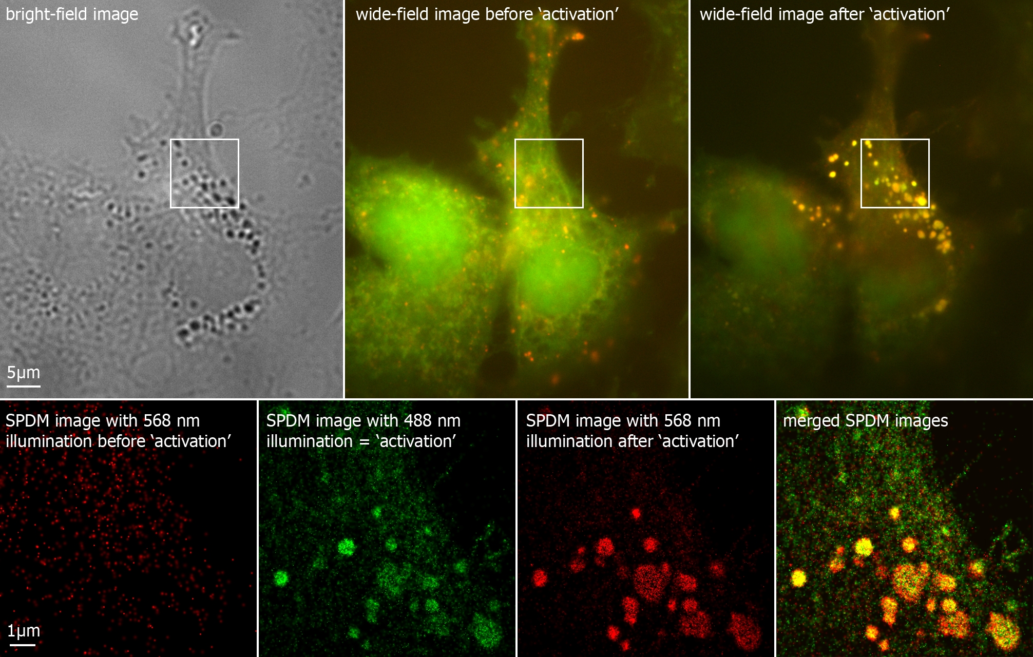
Immunofluorescence Microscopy
Immunofluorescence (IF) is a light microscopy-based technique that allows detection and localization of a wide variety of target biomolecules within a cell or tissue at a quantitative level. The technique utilizes the binding specificity of antibodies and antigens. The specific region an antibody recognizes on an antigen is called an epitope. Several antibodies can recognize the same epitope but differ in their binding affinity. The antibody with the higher affinity for a specific epitope will surpass antibodies with a lower affinity for the same epitope. By conjugating the antibody to a fluorophore, the position of the target biomolecule is visualized by exciting the fluorophore and measuring the emission of light in a specific predefined wavelength using a fluorescence microscope. It is imperative that the binding of the fluorophore to the antibody itself does not interfere with the immunological specificity of the antibody or the binding capacity of its antigen. Immunofluoresc ... [...More Info...] [...Related Items...] OR: [Wikipedia] [Google] [Baidu] |
Blood Vessels In Porcine Skin - SMA A488 - 20x
Blood is a body fluid in the circulatory system of humans and other vertebrates that delivers necessary substances such as nutrients and oxygen to the cells, and transports metabolic waste products away from those same cells. Blood is composed of blood cells suspended in blood plasma. Plasma, which constitutes 55% of blood fluid, is mostly water (92% by volume), and contains proteins, glucose, mineral ions, and hormones. The blood cells are mainly red blood cells (erythrocytes), white blood cells (leukocytes), and (in mammals) platelets (thrombocytes). The most abundant cells are red blood cells. These contain hemoglobin, which facilitates oxygen transport by reversibly binding to it, increasing its solubility. Jawed vertebrates have an adaptive immune system, based largely on white blood cells. White blood cells help to resist infections and parasites. Platelets are important in the clotting of blood. Blood is circulated around the body through blood vessels by the pumping a ... [...More Info...] [...Related Items...] OR: [Wikipedia] [Google] [Baidu] |
Confocal Microscope
Confocal microscopy, most frequently confocal laser scanning microscopy (CLSM) or laser scanning confocal microscopy (LSCM), is an optical imaging technique for increasing optical resolution and contrast of a micrograph by means of using a spatial pinhole to block out-of-focus light in image formation. Capturing multiple two-dimensional images at different depths in a sample enables the reconstruction of three-dimensional structures (a process known as optical sectioning) within an object. This technique is used extensively in the scientific and industrial communities and typical applications are in life sciences, semiconductor inspection and materials science. Light travels through the sample under a conventional microscope as far into the specimen as it can penetrate, while a confocal microscope only focuses a smaller beam of light at one narrow depth level at a time. The CLSM achieves a controlled and highly limited depth of field. Basic concept The principle of co ... [...More Info...] [...Related Items...] OR: [Wikipedia] [Google] [Baidu] |
Super-resolution Microscopy
Super-resolution microscopy is a series of techniques in optical microscopy that allow such images to have Optical resolution, resolutions higher than those imposed by the Diffraction-limited system, diffraction limit, which is due to the diffraction of light. Super-resolution imaging techniques rely on the Electromagnetic radiation#Near and far fields, near-field (photon-tunneling microscopy as well as those that use the Superlens, Pendry Superlens and near field scanning optical microscopy) or on the Near and far field, far-field. Among techniques that rely on the latter are those that improve the resolution only modestly (up to about a factor of two) beyond the diffraction-limit, such as confocal microscopy with closed pinhole or aided by computational methods such as deconvolution or detector-based pixel reassignment (e.g. re-scan microscopy, pixel reassignment), the 4Pi Microscope, 4Pi microscope, and structured-illumination microscopy technologies such as SIM and Vertico SMI ... [...More Info...] [...Related Items...] OR: [Wikipedia] [Google] [Baidu] |
HSP IF IgA
HSP may refer to: Biology, chemistry, and medicine *Hansen solubility parameters *Heat shock protein *Henoch–Schönlein purpura *Hereditary spastic paraplegia *Highly sensitive person, with high sensory processing sensitivity (SPS) Mathematics, software, and technology *Hidden subgroup problem, in mathematics *High Speed Photometer, Hubble Space Telescope instrument *Host signal processing, software emulating hardware *Hot Soup Processor, a programming language *High-Scoring Segment Pair, in the BLAST algorithm * List of Bluetooth profiles#Headset Profile (HSP) Education *Harvard Sussex Program, an inter-university collaboration * Holy Spirit Preparatory School, in Atlanta, Georgia, United States Political parties * Croatian Party of Rights (Croatian: ') * People's Voice Party (Turkish: '), Turkey Other uses *Halal snack pack A halal snack pack is an Australian fast food dish consisting of halal-certified doner kebab meat (lamb, chicken, or beef) and chips. It a ... [...More Info...] [...Related Items...] OR: [Wikipedia] [Google] [Baidu] |
Autofluorescence
Autofluorescence is the natural fluorescence of biological structures such as mitochondria and lysosomes, in contrast to fluorescence originating from artificially added fluorescent markers (fluorophores). The most commonly observed autofluorescencing molecules are NADPH and flavin group, flavins; the extracellular matrix can also contribute to autofluorescence because of the intrinsic properties of collagen and elastin. Generally, proteins containing an increased amount of the amino acids tryptophan, tyrosine, and phenylalanine show some degree of autofluorescence. Autofluorescence also occurs in non-biological materials found in many papers and textiles. Autofluorescence from U.S. paper money has been demonstrated as a means for discerning counterfeit currency from authentic currency. Microscopy Autofluorescence can be problematic in fluorescence microscopy. Light-emitting staining, stains (such as fluorescently labelled antibody, antibodies) are applied to Sample (material ... [...More Info...] [...Related Items...] OR: [Wikipedia] [Google] [Baidu] |
DyLight Fluor
The DyLight Fluor family of fluorescent dyes are produced by Dyomics in collaboration with Thermo Fisher Scientific. DyLight dyes are typically used in biotechnology and research applications as biomolecule, cell and tissue labels for fluorescence microscopy, cell biology or molecular biology. Applications Historically, fluorophores such as fluorescein, rhodamine, Cy3 and Cy5 have been used in a wide variety of applications. These dyes have limitations for use in microscopy and other applications that require exposure to an intense light source such as a laser, because they photobleach quickly (however, bleaching can be reduced at least 10 fold using oxygen scavenging). DyLight Fluors have comparable excitation and emission spectra and are claimed to be more photostable, brighter, and less pH-sensitive. The excitation and emission spectra of the DyLight Fluor series cover much of the visible spectrum and extend into the infrared region, allowing detection using most fluor ... [...More Info...] [...Related Items...] OR: [Wikipedia] [Google] [Baidu] |
Alexa Fluor
The Alexa Fluor family of fluorescent dyes is a series of dyes invented by Molecular Probes, now a part of Thermo Fisher Scientific, and sold under the Invitrogen brand name. Alexa Fluor dyes are frequently used as cell and tissue labels in fluorescence microscopy and cell biology. Alexa Fluor dyes can be conjugated directly to primary antibodies or to secondary antibodies to amplify signal and sensitivity or other biomolecules. The excitation and emission spectra of the Alexa Fluor series cover the visible spectrum and extend into the infrared. The individual members of the family are numbered according roughly to their excitation maxima in nanometers. History Richard and Rosaria Haugland, the founders of Molecular Probes, are well known in biology and chemistry for their research into fluorescent dyes useful in biological research applications. At the time that Molecular Probes was founded, such products were largely unavailable commercially. A number of fluorescent d ... [...More Info...] [...Related Items...] OR: [Wikipedia] [Google] [Baidu] |
Photobleaching
In optics, photobleaching (sometimes termed fading) is the photochemical alteration of a dye or a fluorophore molecule such that it is permanently unable to fluoresce. This is caused by cleaving of covalent bonds or non-specific reactions between the fluorophore and surrounding molecules. Such irreversible modifications in covalent bonds are caused by transition from a singlet state to the triplet state of the fluorophores. The number of excitation cycles to achieve full bleaching varies. In microscopy, photobleaching may complicate the observation of fluorescent molecules, since they will eventually be destroyed by the light exposure necessary to stimulate them into fluorescing. This is especially problematic in time-lapse microscopy. However, photobleaching may also be used prior to applying the (primarily antibody-linked) fluorescent molecules, in an attempt to quench autofluorescence. This can help improve the signal-to-noise ratio. Photobleaching may also be exploited to s ... [...More Info...] [...Related Items...] OR: [Wikipedia] [Google] [Baidu] |
Green Fluorescent Protein
The green fluorescent protein (GFP) is a protein that exhibits green fluorescence when exposed to light in the blue to ultraviolet range. The label ''GFP'' traditionally refers to the protein first isolated from the jellyfish ''Aequorea victoria'' and is sometimes called ''avGFP''. However, GFPs have been found in other organisms including corals, sea anemones, zoanithids, copepods and lancelets. The GFP from ''A. victoria'' has a major excitation peak at a wavelength of 395 nm and a minor one at 475 nm. Its emission peak is at 509 nm, which is in the lower green portion of the visible spectrum. The fluorescence quantum yield (QY) of GFP is 0.79. The GFP from the sea pansy ('' Renilla reniformis'') has a single major excitation peak at 498 nm. GFP makes for an excellent tool in many forms of biology due to its ability to form an internal chromophore without requiring any accessory cofactors, gene products, or enzymes / substrates other than molecular ox ... [...More Info...] [...Related Items...] OR: [Wikipedia] [Google] [Baidu] |
Recombinant Protein
Protein production is the biotechnological process of generating a specific protein. It is typically achieved by the manipulation of gene expression in an organism such that it expresses large amounts of a recombinant gene. This includes the transcription of the recombinant DNA to messenger RNA (mRNA), the translation of mRNA into polypeptide chains, which are ultimately folded into functional proteins and may be targeted to specific subcellular or extracellular locations. Protein production systems (also known as expression systems) are used in the life sciences, biotechnology, and medicine. Molecular biology research uses numerous proteins and enzymes, many of which are from expression systems; particularly DNA polymerase for PCR, reverse transcriptase for RNA analysis, restriction endonucleases for cloning, and to make proteins that are screened in drug discovery as biological targets or as potential drugs themselves. There are also significant applications for expressi ... [...More Info...] [...Related Items...] OR: [Wikipedia] [Google] [Baidu] |


