|
Homonymous Hemianopsia
Hemianopsia, or hemianopia, is a visual field loss on the left or right side of the vertical midline. It can affect one eye but usually affects both eyes. Homonymous hemianopsia (or homonymous hemianopia) is hemianopic visual field loss on the same side of both eyes. Homonymous hemianopsia occurs because the right half of the brain has visual pathways for the left hemifield of both eyes, and the left half of the brain has visual pathways for the right hemifield of both eyes. When one of these pathways is damaged, the corresponding visual field is lost. Signs and symptoms Paris as seen with right homonymous hemianopsia Mobility can be difficult for people with homonymous hemianopsia. "Patients frequently complain of bumping into obstacles on the side of the field loss, thereby bruising their arms and legs." People with homonymous hemianopsia often experience discomfort in crowds. "A patient with this condition may be unaware of what he or she cannot see and frequently bumps ... [...More Info...] [...Related Items...] OR: [Wikipedia] [Google] [Baidu] |
Paris
Paris () is the capital and most populous city of France, with an estimated population of 2,165,423 residents in 2019 in an area of more than 105 km² (41 sq mi), making it the 30th most densely populated city in the world in 2020. Since the 17th century, Paris has been one of the world's major centres of finance, diplomacy, commerce, fashion, gastronomy, and science. For its leading role in the arts and sciences, as well as its very early system of street lighting, in the 19th century it became known as "the City of Light". Like London, prior to the Second World War, it was also sometimes called the capital of the world. The City of Paris is the centre of the Île-de-France region, or Paris Region, with an estimated population of 12,262,544 in 2019, or about 19% of the population of France, making the region France's primate city. The Paris Region had a GDP of €739 billion ($743 billion) in 2019, which is the highest in Europe. According to the Economis ... [...More Info...] [...Related Items...] OR: [Wikipedia] [Google] [Baidu] |
Contralateral
Standard anatomical terms of location are used to unambiguously describe the anatomy of animals, including humans. The terms, typically derived from Latin or Greek roots, describe something in its standard anatomical position. This position provides a definition of what is at the front ("anterior"), behind ("posterior") and so on. As part of defining and describing terms, the body is described through the use of anatomical planes and anatomical axes. The meaning of terms that are used can change depending on whether an organism is bipedal or quadrupedal. Additionally, for some animals such as invertebrates, some terms may not have any meaning at all; for example, an animal that is radially symmetrical will have no anterior surface, but can still have a description that a part is close to the middle ("proximal") or further from the middle ("distal"). International organisations have determined vocabularies that are often used as standard vocabularies for subdisciplines of anatom ... [...More Info...] [...Related Items...] OR: [Wikipedia] [Google] [Baidu] |
Vision Restoration Therapy
Vision restoration therapy (VRT) is a noninvasive form of vision therapy which claims to increase the size of the visual fields in those with hemianopia. It, however, is of unclear benefit as of 2017 and is not part of standardized treatment approaches. Description of therapy Vision restoration therapy (VRT) is a computer-based treatment which claims to help with visual field defects regain visual functions through repetitive light stimulation. As the device used in VRT is similar to the DynaVision 2000 that already exist the Food and Drug Administration The United States Food and Drug Administration (FDA or US FDA) is a federal agency of the Department of Health and Human Services. The FDA is responsible for protecting and promoting public health through the control and supervision of food s ... (FDA) allowed an indication for use "...the diagnosis and improvement of visual functions in patients with impaired vision that may result from trauma, stroke, inflammation, surgical ... [...More Info...] [...Related Items...] OR: [Wikipedia] [Google] [Baidu] |
Blindsight
Blindsight is the ability of people who are cortically blind to respond to visual stimuli that they do not consciously see due to lesions in the primary visual cortex, also known as the striate cortex or Brodmann Area 17. The term was coined by Lawrence Weiskrantz and his colleagues in a paper published in a 1974 issue of ''Brain''. A previous paper studying the discriminatory capacity of a cortically blind patient was published in ''Nature'' in 1973. Type classification The majority of studies on blindsight are conducted on patients who are hemianopic, i.e. blind in one half of their visual field. Following the destruction of the left or right striate cortex, patients are asked to detect, localize, and discriminate amongst visual stimuli that are presented to their blind side, often in a forced-response or guessing situation, even though they may not consciously recognize the visual stimulus. Research shows that such blind patients may achieve a higher accuracy than would be ex ... [...More Info...] [...Related Items...] OR: [Wikipedia] [Google] [Baidu] |
Bitemporal Hemianopsia
Bitemporal hemianopsia, is the medical description of a type of partial blindness where vision is missing in the outer half of both the right and left visual field. It is usually associated with lesions of the optic chiasm, the area where the optic nerves from the right and left eyes cross near the pituitary gland. Causes In bitemporal hemianopsia, vision is missing in the outer (temporal or lateral) half of both the right and left visual fields. Information from the temporal visual field falls on the nasal (medial) retina. The nasal retina is responsible for carrying the information along the optic nerve, and crosses to the other side at the optic chiasm. When there is compression at optic chiasm, the visual impulse from both nasal retina are affected, leading to inability to view the temporal, or peripheral, vision. This phenomenon is known as bitemporal hemianopsia. Knowing the neurocircuitry of visual signal flow through the optic tract is very important in understanding bitem ... [...More Info...] [...Related Items...] OR: [Wikipedia] [Google] [Baidu] |
Binasal Hemianopsia
Binasal hemianopsia is the medical description of a type of partial blindness where vision is missing in the inner half of both the right and left visual field. It is associated with certain lesions of the eye and of the central nervous system, such as congenital hydrocephalus. Causes In binasal hemianopsia, vision is missing in the inner (nasal or medial) half of both the right and left visual fields. Information from the nasal visual field falls on the temporal (lateral) retina. Those lateral retinal nerve fibers do not cross in the optic chiasm. Calcification of the internal carotid arteries can impinge the uncrossed, lateral retinal fibers, leading to loss of vision in the nasal field. Clinical testing of visual fields (by confrontation) can produce a false positive result, particularly in inferior nasal quadrants. Management Etymology The absence of vision in half of a visual field is described as ''hemianopsia''. The absence of visual perception in one quarter of a vis ... [...More Info...] [...Related Items...] OR: [Wikipedia] [Google] [Baidu] |
Sprague Effect
The Sprague effect is the phenomenon where homonymous hemianopia, caused by damage to the visual cortex, gets slightly better when the contralesional superior colliculus is destroyed. The effect is named for its discoverer, James Sprague, who observed this phenomenon in 1966 using a cat model. Several reasons have been thought of for this happening, including mutual inhibition between the two brain hemispheres. For similar reasons of inhibiting an inhibitory structure, damaging the substantia nigra, for instance by using ibotenic acid Ibotenic acid or (''S'')-2-amino-2-(3-hydroxyisoxazol-5-yl)acetic acid, also referred to as ibotenate, is a chemical compound and psychoactive drug which occurs naturally in ''Amanita muscaria'' and related species of mushrooms typically found i ..., can also cause the same improvement. References {{reflist Visual disturbances and blindness Blindness ... [...More Info...] [...Related Items...] OR: [Wikipedia] [Google] [Baidu] |
Peli Lens
The Peli Lens is a mobility aid for people with homonymous hemianopia. It is also known as “EP” or Expansion Prism concept and was developed by Dr. Eli Peli oSchepens Eye Research Institutein 1999. It expands the visual field The visual field is the "spatial array of visual sensations available to observation in introspectionist psychological experiments". Or simply, visual field can be defined as the entire area that can be seen when an eye is fixed straight at a point ... by 20 degrees. He tested this concept on several patients in his private practice with great success using 40Δ Fresnel press-on prisms (Peli 2000). Development of the lens and clinical trials were funded by NEI-NIH Grant EY014723 awarded to Chadwick Optical. The results of the multi-center clinical trials were published in 2008 reporting a 74% patient acceptance rate.Bowers A, Keeney K, Peli E. (2008) Community-Based Trial of Peripheral Prism Visual Field Expansion Device for Hemianopia. Archives of Opht ... [...More Info...] [...Related Items...] OR: [Wikipedia] [Google] [Baidu] |
Posterior Cerebral Artery
The posterior cerebral artery (PCA) is one of a pair of cerebral arteries that supply oxygenated blood to the occipital lobe, part of the back of the human brain. The two arteries originate from the distal end of the basilar artery, where it bifurcates into the left and right posterior cerebral arteries. These anastomose with the middle cerebral arteries and internal carotid arteries via the posterior communicating arteries. Structure The posterior cerebral artery is subdivided into 4 sections P1: pre-communicating segment * Originated at the termination of the basilar artery * May give rise to the artery of Percheron if present P2: post-communicating segment * From the PCOM around the midbrain * Terminates as it enters the quadrigeminal ganglion * Gives rise to the choroidal branches (medial and lateral posterior choroidal arteries) P3: quadrigeminal segment * Courses posteromedially through the quadrigeminal cistern * Terminates as it enters the sulk of the occipi ... [...More Info...] [...Related Items...] OR: [Wikipedia] [Google] [Baidu] |
Cerebral Cancer
A brain tumor occurs when abnormal cells form within the brain. There are two main types of tumors: malignant tumors and benign (non-cancerous) tumors. These can be further classified as primary tumors, which start within the brain, and secondary tumors, which most commonly have spread from tumors located outside the brain, known as brain metastasis tumors. All types of brain tumors may produce symptoms that vary depending on the size of the tumor and the part of the brain that is involved. Where symptoms exist, they may include headaches, seizures, problems with vision, vomiting and mental changes. Other symptoms may include difficulty walking, speaking, with sensations, or unconsciousness. The cause of most brain tumors is unknown. Uncommon risk factors include exposure to vinyl chloride, Epstein–Barr virus, ionizing radiation, and inherited syndromes such as neurofibromatosis, tuberous sclerosis, and von Hippel-Lindau Disease. Studies on mobile phone exposure have not sh ... [...More Info...] [...Related Items...] OR: [Wikipedia] [Google] [Baidu] |
Scintillating Scotoma
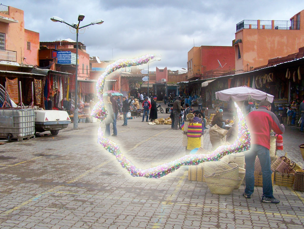
Scintillating scotoma is a common visual aura that was first described by 19th-century physician Hubert Airy (1838–1903). Originating from the brain, it may precede a migraine headache, but can also occur acephalgically (without headache), also known as visual migraine or migraine aura. It is often confused with retinal migraine, which originates in the eyeball or socket. Signs and symptoms Many variations occur, but scintillating scotoma usually begins as a spot of flickering light near or in the center of the visual field, which prevents vision within the scotoma area. It typically affects both eyes, as it is not a problem specific to one eye. The affected area flickers but is not dark. It then gradually expands outward from the initial spot. Vision remains normal beyond the borders of the expanding scotoma(s), with objects melting into the scotoma area background similarly to the physiological blind spot, which means that objects may be seen better by not looking directly ... [...More Info...] [...Related Items...] OR: [Wikipedia] [Google] [Baidu] |


