|
Hypothalamospinal Tract
The hypothalamospinal tract is an unmyelinated non-decussated descending nerve tract that arises in the hypothalamus and projects to the brainstem and spinal cord to synapse with pre-ganglionic autonomic (both sympathetic and parasympathetic) neurons. The direct autonomic projections of the hypothalamospinal tract represent a minority of the autonomic output of the hypothalamus; most is thought to project to various relay structures. Anatomy Origin The tract originates mainly from the paraventricular nucleus of hypothalamus, with minor contributions from the dorsomedial, ventromedial, and posterior nuclei of hypothalamus, and lateral hypothalamus. The neurons of the hypothalamospinal tract receive direct afferents from the ascending nociceptive sensory spinohypothalamic tract to mediate the autonomic response to painful stimuli. The tract terminates upon pre-ganglionic autonomic neurons in the brainstem, and spinal segments T1-L3 ( sympathetic outflow), and S2-S4 (par ... [...More Info...] [...Related Items...] OR: [Wikipedia] [Google] [Baidu] |
Unmyelinated
Myelin Sheath ( ) is a lipid-rich material that in most vertebrates surrounds the axons of neurons to Insulator (electricity), insulate them and increase the rate at which electrical impulses (called action potentials) pass along the axon. The myelinated axon can be likened to an electrical wire (the axon) with insulating material (myelin) around it. However, unlike the plastic covering on an electrical wire, myelin does not form a single long sheath over the entire length of the axon. Myelin ensheaths part of an axon known as an internodal segment, in multiple myelin layers of a tightly regulated internodal length. The ensheathed segments are separated at regular short unmyelinated intervals, called Node of Ranvier, nodes of Ranvier. Each node of Ranvier is around one micrometre long. Nodes of Ranvier enable a much faster rate of Conduction of electricity, conduction known as saltatory conduction where the action potential recharges at each node to jump over to the next node, an ... [...More Info...] [...Related Items...] OR: [Wikipedia] [Google] [Baidu] |
Dorsal Longitudinal Fasciculus
The dorsal longitudinal fasciculus (DLF) is a distinctive nerve tract in the midbrain. It extends from the hypothalamus rostrally to the spinal cord caudally, and contains both descending and ascending fibers. Descending fibers arise in the hypothalamus to project directly or indirectly onto autonomic nuclei and lower motor neurons of the brainstem and spinal cord; the descending component is involved in controlling chewing, swallowing, salivation and gastrointestinal secretory function, and shivering. Among the ascending fibers is a serotonin pathway arising in the raphe nuclei. Anatomy Ascending fibers Fibres arising from the nuclei of the reticular formation ascend in the DLF to terminate in the hypothalamus. It conveys visceral information to the brain. Fibers arising from the parabrachial area pass in the DLF to convey taste and general visceral sensation from the nucleus tractus solitarii to the posterior nucleus and periventricular nuclei of the hypothalamus. A ... [...More Info...] [...Related Items...] OR: [Wikipedia] [Google] [Baidu] |
Horner's Syndrome
Horner's syndrome, also known as oculosympathetic paresis, is a combination of symptoms that arises when a group of nerves known as the sympathetic trunk is damaged. The signs and symptoms occur on the same side (ipsilateral) as it is a lesion of the sympathetic trunk. It is characterized by miosis (a constricted pupil), partial ptosis (a weak, droopy eyelid), apparent anhidrosis (decreased sweating), with apparent enophthalmos (inset eyeball). The nerves of the sympathetic trunk arise from the spinal cord in the chest, and from there ascend to the neck and face. The nerves are part of the sympathetic nervous system, a division of the autonomic (or involuntary) nervous system. Once the syndrome has been recognized, medical imaging and response to particular eye drops may be required to identify the location of the problem and the underlying cause. Signs and symptoms Signs that are found in people with Horner's syndrome on the affected side of the face include the following ... [...More Info...] [...Related Items...] OR: [Wikipedia] [Google] [Baidu] |
Oxytocin
Oxytocin is a peptide hormone and neuropeptide normally produced in the hypothalamus and released by the posterior pituitary. Present in animals since early stages of evolution, in humans it plays roles in behavior that include Human bonding, social bonding, love, reproduction, childbirth, and the Postpartum period, period after childbirth. Oxytocin is released into the bloodstream as a hormone in response to Human sexual activity, sexual activity and during childbirth. It is also available in Oxytocin (medication), pharmaceutical form. In either form, oxytocin stimulates uterine contractions to speed up the process of childbirth. In its natural form, it also plays a role in maternal bonding and lactation, milk production. Production and secretion of oxytocin is controlled by a positive feedback mechanism, where its initial release stimulates production and release of further oxytocin. For example, when oxytocin is released during a contraction of the uterus at the start of c ... [...More Info...] [...Related Items...] OR: [Wikipedia] [Google] [Baidu] |
Pupillary Reflex
Pupillary reflex refers to one of the reflexes associated with pupillary function. These include the pupillary light reflex and accommodation reflex. Although the pupillary response, in which the pupil dilates or constricts due to light is not usually called a "reflex", it is still usually considered a part of this topic. Adjustment to close-range vision is known as "the near response", while relaxation of the ciliary muscle to view distant objects is known as the "far response". In "the near response" there are three processes that occur to focus an image on the retina. Convergence of the eyes, or the orientation of the visual axis of each eye towards an object in order to focus its image on each fovea, is the first of the three responses. This can be observed by the cross-eyed movement of the eyes when a finger is held up in front of a face and moved towards the face. Next, constriction of the pupil occurs. Because the lens cannot refract In physics, refraction is the r ... [...More Info...] [...Related Items...] OR: [Wikipedia] [Google] [Baidu] |
Pupillary Dilatation
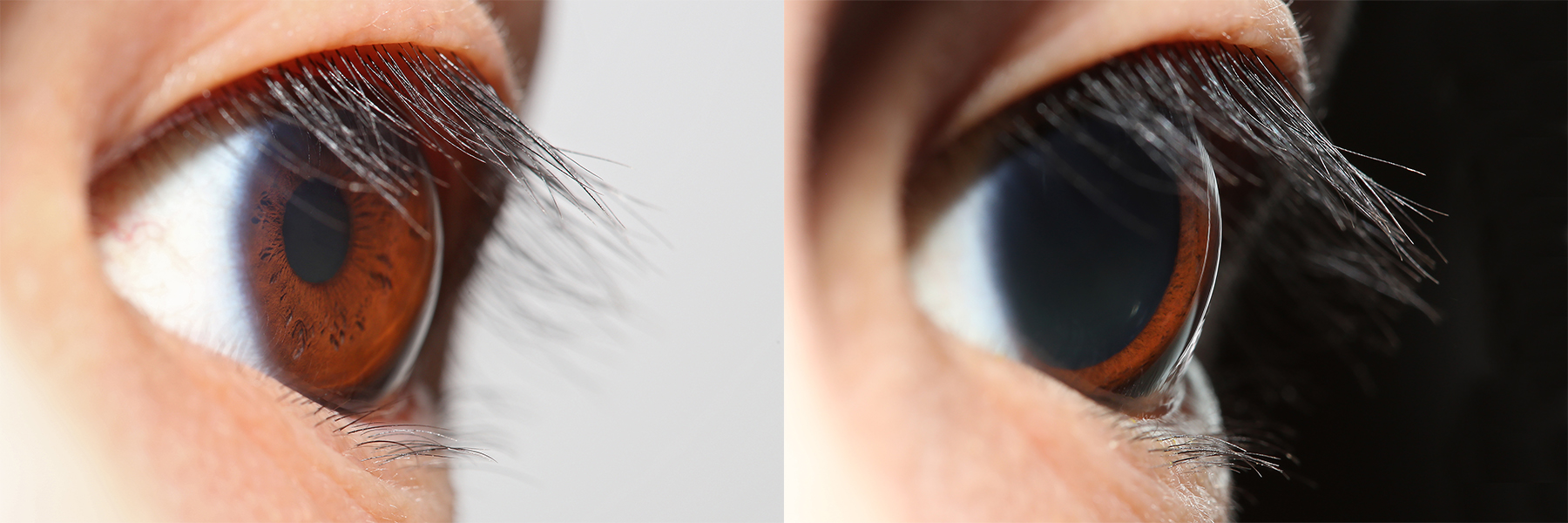
Mydriasis is the dilation of the pupil, usually having a non-physiological cause, or sometimes a physiological pupillary response. Non-physiological causes of mydriasis include disease, trauma, or the use of certain types of drugs. It may also be of unknown cause. Normally, as part of the pupillary light reflex, the pupil dilates in the dark and constricts in the light to respectively improve vividity at night and to protect the retina from sunlight damage during the day. A ''mydriatic'' pupil will remain excessively large even in a bright environment. The excitation of the radial fibres of the iris which increases the pupillary aperture is referred to as a mydriasis. More generally, mydriasis also refers to the natural dilation of pupils, for instance in low light conditions or under sympathetic stimulation. Mydriasis is frequently induced by drugs for certain ophthalmic examinations and procedures, particularly those requiring visual access to the retina. Fixed, unilateral ... [...More Info...] [...Related Items...] OR: [Wikipedia] [Google] [Baidu] |
Ciliospinal Center
The ciliospinal center (also known as Budge's center) is a cluster of pre-ganglionic sympathetic neuron cell bodies located in the intermediolateral cell column (of the cornu laterale) at spinal cord segment (C8: ''Anatomic variation'') T1-T2 It receives afferents from (the posterior part of) the hypothalamus via the (ipsilateral) hypothalamospinal tract which synapse with the center's pre-ganglionic sympathetic neurons. The efferent, pre-ganglionic axons then leave the spinal cord to enter and ascend in the sympathetic trunk to reach the superior cervical ganglion (SCG) where they synapse with post-ganglionic sympathetic neurons. The post-ganglionic neurons of the SCG then join the internal carotid nerve plexus of the internal carotid artery, accompanying first this artery and subsequently its branches to reach the orbit. In the orbit, they join the long ciliary nerves and short ciliary nerves to reach and innervate the dilator pupillae muscle to mediate pupillary dilatatio ... [...More Info...] [...Related Items...] OR: [Wikipedia] [Google] [Baidu] |
Hypothalamus
The hypothalamus (: hypothalami; ) is a small part of the vertebrate brain that contains a number of nucleus (neuroanatomy), nuclei with a variety of functions. One of the most important functions is to link the nervous system to the endocrine system via the pituitary gland. The hypothalamus is located below the thalamus and is part of the limbic system. It forms the Basal (anatomy), basal part of the diencephalon. All vertebrate brains contain a hypothalamus. In humans, it is about the size of an Almond#Nut, almond. The hypothalamus has the function of regulating certain metabolic biological process, processes and other activities of the autonomic nervous system. It biosynthesis, synthesizes and secretes certain neurohormones, called releasing hormones or hypothalamic hormones, and these in turn stimulate or inhibit the secretion of hormones from the pituitary gland. The hypothalamus controls thermoregulation, body temperature, hunger (physiology), hunger, important aspects o ... [...More Info...] [...Related Items...] OR: [Wikipedia] [Google] [Baidu] |
Superior Cervical Ganglion
The superior cervical ganglion (SCG) is the upper-most and largest of the cervical sympathetic ganglia of the sympathetic trunk. It probably formed by the union of four sympathetic ganglia of the cervical spinal nerves C1–C4. It is the only ganglion of the sympathetic nervous system that innervates the head and neck. The SCG innervates numerous structures of the head and neck. Structure The superior cervical ganglion is reddish-gray color, and usually shaped like a spindle with tapering ends. It measures about 3 cm in length. Sometimes the SCG is broad and flattened, and occasionally constricted at intervals. It formed by the coalescence of four ganglia, corresponding to the four upper-most cervical nerves C1–C4. The bodies of its preganglionic sympathetic afferent neurons are located in the lateral horn of the spinal cord. Their axons enter the SCG to synapse with postganglionic neurons whose axons then exit the rostral end of the SCG and proceed to innervate their targ ... [...More Info...] [...Related Items...] OR: [Wikipedia] [Google] [Baidu] |
Lateral Funiculus
The most lateral of the bundles of the anterior nerve roots is generally taken as a dividing line that separates the anterolateral system into two parts. These are the anterior funiculus, between the anterior median fissure and the most lateral of the anterior nerve roots, and the lateral funiculus between the exit of these roots and the posterolateral sulcus. The lateral funiculus transmits the contralateral corticospinal and spinothalamic tracts. A lateral cutting of the spinal cord results in the transection of both ipsilateral posterior column and lateral funiculus and this produces Brown-Séquard syndrome.Kaplan Qbook - USMLE Step 1 - 5th edition - page See also * Funiculus (neuroanatomy) * Anterior funiculus * Posterior funiculus References Central nervous system {{Neuroanatomy-stub ... [...More Info...] [...Related Items...] OR: [Wikipedia] [Google] [Baidu] |
Medulla Oblongata
The medulla oblongata or simply medulla is a long stem-like structure which makes up the lower part of the brainstem. It is anterior and partially inferior to the cerebellum. It is a cone-shaped neuronal mass responsible for autonomic (involuntary) functions, ranging from vomiting to sneezing. The medulla contains the cardiovascular center, the respiratory center, vomiting and vasomotor centers, responsible for the autonomic functions of breathing, heart rate and blood pressure as well as the sleep–wake cycle. "Medulla" is from Latin, ‘pith or marrow’. And "oblongata" is from Latin, ‘lengthened or longish or elongated'. During embryonic development, the medulla oblongata develops from the myelencephalon. The myelencephalon is a secondary brain vesicle which forms during the maturation of the rhombencephalon, also referred to as the hindbrain. The bulb is an archaic term for the medulla oblongata. In modern clinical usage, the word bulbar (as in bulbar palsy) is r ... [...More Info...] [...Related Items...] OR: [Wikipedia] [Google] [Baidu] |
Pons
The pons (from Latin , "bridge") is part of the brainstem that in humans and other mammals, lies inferior to the midbrain, superior to the medulla oblongata and anterior to the cerebellum. The pons is also called the pons Varolii ("bridge of Varolius"), after the Italian anatomist and surgeon Costanzo Varolio (1543–75). This region of the brainstem includes neural pathways and tracts that conduct signals from the brain down to the cerebellum and medulla, and tracts that carry the sensory signals up into the thalamus. Structure The pons in humans measures about in length. It is the part of the brainstem situated between the midbrain and the medulla oblongata. The horizontal ''medullopontine sulcus'' demarcates the boundary between the pons and medulla oblongata on the ventral aspect of the brainstem, and the roots of cranial nerves VI/VII/VIII emerge from the brainstem along this groove. The junction of pons, medulla oblongata, and cerebellum forms the cerebellopontine ... [...More Info...] [...Related Items...] OR: [Wikipedia] [Google] [Baidu] |


