|
Hemispherectomy
Hemispherectomy is a surgery that is performed by a Neurosurgery, neurosurgeon where an unhealthy Cerebral hemisphere, hemisphere of the brain is disconnected or removed. There are two types of hemispherectomy. ''Functional'' ''hemispherectomy'' refers to when the diseased brain is simply disconnected so that it can no longer send signals to the rest of the brain and body. ''Anatomical hemispherectomy'' refers to when not only is there disconnection, but also the diseased brain is physically removed from the skull. This surgery is mostly used as a treatment for medically intractable epilepsy, which is the term used when Anticonvulsant, anti-seizure medications are unable to control seizures. History The first anatomical hemispherectomy was performed and described in 1928 by Walter Dandy. This was done as an attempt to treat glioma, a brain tumor. The first known anatomical hemispherectomy performed as a treatment for intractable epilepsy was in 1938 by Kenneth McKenzie, a Canadi ... [...More Info...] [...Related Items...] OR: [Wikipedia] [Google] [Baidu] [Amazon] |
Rasmussen's Encephalitis
Rasmussen syndrome, also known as Rasmussen's encephalitis, is a rare progressive autoimmune Neurology, neurological disease. It is characterized by frequent and severe Focal seizure, focal seizures, progressive neurological decline, hemiparesis (weakness on one side of the body), encephalitis, and unilateral cerebral atrophy. The disease primarily affects children under the age of 15, though adult cases have been reported. Originally described as a form of chronic focal motor epilepsy by Dr. Aleksei Kozhevnikov, A. Ya. Kozhevnikov in the 1880s and separately identified as focal seizures due to chronic localized encephalitis in the 1950s by Dr. Theodore Rasmussen. It is now classified to be a cytotoxic T-cell–mediated encephalitis. Signs and symptoms The hallmark symptoms are focal seizures, particularly Epilepsia partialis continua, Epilepsia Partialis Continua (EPC) a form of epilepsy characterized by continuous or near-continuous clonic movements in a localized body part. Th ... [...More Info...] [...Related Items...] OR: [Wikipedia] [Google] [Baidu] [Amazon] |
Sturge–Weber Syndrome
Sturge–Weber syndrome, sometimes referred to as encephalotrigeminal angiomatosis, is a rare congenital neurocutaneous disorder (also known as phakomatoses). It is often associated with port-wine stains of the face, glaucoma, seizures, intellectual disability, and ipsilateral leptomeningeal angioma (cerebral malformations and tumors). Sturge–Weber syndrome can be classified into three different types. Type 1 includes facial and leptomeningeal angiomas as well as the possibility of glaucoma or choroidal lesions. Normally, only one side of the brain is affected. This type is the most common. Type 2 involvement includes a facial angioma (port wine stain) with a possibility of glaucoma developing. There is no evidence of brain involvement. Symptoms can show at any time beyond the initial diagnosis of the facial angioma. The symptoms can include glaucoma, cerebral blood flow abnormalities and headaches. More research is needed on this type of Sturge–Weber syndrome. Type 3 has l ... [...More Info...] [...Related Items...] OR: [Wikipedia] [Google] [Baidu] [Amazon] |
Neurosurgery
Neurosurgery or neurological surgery, known in common parlance as brain surgery, is the specialty (medicine), medical specialty that focuses on the surgical treatment or rehabilitation of disorders which affect any portion of the nervous system including the Human brain, brain, spinal cord, peripheral nervous system, and cerebrovascular system. Neurosurgery as a medical specialty also includes non-surgical management of some neurological conditions. Education and context In different countries, there are different requirements for an individual to legally practice neurosurgery, and there are varying methods through which they must be educated. In most countries, neurosurgeon training requires a minimum period of seven years after graduating from medical school. United Kingdom In the United Kingdom, students must gain entry into medical school. The MBBS qualification (Bachelor of Medicine, Bachelor of Surgery) takes four to six years depending on the student's route. The newly qu ... [...More Info...] [...Related Items...] OR: [Wikipedia] [Google] [Baidu] [Amazon] |
Seizure
A seizure is a sudden, brief disruption of brain activity caused by abnormal, excessive, or synchronous neuronal firing. Depending on the regions of the brain involved, seizures can lead to changes in movement, sensation, behavior, awareness, or consciousness. Symptoms vary widely. Some seizures involve subtle changes, such as brief lapses in attention or awareness (as seen in absence seizures), while others cause generalized convulsions with loss of consciousness ( tonic–clonic seizures). Most seizures last less than two minutes and are followed by a postictal period of confusion, fatigue, or other symptoms. A seizure lasting longer than five minutes is a medical emergency known as status epilepticus. Seizures are classified as provoked, when triggered by a known cause such as fever, head trauma, or metabolic imbalance, or unprovoked, when no immediate trigger is identified. Recurrent unprovoked seizures define the neurological condition epilepsy. Clinical features Seizur ... [...More Info...] [...Related Items...] OR: [Wikipedia] [Google] [Baidu] [Amazon] |
Walter Dandy
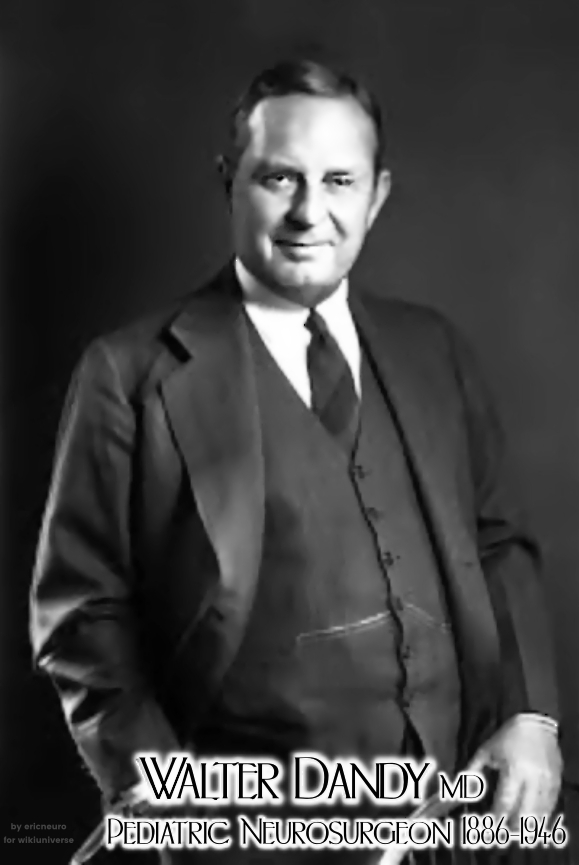
Walter Edward Dandy (April 6, 1886 – April 19, 1946) was an American neurosurgeon and scientist. He is considered one of the founding fathers of neurosurgery, along with Victor Horsley and Harvey Cushing. Dandy is credited with numerous neurosurgical discoveries and innovations, including the description of the circulation of cerebrospinal fluid in the brain, surgical treatment of hydrocephalus, the invention of air ventriculography and pneumoencephalography, the description of brain endoscopy, the establishment of the first intensive care unit, and the first clipping of an intracranial aneurysm, which marked the birth of cerebrovascular neurosurgery. During his 40-year medical career, Dandy published five books and more than 160 peer-reviewed articles while conducting a full-time, ground-breaking neurosurgical practice in which he performed during his peak years about 1000 operations per year. He was recognized at the time as a remarkably fast and particularly dexterous surgeo ... [...More Info...] [...Related Items...] OR: [Wikipedia] [Google] [Baidu] [Amazon] |
Focal Cortical Dysplasia
Focal cortical dysplasia (FCD) is a congenital abnormality of brain development where the neurons in an area of the brain failed to migrate in the proper formation ''in utero''. ''Focal'' means that it is limited to a focal zone in any lobe. Focal cortical dysplasia is a common cause of intractable epilepsy in children and is a frequent cause of epilepsy in adults. There are three types of FCD with subtypes, including type 1a, 1b, 1c, 2a, 2b, 3a, 3b, 3c, and 3d, each with distinct histopathological features. All forms of focal cortical dysplasia lead to disorganization of the normal structure of the cerebral cortex: * Type 1 FCD exhibits subtle alterations in cortical lamination. * Type 2a FCD exhibits neurons that are larger than normal that are called dysmorphic neurons (DN). FCD type 2b exhibits complete loss of laminar structure, and the presence of DN and enlarged cells are called balloon cells (BC) for their large elliptical cell body shape, laterally displaced nucleus, an ... [...More Info...] [...Related Items...] OR: [Wikipedia] [Google] [Baidu] [Amazon] |
Hemimegalencephaly
Hemimegalencephaly (HME), or unilateral megalencephaly, is a rare congenital disorder affecting all or a part of a cerebral hemisphere. It causes severe seizures, which are often frequent and hard to control. A minority might have seizure control with medicines, but most will need removal or disconnection of the affected hemisphere as the best chance. Uncontrolled, they often cause progressive intellectual disability and brain damage and stop development. Symptoms and signs Seizures are the main symptom. There can be as many as hundreds of seizures a day. Seizures tend to begin soon after birth, but may sometimes commence during later infancy or, rarely, during early childhood. Other symptoms * Asymmetrical or enlarged head * Developmental delay * Progressive weakness of half the body * Progressive blindness of half the body Genetics Somatic activation of '' AKT3'' causes hemispheric developmental brain malformations. Pathophysiology It is a disorder related to excessive ne ... [...More Info...] [...Related Items...] OR: [Wikipedia] [Google] [Baidu] [Amazon] |
Cerebral Hemisphere
The vertebrate cerebrum (brain) is formed by two cerebral hemispheres that are separated by a groove, the longitudinal fissure. The brain can thus be described as being divided into left and right cerebral hemispheres. Each of these hemispheres has an outer layer of grey matter, the cerebral cortex, that is supported by an inner layer of white matter. In eutherian (placental) mammals, the hemispheres are linked by the corpus callosum, a very large bundle of axon, nerve fibers. Smaller commissures, including the anterior commissure, the posterior commissure and the fornix (neuroanatomy), fornix, also join the hemispheres and these are also present in other vertebrates. These commissures transfer information between the two hemispheres to coordinate localized functions. There are three known poles of the cerebral hemispheres: the ''occipital lobe, occipital pole'', the ''frontal lobe, frontal pole'', and the ''temporal lobe, temporal pole''. The central sulcus is a prominent fissu ... [...More Info...] [...Related Items...] OR: [Wikipedia] [Google] [Baidu] [Amazon] |
Corpus Callosum
The corpus callosum (Latin for "tough body"), also callosal commissure, is a wide, thick nerve tract, consisting of a flat bundle of commissural fibers, beneath the cerebral cortex in the brain. The corpus callosum is only found in placental mammals. It spans part of the longitudinal fissure, connecting the left and right cerebral hemispheres, enabling communication between them. It is the largest white matter structure in the human brain, about in length and consisting of 200–300 million axonal projections. A number of separate nerve tracts, classed as subregions of the corpus callosum, connect different parts of the hemispheres. The main ones are known as the genu, the rostrum, the trunk or body, and the splenium. Structure The corpus callosum forms the floor of the longitudinal fissure that separates the two cerebral hemispheres. Part of the corpus callosum forms the roof of the lateral ventricles. The corpus callosum has four main parts – individual nerv ... [...More Info...] [...Related Items...] OR: [Wikipedia] [Google] [Baidu] [Amazon] |
Anesthesia
Anesthesia (American English) or anaesthesia (British English) is a state of controlled, temporary loss of sensation or awareness that is induced for medical or veterinary purposes. It may include some or all of analgesia (relief from or prevention of pain), paralysis (muscle relaxation), amnesia (loss of memory), and unconsciousness. An individual under the effects of anesthetic drugs is referred to as being anesthetized. Anesthesia enables the painless performance of procedures that would otherwise require physical restraint in a non-anesthetized individual, or would otherwise be technically unfeasible. Three broad categories of anesthesia exist: * ''General anesthesia'' suppresses central nervous system activity and results in unconsciousness and total lack of Sensation (psychology), sensation, using either injected or inhaled drugs. * ''Sedation'' suppresses the central nervous system to a lesser degree, inhibiting both anxiolysis, anxiety and creation of long-term memory, ... [...More Info...] [...Related Items...] OR: [Wikipedia] [Google] [Baidu] [Amazon] |
Magnetic Resonance Imaging
Magnetic resonance imaging (MRI) is a medical imaging technique used in radiology to generate pictures of the anatomy and the physiological processes inside the body. MRI scanners use strong magnetic fields, magnetic field gradients, and radio waves to form images of the organs in the body. MRI does not involve X-rays or the use of ionizing radiation, which distinguishes it from computed tomography (CT) and positron emission tomography (PET) scans. MRI is a medical application of nuclear magnetic resonance (NMR) which can also be used for imaging in other NMR applications, such as NMR spectroscopy. MRI is widely used in hospitals and clinics for medical diagnosis, staging and follow-up of disease. Compared to CT, MRI provides better contrast in images of soft tissues, e.g. in the brain or abdomen. However, it may be perceived as less comfortable by patients, due to the usually longer and louder measurements with the subject in a long, confining tube, although ... [...More Info...] [...Related Items...] OR: [Wikipedia] [Google] [Baidu] [Amazon] |


