|
Heel
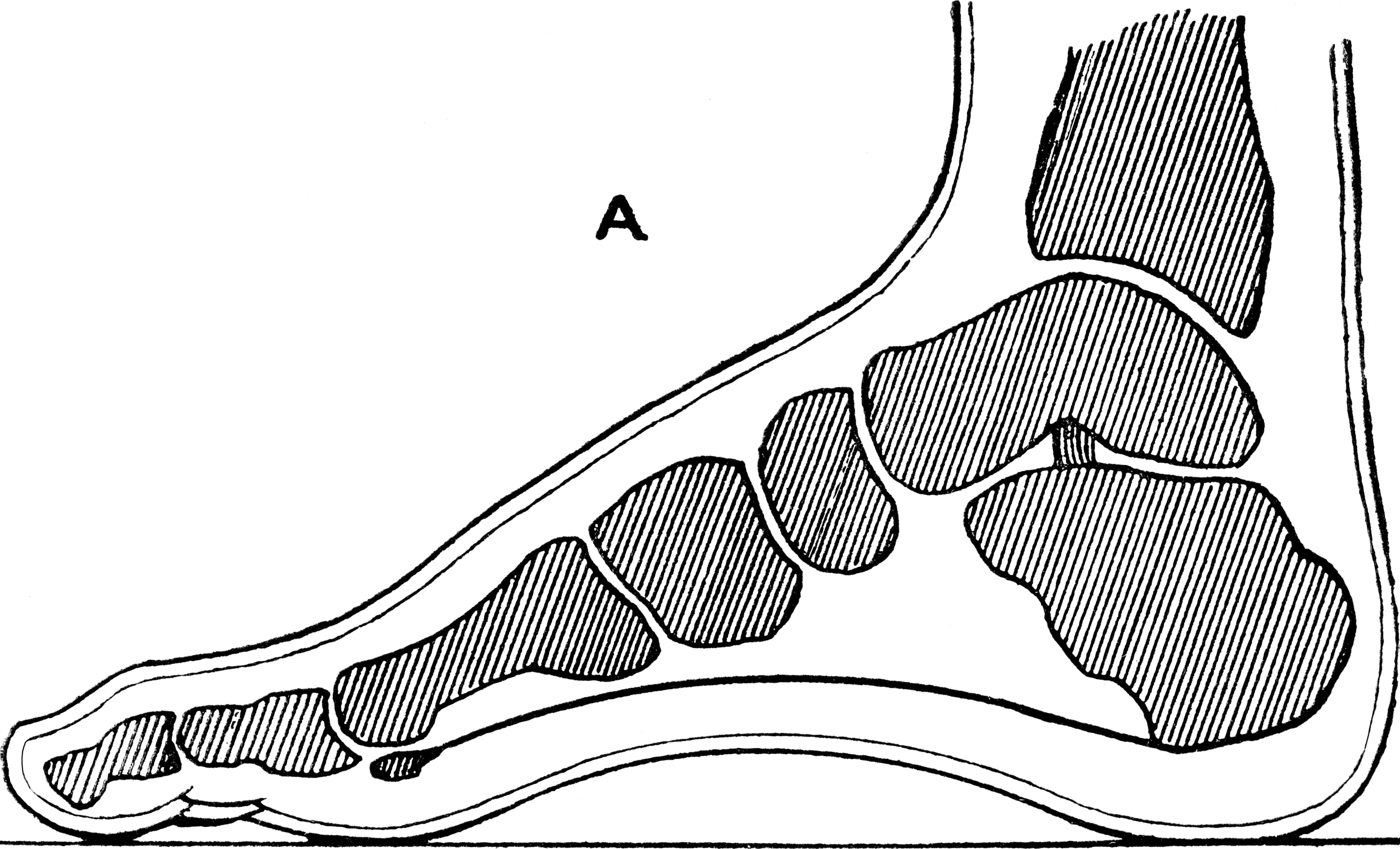
The heel is the prominence at the posterior end of the foot. It is based on the projection of one bone, the calcaneus or heel bone, behind the articulation of the bones of the lower leg. Structure To distribute the compressive forces exerted on the heel during gait, and especially the stance phase when the heel contacts the ground, the sole of the foot is covered by a layer of subcutaneous connective tissue up to 2 cm thick (under the heel). This tissue has a system of pressure chambers that both acts as a shock absorber and stabilises the sole. Each of these chambers contains fibrofatty tissue covered by a layer of tough connective tissue made of collagen fibers. These septa ("walls") are firmly attached both to the plantar aponeurosis above and the sole's skin below. The sole of the foot is one of the most highly vascularized regions of the body surface, and the dense system of blood vessels further stabilize the septa. The Achilles tendon is the muscle tendon of t ... [...More Info...] [...Related Items...] OR: [Wikipedia] [Google] [Baidu] |
Achilles' Heel
An Achilles' heel (or Achilles heel) is a weakness despite overall strength, which can lead to downfall. While the mythological origin refers to a physical vulnerability, idiomatic references to other attributes or qualities that can lead to downfall are common. The classical myth Although the death of Achilles was predicted by Hector in Homer's ''Iliad'', it does not actually occur in the ''Iliad,'' but was described in later Greek and Roman poetry and drama concerning events after the ''Iliad'', later in the Trojan War. In the myths surrounding the war, Achilles was said to have died from a wound to his heel, ankle, or torso, which was the result of an arrow—possibly poisoned—shot by Paris (mythology), Paris. The ''Iliad'' may have purposefully suppressed the myth to emphasise Achilles' human mortality and the stark chasm between gods and heroes. Some later Hellenistic-era myths record Thetis trying to make her son immortal by anointing him with ambrosia and burning a ... [...More Info...] [...Related Items...] OR: [Wikipedia] [Google] [Baidu] |
Foot
The foot (: feet) is an anatomical structure found in many vertebrates. It is the terminal portion of a limb which bears weight and allows locomotion. In many animals with feet, the foot is an organ at the terminal part of the leg made up of one or more segments or bones, generally including claws and/or nails. Etymology The word "foot", in the sense of meaning the "terminal part of the leg of a vertebrate animal" comes from Old English ''fot'', from Proto-Germanic *''fot'' (source also of Old Frisian ''fot'', Old Saxon ''fot'', Old Norse ''fotr'', Danish ''fod'', Swedish ''fot'', Dutch ''voet'', Old High German ''fuoz'', German ''Fuß'', Gothic ''fotus'', all meaning "foot"), from PIE root *''ped-'' "foot". The plural form ''feet'' is an instance of i-mutation. Structure The human foot is a strong and complex mechanical structure containing 26 bones, 33 joints (20 of which are actively articulated), and more than a hundred muscles, tendons, and ligaments.Podiatry Chan ... [...More Info...] [...Related Items...] OR: [Wikipedia] [Google] [Baidu] |
Achilles Tendon
The Achilles tendon or heel cord, also known as the calcaneal tendon, is a tendon at the back of the lower leg, and is the thickest in the human body. It serves to attach the plantaris, gastrocnemius (calf) and soleus muscles to the calcaneus (heel) bone. These muscles, acting via the tendon, cause plantar flexion of the foot at the ankle joint, and (except the soleus) flexion at the knee. Abnormalities of the Achilles tendon include inflammation ( Achilles tendinitis), degeneration, rupture, and becoming embedded with cholesterol deposits ( xanthomas). The Achilles tendon was named in 1693 after the Greek hero Achilles. History The oldest-known written record of the tendon being named after Achilles is in 1693 by the Flemish/Dutch anatomist Philip Verheyen. In his widely used text he described the tendon's location and said that it was commonly called "the cord of Achilles." The tendon has been described as early as the time of Hippocrates, who described it as th ... [...More Info...] [...Related Items...] OR: [Wikipedia] [Google] [Baidu] |
Calcaneus
In humans and many other primates, the calcaneus (; from the Latin ''calcaneus'' or ''calcaneum'', meaning heel; : calcanei or calcanea) or heel bone is a bone of the Tarsus (skeleton), tarsus of the foot which constitutes the heel. In some other animals, it is the point of the hock (anatomy), hock. Structure In humans, the calcaneus is the largest of the tarsal bones and the largest bone of the foot. Its long axis is pointed forwards and laterally. The talus bone, calcaneus, and navicular bone are considered the proximal row of tarsal bones. In the calcaneus, several important structures can be distinguished:Platzer (2004), p 216 There is a large calcaneal tuberosity located posteriorly on plantar surface with medial and lateral tubercles on its surface. Besides, there is another peroneal tubercle on its lateral surface. On its lower edge on either side are its lateral and medial processes (serving as the origins of the Abductor hallucis muscle, abductor hallucis and Abductor di ... [...More Info...] [...Related Items...] OR: [Wikipedia] [Google] [Baidu] |
Plantigrade
151px, Portion of a human skeleton, showing plantigrade habit In terrestrial animals, plantigrade locomotion means walking with the toes and metatarsals flat on the ground. It is one of three forms of locomotion adopted by terrestrial mammals. The other options are digitigrade, walking on the toes and fingers with the heel and wrist permanently raised, and unguligrade, walking on the nail or nails of the toes (the hoof) with the heel/wrist and the digits permanently raised. The leg of a plantigrade mammal includes the bones of the upper leg (femur/humerus) and lower leg (tibia and fibula/radius and ulna). The leg of a digitigrade mammal also includes the metatarsals/metacarpals, the bones that in a human compose the arch of the foot and the palm of the hand. The leg of an unguligrade mammal also includes the phalanges, the finger and toe bones. Among extinct animals, most early mammals such as pantodonts were plantigrade. A plantigrade foot is the primitive condition ... [...More Info...] [...Related Items...] OR: [Wikipedia] [Google] [Baidu] |
Plantar Aponeurosis
The plantar fascia or plantar aponeurosis is the thick connective tissue aponeurosis which supports the arch on the bottom (plantar side) of the foot. Recent studies suggest that the plantar fascia is actually an aponeurosis rather than true fascia. It runs from the tuberosity of the calcaneus (heel bone) forward to the heads of the metatarsal bones (the bone between each toe and the bones of the mid-foot). Anatomy The plantar fascia is the thick central portion of the fascia investing the plantar muscles. It extends between the medial process of the and the proximal phalanges of the toes. It provides some attachment to the flexor muscles of the toes. Distally, the plantar fascia becomes continuous with the fibrous sheaths enveloping the flexor tendons passing to the toes. At the anterior extremity of the sole inferior to the heads of the metatarsal bones the plantar aponeurosis forms the superficial transverse metatarsal ligament. Structure The plantar fascia is made u ... [...More Info...] [...Related Items...] OR: [Wikipedia] [Google] [Baidu] |
Ball (anatomy)
The ball of the foot is the padded portion of the sole between the toes and the arch, underneath the heads of the metatarsal bones. In comparative foot morphology, the ball is most analogous to the metacarpal (forepaw) or metatarsal (hindpaw) pad in many mammals with paws, and serves mostly the same functions. The ball is a common area in which people develop pain, known as metatarsalgia. People who frequently wear high heels often develop pain in the balls of their feet from the immense amount of pressure that is placed on them for long periods of time, due to the inclination of the shoes. To remedy this, there is a market for ball-of-foot or general foot cushions that are placed into shoes to relieve some of the pressure. Alternately, people can have a procedure done in which a dermal filler is injected into the balls of the feet to add cushioning. See also * Fat pad * Metatarsophalangeal joint The metatarsophalangeal joints (MTP joints) are the joints between the met ... [...More Info...] [...Related Items...] OR: [Wikipedia] [Google] [Baidu] |
Arches Of The Foot
The arches of the foot, formed by the tarsal and metatarsal bones, strengthened by ligaments and tendons, allow the foot to support the weight of the body in the erect posture with the least weight. They are categorized as longitudinal and transverse arches. Structure Longitudinal arches The longitudinal arches of the foot can be divided into medial and lateral arches. Medial arch The medial arch is higher than the lateral longitudinal arch. It is made up by the calcaneus, the talus, the navicular, the three cuneiforms (medial, intermediate, and lateral), and the first, second, and third metatarsals. Its summit is at the superior articular surface of the talus, and its two extremities or piers, on which it rests in standing, are the tuberosity on the plantar surface of the calcaneus posteriorly and the heads of the first, second, and third metatarsal bones anteriorly. The chief characteristic of this arch is its elasticity, due to its height and to the number of ... [...More Info...] [...Related Items...] OR: [Wikipedia] [Google] [Baidu] |
Gastrocnemius Muscle
The gastrocnemius muscle (plural ''gastrocnemii'') is a superficial two-headed muscle that is in the back part of the lower leg of humans. It is located superficial to the soleus in the posterior (back) compartment of the leg. It runs from its two heads just above the knee to the heel, extending across a total of three joints (knee, ankle and subtalar joints). The muscle is named via Latin, from Greek γαστήρ (''gaster'') 'belly' or 'stomach' and κνήμη (''knḗmē'') 'leg', meaning 'stomach of the leg' (referring to the bulging shape of the calf). Structure Origin/proximal attachment The lateral head originates from the lateral condyle of the femur, while the medial head originates from the medial condyle of the femur. Insertion/distal attachment Its other end forms a common tendon with the soleus muscle; this tendon is known as the calcaneal tendon or Achilles tendon and inserts onto the posterior surface of the calcaneus, or heel bone. Relations The ga ... [...More Info...] [...Related Items...] OR: [Wikipedia] [Google] [Baidu] |
Soleus Muscle
In humans and some other mammals, the soleus is a powerful muscle in the back part of the lower leg (the calf). It runs from just below the knee to the heel and is involved in standing and walking. It is closely connected to the gastrocnemius muscle, and some anatomists consider this combination to be a single muscle, the triceps surae. Its name is derived from the Latin word "solea", meaning "sandal". Structure The soleus is located in the superficial posterior compartment of the leg. The soleus exhibits significant morphological differences across species. It is unipennate in many species. In some animals, such as the rabbit, it is fused for much of its length with the gastrocnemius muscle. The soleus is a complex, multi-pennate muscle in humans, normally having a separate (posterior) aponeurosis from the gastrocnemius muscle. Most soleus muscle fibers originate from each side of the anterior aponeurosis, attached to the tibia and fibula. Other fibers originate from the po ... [...More Info...] [...Related Items...] OR: [Wikipedia] [Google] [Baidu] |
Ankle Bone
The talus (; Latin for ankle or ankle bone; : tali), talus bone, astragalus (), or ankle bone is one of the group of foot bones known as the tarsus. The tarsus forms the lower part of the ankle joint. It transmits the entire weight of the body from the lower legs to the foot.Platzer (2004), p 216 The talus has joints with the two bones of the lower leg, the tibia and thinner fibula. These leg bones have two prominences (the lateral and medial malleoli) that articulate with the talus. At the foot end, within the tarsus, the talus articulates with the calcaneus (heel bone) below, and with the curved navicular bone in front; together, these foot articulations form the ball-and-socket-shaped talocalcaneonavicular joint. The talus is the second largest of the tarsal bones; it is also one of the bones in the human body with the highest percentage of its surface area covered by articular cartilage. It is also unusual in that it has a retrograde blood supply, i.e. arterial bloo ... [...More Info...] [...Related Items...] OR: [Wikipedia] [Google] [Baidu] |
Navicular Bone
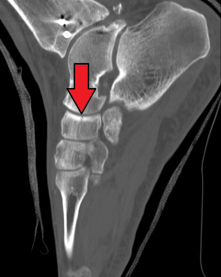
The navicular bone is a small bone found in the feet of most mammals. Human anatomy The navicular bone in humans is one of the tarsus (skeleton), tarsal bones, found in the foot. Its name derives from the human bone's resemblance to a small boat, caused by the strongly concave Anatomical terms of location#Proximal and distal, proximal joint, articular surface. The term ''navicular bone'' or ''hand navicular bone'' was formerly used for the scaphoid bone, one of the Carpal bones, carpal bones of the wrist. The navicular bone in humans is located on the Anatomical terms of location#Relative directions, medial side of the foot, and articulates proximally with the Talus bone, talus, Anatomical terms of location#Relative directions, distally with the three cuneiform bones, and Anatomical terms of location#Relative directions, laterally with the Cuboid bone, cuboid. It is the last of the foot bones to start ossification and does not tend to do so until the end of the third year in ... [...More Info...] [...Related Items...] OR: [Wikipedia] [Google] [Baidu] |


