|
Hamstring Muscles
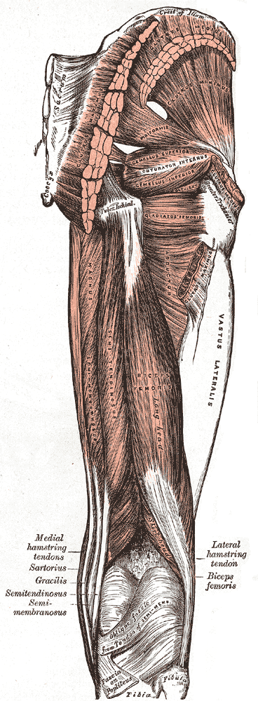
A hamstring () is any one of the three posterior thigh muscles in human anatomy between the hip and the knee: from medial to lateral, the semimembranosus, semitendinosus and biceps femoris. Etymology The word "ham" is derived from the Old English “ham” or “hom” meaning the hollow or bend of the knee, from a Germanic base where it meant "crooked". It gained the meaning of the leg of an animal around the 15th century. ''String'' refers to tendons, and thus the hamstrings' string-like tendons felt on either side of the back of the knee. Criteria The common criteria of any hamstring muscles are: # Muscles should originate from ischial tuberosity. # Muscles should be inserted over the knee joint, in the tibia or in the fibula. # Muscles will be innervated by the tibial branch of the sciatic nerve. # Muscle will participate in flexion of the knee joint and extension of the hip joint. Those muscles which fulfill all of the four criteria are called true hamstrings. The adduct ... [...More Info...] [...Related Items...] OR: [Wikipedia] [Google] [Baidu] |
Tuberosity Of The Ischium
The ischial tuberosity (or tuberosity of the ischium, tuber ischiadicum), also known colloquially as the sit bones or sitz bones, or as a pair the sitting bones, is a large posterior (anatomy), posterior bone, bony protuberance on the superior ramus of the ischium, superior ramus of the ischium. It marks the lateral boundary of the pelvic outlet. When sitting, the weight is frequently placed upon the ischial tuberosity. The Gluteus maximus muscle, gluteus maximus provides cover in the upright posture, but leaves it free in the seated position.Platzer (2004), p 236 The distance between a cyclist's ischial tuberosities is one of the factors in the choice of a bicycle saddle. Divisions The tuberosity is divided into two portions: a lower, rough, somewhat triangular part, and an upper, smooth, quadrilateral portion. * The ''lower portion'' is subdivided by a prominent longitudinal ridge, passing from base to apex, into two parts: ** The outer gives attachment to the adductor magnus ... [...More Info...] [...Related Items...] OR: [Wikipedia] [Google] [Baidu] |
Ischial Tuberosity
The ischial tuberosity (or tuberosity of the ischium, tuber ischiadicum), also known colloquially as the sit bones or sitz bones, or as a pair the sitting bones, is a large posterior bony protuberance on the superior ramus of the ischium. It marks the lateral boundary of the pelvic outlet. When sitting, the weight is frequently placed upon the ischial tuberosity. The gluteus maximus provides cover in the upright posture, but leaves it free in the seated position.Platzer (2004), p 236 The distance between a cyclist's ischial tuberosities is one of the factors in the choice of a bicycle saddle. Divisions The tuberosity is divided into two portions: a lower, rough, somewhat triangular part, and an upper, smooth, quadrilateral portion. * The ''lower portion'' is subdivided by a prominent longitudinal ridge, passing from base to apex, into two parts: ** The outer gives attachment to the adductor magnus ** The inner to the sacrotuberous ligament * The ''upper portion'' is subdiv ... [...More Info...] [...Related Items...] OR: [Wikipedia] [Google] [Baidu] |
Antagonist (muscle)
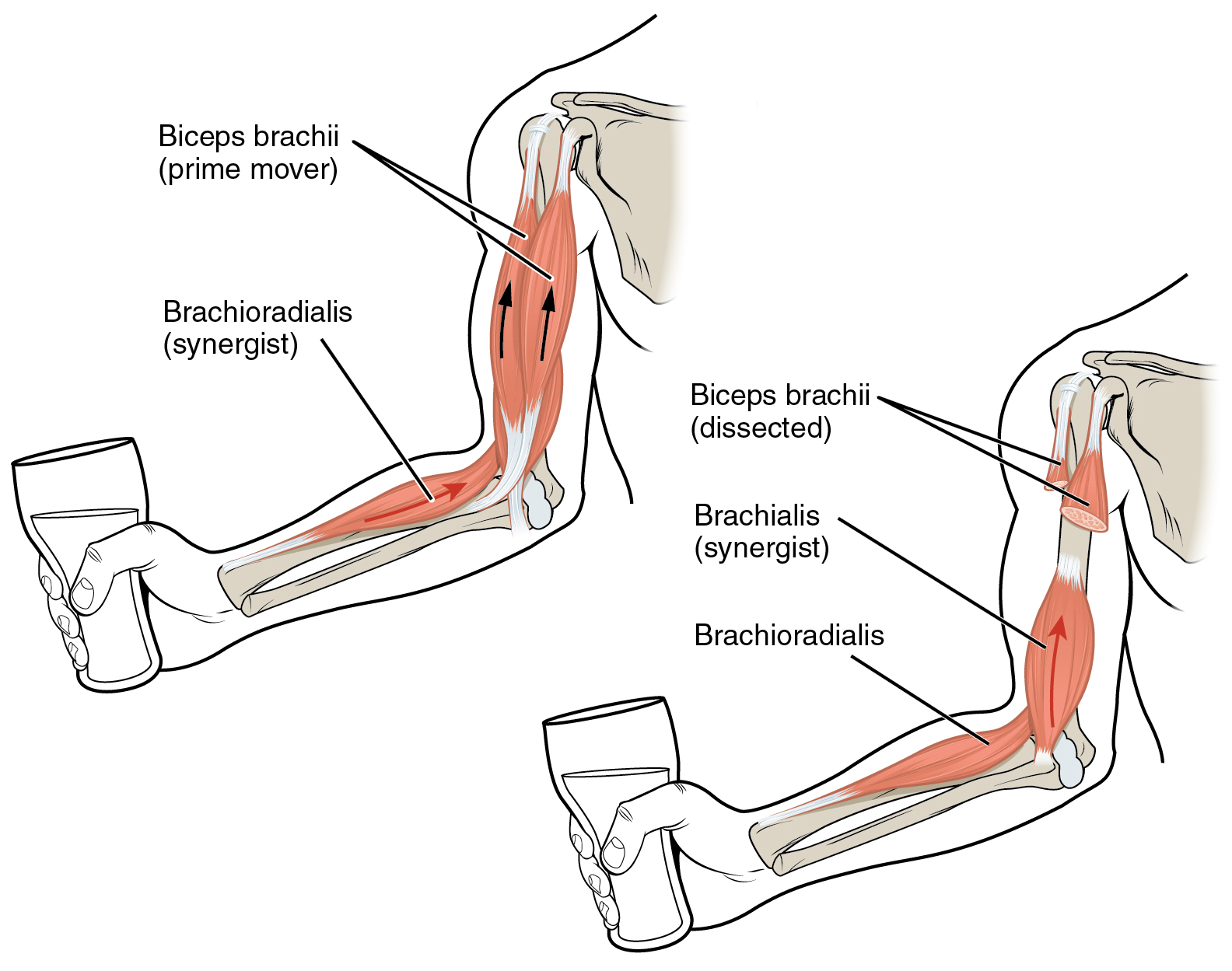
Anatomical terminology is used to uniquely describe aspects of skeletal muscle, cardiac muscle, and smooth muscle such as their actions, structure, size, and location. Types There are three types of muscle tissue in the body: skeletal, smooth, and cardiac. Skeletal muscle Skeletal muscle, or "voluntary muscle", is a striated muscle tissue that primarily joins to bone with tendons. Skeletal muscle enables movement of bones, and maintains posture. The widest part of a muscle that pulls on the tendons is known as the belly. Muscle slip A muscle slip is a slip of muscle that can either be an anatomical variant, or a branching of a muscle as in rib connections of the serratus anterior muscle. Smooth muscle Smooth muscle is involuntary and found in parts of the body where it conveys action without conscious intent. The majority of this type of muscle tissue is found in the digestive and urinary systems where it acts by propelling forward food, chyme, and feces in the former and ... [...More Info...] [...Related Items...] OR: [Wikipedia] [Google] [Baidu] |
Gluteal Muscles
The gluteal muscles, often called glutes, are a group of three muscles which make up the gluteal region commonly known as the buttocks: the gluteus maximus muscle, gluteus maximus, gluteus medius muscle, gluteus medius and gluteus minimus muscle, gluteus minimus. The three muscles originate from the ilium bone, ilium and sacrum and insert on the femur. The functions of the muscles include Extension (kinesiology), extension, Abduction (kinesiology), abduction, external rotation, and internal rotation of the hip joint. Structure The gluteus maximus is the largest and most wiktionary:superficial, superficial of the three gluteal muscles. It makes up a large part of the shape and appearance of the hips. It is a narrow and thick fleshy mass of a quadrilateral shape, and forms the prominence of the buttocks. The gluteus medius is a broad, thick, radiating muscle, situated on the outer surface of the pelvis. It lies profound to the gluteus maximus and its posterior third is covered by ... [...More Info...] [...Related Items...] OR: [Wikipedia] [Google] [Baidu] |
Biarticular Muscle
Biarticular muscles are muscles that cross two joints rather than just one, such as the hamstrings which cross both the hip and the knee. The function of these muscles is complex and often depends upon both their anatomy and the activity of other muscles at the joints in question. Their role in movement is poorly understood. Anatomy Biarticular muscles cross two joints in series, usually in a limb. The details of the origin (proximal attachment) and insertion (distal attachment) can play a large role in determining muscle function. For instance, the human gastrocnemius technically spans both the knee and ankle joints. However, the origin point of the muscle is so close to the axis of rotation of the knee joint that the muscle's effective lever arm would be very small, especially compared to its large lever arm at the ankle. As a result, even though it spans two joints, the strong bias in lever arms allows it to function primarily as an ankle plantar flexor. Other muscles, such ... [...More Info...] [...Related Items...] OR: [Wikipedia] [Google] [Baidu] |
Common Peroneal Nerve
The common fibular nerve (also known as the common peroneal nerve, external popliteal nerve, or lateral popliteal nerve) is a nerve in the lower leg that provides sensation over the posterolateral part of the leg and the knee joint. It divides at the knee into two terminal branches: the superficial fibular nerve and deep fibular nerve, which innervate the muscles of the lateral and anterior compartments of the leg respectively. When the common fibular nerve is damaged or compressed, foot drop can ensue. Structure The common fibular nerve is the smaller terminal branch of the sciatic nerve. The common fibular nerve has root values of L4, L5, S1, and S2. It arises from the superior angle of the popliteal fossa and extends to the lateral angle of the popliteal fossa, along the medial border of the biceps femoris. It then winds around the neck of the fibula to pierce the fibularis longus and divides into terminal branches of the superficial fibular nerve and the deep fibular nerve ... [...More Info...] [...Related Items...] OR: [Wikipedia] [Google] [Baidu] |
Lateral Supracondylar Line Of Femur
The femur (; : femurs or femora ), or thigh bone is the only bone in the thigh — the region of the lower limb between the hip and the knee. In many four-legged animals the femur is the upper bone of the hindleg. The top of the femur fits into a socket in the pelvis called the hip joint, and the bottom of the femur connects to the shinbone (tibia) and kneecap (patella) to form the knee. In humans the femur is the largest and thickest bone in the body. Structure The femur is the only bone in the upper leg. The two femurs converge medially toward the knees, where they articulate with the proximal ends of the tibiae. The angle at which the femora converge is an important factor in determining the femoral-tibial angle. In females, thicker pelvic bones cause the femora to converge more than in males. In the condition ''genu valgum'' (knock knee), the femurs converge so much that the knees touch. The opposite condition, ''genu varum'' (bow-leggedness), occurs when the femurs ... [...More Info...] [...Related Items...] OR: [Wikipedia] [Google] [Baidu] |
Head Of The Fibula
The fibula (: fibulae or fibulas) or calf bone is a leg bone on the lateral side of the tibia, to which it is connected above and below. It is the smaller of the two bones and, in proportion to its length, the most slender of all the long bones. Its upper extremity is small, placed toward the back of the head of the tibia, below the knee joint and excluded from the formation of this joint. Its lower extremity inclines a little forward, so as to be on a plane anterior to that of the upper end; it projects below the tibia and forms the lateral part of the ankle joint. Structure The bone has the following components: * Lateral malleolus * Interosseous membrane connecting the fibula to the tibia, forming a syndesmosis joint * The superior tibiofibular articulation is an arthrodial joint between the lateral condyle of the tibia and the head of the fibula. * The inferior tibiofibular articulation (tibiofibular syndesmosis) is formed by the rough, convex surface of the medial side of ... [...More Info...] [...Related Items...] OR: [Wikipedia] [Google] [Baidu] |
Medial Tibial Condyle
The medial condyle is the medial (or inner) portion of the upper extremity of tibia. It is the site of insertion for the semimembranosus muscle. See also * Lateral condyle of tibia * Medial collateral ligament The medial collateral ligament (MCL), also called the superficial medial collateral ligament (sMCL) or tibial collateral ligament (TCL), is one of the major ligaments of the knee. It is on the medial (inner) side of the knee joint and occurs in ... Additional images File:Gray258.png, Bones of the right leg. Anterior surface. File:Gray259.png, Bones of the right leg. Posterior surface. File:Slide2bib.JPG, Right knee in extension. Deep dissection. Posterior view. File:Slide2cocc.JPG, Right knee in extension. Deep dissection. Posterior view. References External links * * * () Bones of the lower limb Tibia {{musculoskeletal-stub ... [...More Info...] [...Related Items...] OR: [Wikipedia] [Google] [Baidu] |
Flexion
Motion, the process of movement, is described using specific anatomical terminology, anatomical terms. Motion includes movement of Organ (anatomy), organs, joints, Limb (anatomy), limbs, and specific sections of the body. The terminology used describes this motion according to its direction relative to the anatomical position of the body parts involved. Anatomy, Anatomists and others use a unified set of terms to describe most of the movements, although other, more specialized terms are necessary for describing unique movements such as those of the hands, feet, and eyes. In general, motion is classified according to the anatomical plane it occurs in. ''Flexion'' and ''extension'' are examples of ''angular'' motions, in which two axes of a joint are brought closer together or moved further apart. ''Rotational'' motion may occur at other joints, for example the shoulder, and are described as ''internal'' or ''external''. Other terms, such as ''elevation'' and ''depression'', descri ... [...More Info...] [...Related Items...] OR: [Wikipedia] [Google] [Baidu] |
Epicondyle
An epicondyle () is a rounded eminence on a bone that lies upon a condyle ('' epi-'', "upon" + ''condyle'', from a root meaning "knuckle" or "rounded articular area"). There are various epicondyles in the human skeleton The human skeleton is the internal framework of the human body. It is composed of around 270 bones at birth – this total decreases to around 206 bones by adulthood after some bones get fused together. The bone mass in the skeleton makes up ab ..., each named by its anatomic site. They include the following: Skeletal system {{musculoskeletal-stub ... [...More Info...] [...Related Items...] OR: [Wikipedia] [Google] [Baidu] |


