|
Great Trochanter
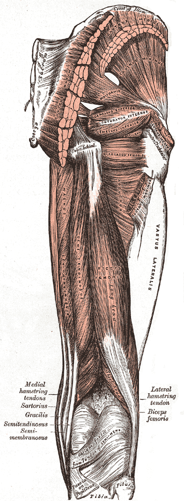
The greater trochanter of the femur is a large, irregular, quadrilateral eminence and a part of the skeletal system. It is directed lateral and medially and slightly posterior. In the adult it is about 2–4 cm lower than the femoral head.Standring, Susan, editor. ''Gray’s Anatomy: The Anatomical Basis of Clinical Practice''. Forty-First edition, Elsevier Limited, 2016, p. 1327. Because the pelvic outlet in the female is larger than in the male, there is a greater distance between the greater trochanters in the female. It has two surfaces and four borders. It is a traction epiphysis. Surfaces The ''lateral surface'', quadrilateral in form, is broad, rough, convex, and marked by a diagonal impression, which extends from the postero-superior to the antero-inferior angle, and serves for the insertion of the tendon of the gluteus medius. Above the impression is a triangular surface, sometimes rough for part of the tendon of the same muscle, sometimes smooth for the interpos ... [...More Info...] [...Related Items...] OR: [Wikipedia] [Google] [Baidu] |
Femur
The femur (; ), or thigh bone, is the proximal bone of the hindlimb in tetrapod vertebrates. The head of the femur articulates with the acetabulum in the pelvic bone forming the hip joint, while the distal part of the femur articulates with the tibia (shinbone) and patella (kneecap), forming the knee joint. By most measures the two (left and right) femurs are the strongest bones of the body, and in humans, the largest and thickest. Structure The femur is the only bone in the upper leg. The two femurs converge medially toward the knees, where they articulate with the proximal ends of the tibiae. The angle of convergence of the femora is a major factor in determining the femoral-tibial angle. Human females have thicker pelvic bones, causing their femora to converge more than in males. In the condition ''genu valgum'' (knock knee) the femurs converge so much that the knees touch one another. The opposite extreme is ''genu varum'' (bow-leggedness). In the general popu ... [...More Info...] [...Related Items...] OR: [Wikipedia] [Google] [Baidu] |
Epiphysis
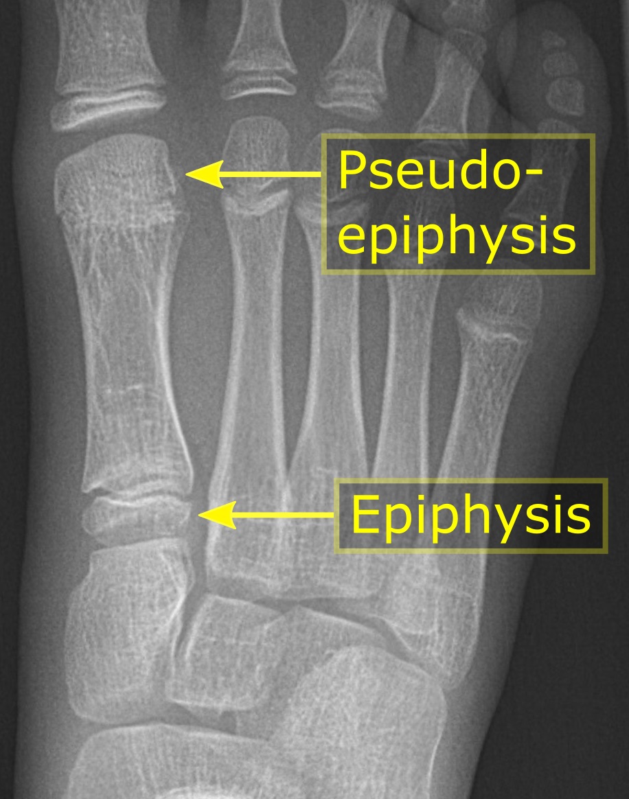
The epiphysis () is the rounded end of a long bone, at its joint with adjacent bone(s). Between the epiphysis and diaphysis (the long midsection of the long bone) lies the metaphysis, including the epiphyseal plate (growth plate). At the joint, the epiphysis is covered with articular cartilage; below that covering is a zone similar to the epiphyseal plate, known as subchondral bone. The epiphysis is filled with red bone marrow, which produces erythrocytes (red blood cells). Structure There are four types of epiphysis: # Pressure epiphysis: The region of the long bone that forms the joint is a pressure epiphysis (e.g. the head of the femur, part of the hip joint complex). Pressure epiphyses assist in transmitting the weight of the human body and are the regions of the bone that are under pressure during movement or locomotion. Another example of a pressure epiphysis is the head of the humerus which is part of the shoulder complex. condyles of femur and tibia also comes un ... [...More Info...] [...Related Items...] OR: [Wikipedia] [Google] [Baidu] |
Third Trochanter
In human anatomy, the third trochanter is a bony projection occasionally present on the proximal femur near the superior border of the gluteal tuberosity. When present, it is oblong, rounded, or conical in shape and sometimes continuous with the gluteal ridge. It generally occurs bilaterally without significant side to side dimorphism. A structure of minor importance in humans, the incidence of the third trochanter varies from 17 to 72% between ethnic groups and it is frequently reported as more common in females than in males. Structures analogous to the third trochanter are present in other mammals, including some primates. It is called the third trochanter in reference to the greater and lesser trochanters that are always present on the femur. Function Its function is to provide an attachment for the ascending tendon of the gluteus maximus muscle. It may function as (1) a reinforcement mechanism for the proximal femoral diaphysis in response to increased ground reaction ... [...More Info...] [...Related Items...] OR: [Wikipedia] [Google] [Baidu] |
Inferior Gemellus Muscle
The gemelli muscles are the inferior gemellus muscle and the superior gemellus muscle, two small accessory fasciculi to the tendon of the internal obturator muscle. The gemelli muscles belong to the lateral rotator group of six muscles of the hip that rotate the femur in the hip joint. Superior gemellus muscle The gemelli muscles are two small muscular fasciculi, accessories to the tendon of the internal obturator muscle which is received into a groove between them. The superior gemellus muscle is the higher placed gemellus muscle that arises from the outer (gluteal) surface of the ischial spine, and blends with the upper part of the tendon of the internal obturator. It is smaller than the inferior gemellus. In some people, the fibres of the gemellus superior extend further than average, and are prolonged onto the medial surface of the greater trochanter of the femur. The superior and inferior gemelli are supplied by the inferior gluteal artery. Nerve supply to the superior ... [...More Info...] [...Related Items...] OR: [Wikipedia] [Google] [Baidu] |
Superior Gemellus Muscle
The gemelli muscles are the inferior gemellus muscle and the superior gemellus muscle, two small accessory fasciculi to the tendon of the internal obturator muscle. The gemelli muscles belong to the lateral rotator group of six muscles of the hip that rotate the femur in the hip joint. Superior gemellus muscle The gemelli muscles are two small muscular fasciculi, accessories to the tendon of the internal obturator muscle which is received into a groove between them. The superior gemellus muscle is the higher placed gemellus muscle that arises from the outer (gluteal) surface of the ischial spine, and blends with the upper part of the tendon of the internal obturator. It is smaller than the inferior gemellus. In some people, the fibres of the gemellus superior extend further than average, and are prolonged onto the medial surface of the greater trochanter of the femur. The superior and inferior gemelli are supplied by the inferior gluteal artery. Nerve supply to the superior ... [...More Info...] [...Related Items...] OR: [Wikipedia] [Google] [Baidu] |
Obturator Externus
The external obturator muscle, obturator externus muscle (; OE) is a flat, triangular muscle, which covers the outer surface of the anterior wall of the pelvis. It is sometimes considered part of the medial compartment of thigh, and sometimes considered part of the gluteal region. Structure It arises from the margin of bone immediately around the medial side of the obturator membrane and surrounding bone, viz., from the inferior pubic ramus, and the ramus of the ischium; it also arises from the medial two-thirds of the outer surface of the obturator membrane, and from the tendinous arch which completes the canal for the passage of the obturator vessels and nerves. The fibers springing from the pubic arch extend on to the inner surface of the bone, where they obtain a narrow origin between the margin of the foramen and the attachment of the obturator membrane. The fibers converge and pass posterolateral and upward, and end in a tendon which runs across the back of the neck of ... [...More Info...] [...Related Items...] OR: [Wikipedia] [Google] [Baidu] |
Trochanteric Fossa
In mammals including humans, the medial surface of the greater trochanter has at its base a deep depression bounded posteriorly by the intertrochanteric crest, called the trochanteric fossa. This fossa is the point of insertion of four muscles. Moving from the inferior-most to the superior-most, they are: the tendon of the obturator externus muscle, the obturator internus, the superior gemellus and inferior gemellus. The width and depth of the trochanteric fossa varies taxonomically.Gray's AnatomyNetter 2003Romer 1956 In reptiliomorphs such as Seymouria or Diadectes and basal reptiles such as Pareiasaurus, the trochanteric fossa (also known as the intertrochanteric fossa) is a very large depression on the ventral/posterior side of the femur. It is bounded medially by the internal trochanter (also known as the lesser trochanter), laterally by the posterior branch of the ventral ridge, and inferiorly by the convergence of the anterior and posterior branches of the ventral ridge ... [...More Info...] [...Related Items...] OR: [Wikipedia] [Google] [Baidu] |
Bursa (anatomy)
( grc-gre, Προῦσα, Proûsa, Latin: Prusa, ota, بورسه, Arabic:بورصة) is a city in northwestern Turkey and the administrative center of Bursa Province. The fourth-most populous city in Turkey and second-most populous in the Marmara Region, Bursa is one of the industrial centers of the country. Most of Turkey's automotive production takes place in Bursa. As of 2019, the Metropolitan Province was home to 3,056,120 inhabitants, 2,161,990 of whom lived in the 3 city urban districts (Osmangazi, Yildirim and Nilufer) plus Gursu and Kestel, largely conurbated. Bursa was the first major and second overall capital of the Ottoman State between 1335 and 1363. The city was referred to as (, meaning "God's Gift" in Ottoman Turkish, a name of Persian origin) during the Ottoman period, while a more recent nickname is ("") in reference to the parks and gardens located across its urban fabric, as well as to the vast and richly varied forests of the surrounding region ... [...More Info...] [...Related Items...] OR: [Wikipedia] [Google] [Baidu] |
Gluteus Maximus
The gluteus maximus is the main extensor muscle of the hip. It is the largest and outermost of the three gluteal muscles and makes up a large part of the shape and appearance of each side of the hips. It is the single largest muscle in the human body. Its thick fleshy mass, in a quadrilateral shape, forms the prominence of the buttocks. The other gluteal muscles are the medius and minimus, and sometimes informally these are collectively referred to as the glutes. Its large size is one of the most characteristic features of the muscular system in humans,Norman Eizenberg et al., ''General Anatomy: Principles and Applications'' (2008), p. 17. connected as it is with the power of maintaining the trunk in the erect posture. Other primates have much flatter hips and cannot sustain standing erectly. The muscle is made up of muscle fascicles lying parallel with one another, and are collected together into larger bundles separated by fibrous septa. Structure The gluteus maximus i ... [...More Info...] [...Related Items...] OR: [Wikipedia] [Google] [Baidu] |
Trochanter
A trochanter is a tubercle of the femur near its joint with the hip bone. In humans and most mammals, the trochanters serve as important muscle attachment sites. Humans are known to have three trochanters, though the anatomic "normal" includes only the greater and lesser trochanters. (The third trochanter is not present in all specimens.) Etymology "Trokhos" (Greek) = "wheel", with reference to the spherical femoral head which was first named "trokhanter". Later usage came to include the femoral neck. Structure In human anatomy, the trochanter is a part of the femur. It can refer to: * Greater trochanter * Lesser trochanter * Third trochanter, which is occasionally present Other animals * Fourth trochanter, of archosaur leg bones * Trochanter (arthropod leg), a segment of the arthropod leg See also * Intertrochanteric crest * Intertrochanteric line The intertrochanteric line (or ''spiral line of the femur''White (2005), p 256 ) is a line located on the anterior side o ... [...More Info...] [...Related Items...] OR: [Wikipedia] [Google] [Baidu] |
Vastus Lateralis
The vastus lateralis (), also called the vastus externus, is the largest and most powerful part of the quadriceps femoris, a muscle in the thigh. Together with other muscles of the quadriceps group, it serves to extend the knee joint, moving the lower leg forward. It arises from a series of flat, broad tendons attached to the femur, and attaches to the outer border of the patella. It ultimately joins with the other muscles that make up the quadriceps in the quadriceps tendon, which travels over the knee to connect to the tibia. The vastus lateralis is the recommended site for intramuscular injection in infants less than 7 months old and those unable to walk, with loss of muscular tone.Mann, E. (2016). ''Injection (Intramuscular): Clinician Information.'' The Johanna Briggs Institute. Structure The vastus lateralis muscle arises from several areas of the femur, including the upper part of the intertrochanteric line; the lower, anterior borders of the greater trochanter, to the ... [...More Info...] [...Related Items...] OR: [Wikipedia] [Google] [Baidu] |
Femoral Head
The femoral head (femur head or head of the femur) is the highest part of the thigh bone (femur). It is supported by the femoral neck. Structure The head is globular and forms rather more than a hemisphere, is directed upward, medialward, and a little forward, the greater part of its convexity being above and in front. The femoral head's surface is smooth. It is coated with cartilage in the fresh state, except over an ovoid depression, the fovea capitis, which is situated a little below and behind the center of the femoral head, and gives attachment to the ligament of head of femur. The thickest region of the articular cartilage is at the centre of the femoral head, measuring up to 2.8 mm. The diameter of the femoral head is usually larger in men than in women. Fovea capitis The fovea capitis is a small, concave depression within the head of the femur that serves as an attachment point for the ligamentum teres (Saladin). It is slightly ovoid in shape and is oriented "superior- ... [...More Info...] [...Related Items...] OR: [Wikipedia] [Google] [Baidu] |


