|
Giemsa
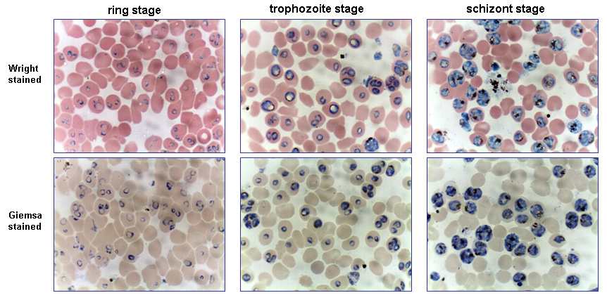
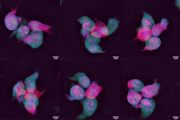
Giemsa stain (), named after German chemist and bacteriologist Gustav Giemsa, is a nucleic acid stain used in cytogenetics and for the histopathological diagnosis of malaria and other parasites. Uses It is specific for the phosphate groups of DNA and attaches itself to regions of DNA where there are high amounts of adenine-thymine bonding. Giemsa stain is used in Giemsa banding, commonly called G-banding, to stain chromosomes and often used to create a karyogram (chromosome map). It can identify chromosomal aberrations such as translocations and rearrangements. It stains the trophozoite ''Trichomonas vaginalis'', which presents with greenish discharge and motile cells on wet prep. Giemsa stain is also a differential stain, such as when it is combined with Wright stain to form Wright-Giemsa stain. It can be used to study the adherence of pathogenic bacteria to human cells. It differentially stains human and bacterial cells purple and pink respectively. It can be used for ... [...More Info...] [...Related Items...] OR: [Wikipedia] [Google] [Baidu] |
Karyotype
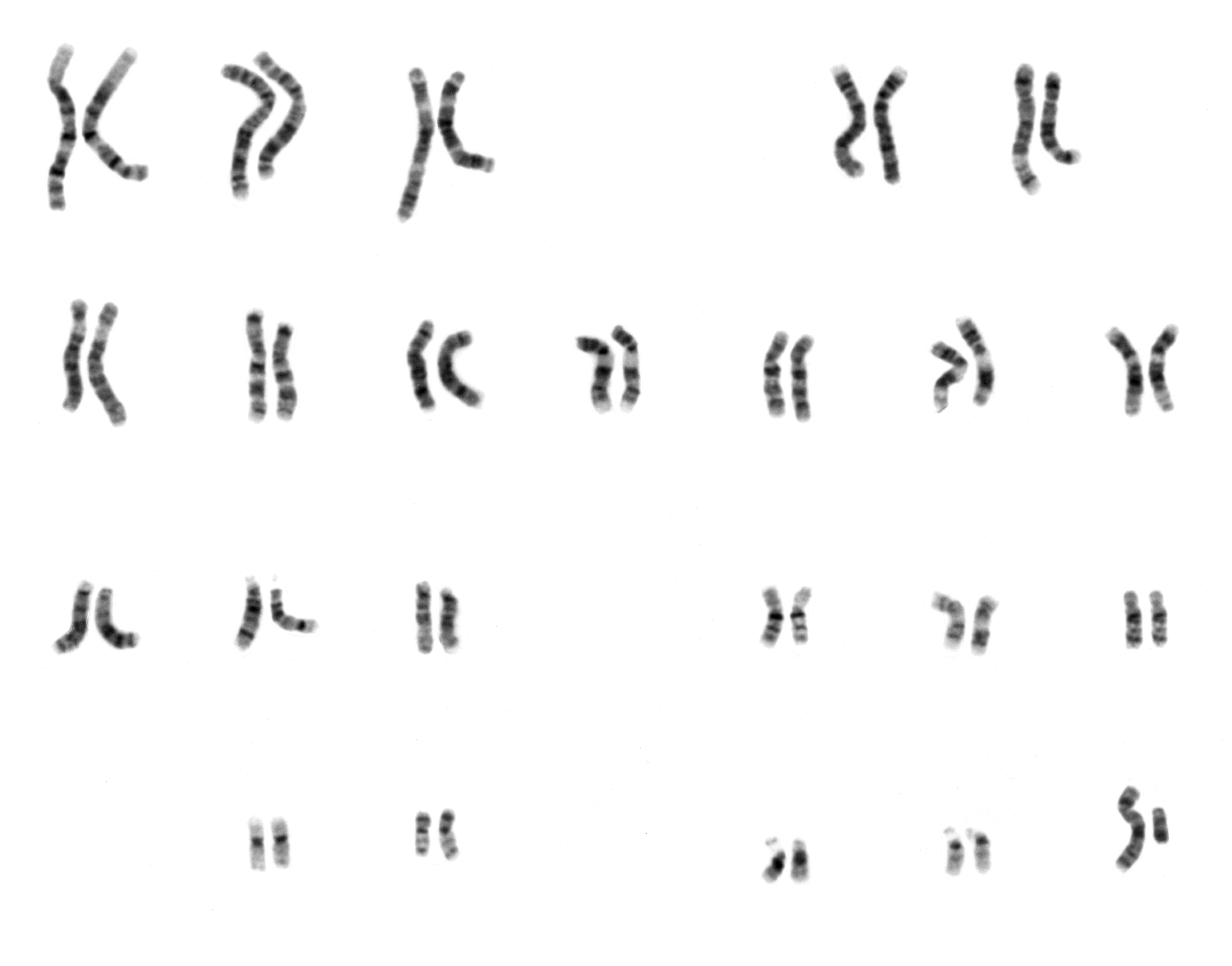
A karyotype is the general appearance of the complete set of metaphase chromosomes in the cells of a species or in an individual organism, mainly including their sizes, numbers, and shapes. Karyotyping is the process by which a karyotype is discerned by determining the chromosome complement of an individual, including the number of chromosomes and any abnormalities. A karyogram or idiogram is a graphical depiction of a karyotype, wherein chromosomes are organized in pairs, ordered by size and position of centromere for chromosomes of the same size. Karyotyping generally combines light microscopy and photography, and results in a photomicrographic (or simply micrographic) karyogram. In contrast, a schematic karyogram is a designed graphic representation of a karyotype. In schematic karyograms, just one of the sister chromatids of each chromosome is generally shown for brevity, and in reality they are generally so close together that they look as one on photomicrographs as well ... [...More Info...] [...Related Items...] OR: [Wikipedia] [Google] [Baidu] |
Gustav Giemsa
Gustav Giemsa (; November 20, 1867 – June 10, 1948) was a German chemist and bacteriologist who was a native of Medar-Blechhammer (now part of the city Kędzierzyn-Koźle). He is remembered for creating a dye solution commonly known as "Giemsa stain". This dye is used for the histopathological diagnosis of malaria and parasites such as ''Plasmodium'', ''Trypanosoma'', and ''Chlamydia''. Giemsa studied pharmacy and mineralogy at the University of Leipzig, and chemistry and bacteriology at the University of Berlin. Between 1895 and 1898 he worked as a pharmacist in German East Africa. He was an early assistant to Bernhard Nocht at the '' Institut für Tropenmedizin'' in Hamburg, where in 1900 he became head of the Department of Chemistry. In 1904 Giemsa published an essay on the staining procedure for flagellates, blood cells, and bacteria. Giemsa improved the Romanowsky stain (Eosin Y and Methylene Blue) by stabilizing this dye solution with glycerol. This allowed for re ... [...More Info...] [...Related Items...] OR: [Wikipedia] [Google] [Baidu] |
G-banding
G-banding, G banding or Giemsa banding is a technique used in cytogenetics to produce a visible karyotype by staining condensed chromosomes. It is the most common chromosome banding method. It is useful for identifying genetic diseases through the photographic representation of the entire chromosome complement.Speicher, Michael R. and Nigel P. Carter. "The New Cytogenetics: Blurring the Boundaries with Molecular Biology." ''Nature'' Reviews Genetics, Vol 6. Oct 2005. The metaphase chromosomes are treated with trypsin (to partially digest the chromosome) and stained with Giemsa stain. Heterochromatic regions, which tend to be rich with adenine and thymine (AT-rich) DNA and relatively gene-poor, stain more darkly in G-banding. In contrast, less condensed chromatin (Euchromatin)—which tends to be rich with guanine and cytosine ( GC-rich) and more transcriptionally active—incorporates less Giemsa stain, and these regions appear as light bands in G-banding. The pattern of bands ar ... [...More Info...] [...Related Items...] OR: [Wikipedia] [Google] [Baidu] |
Peripheral Blood Smear
A blood smear, peripheral blood smear or blood film is a thin layer of blood smeared on a glass microscope slide and then stained in such a way as to allow the various blood cells to be examined microscopically. Blood smears are examined in the investigation of hematological (blood) disorders and are routinely employed to look for blood parasites, such as those of malaria and filariasis. Preparation A blood smear is made by placing a drop of blood on one end of a slide, and using a ''spreader slide'' to disperse the blood over the slide's length. The aim is to get a region, called a monolayer, where the cells are spaced far enough apart to be counted and differentiated. The monolayer is found in the "feathered edge" created by the spreader slide as it draws the blood forward. The slide is left to air dry, after which the blood is fixed to the slide by immersing it briefly in methanol. The fixative is essential for good staining and presentation of cellular detail. After fixa ... [...More Info...] [...Related Items...] OR: [Wikipedia] [Google] [Baidu] |
Blood Film
A blood smear, peripheral blood smear or blood film is a thin layer of blood smeared on a glass microscope slide and then stained in such a way as to allow the various blood cells to be examined microscopically. Blood smears are examined in the investigation of hematological (blood) disorders and are routinely employed to look for blood parasites, such as those of malaria and filariasis. Preparation A blood smear is made by placing a drop of blood on one end of a slide, and using a ''spreader slide'' to disperse the blood over the slide's length. The aim is to get a region, called a monolayer, where the cells are spaced far enough apart to be counted and differentiated. The monolayer is found in the "feathered edge" created by the spreader slide as it draws the blood forward. The slide is left to air dry, after which the blood is fixed to the slide by immersing it briefly in methanol. The fixative is essential for good staining and presentation of cellular detail. After fixa ... [...More Info...] [...Related Items...] OR: [Wikipedia] [Google] [Baidu] |
Wright's Stain
Wright's stain is a hematologic stain that facilitates the differentiation of blood cell types. It is classically a mixture of eosin (red) and methylene blue dyes. It is used primarily to stain peripheral blood smears, urine samples, and bone marrow aspirates, which are examined under a light microscope. In cytogenetics, it is used to stain chromosomes to facilitate diagnosis of syndromes and diseases. It is named for James Homer Wright, who devised the stain, a modification of the Romanowsky stain, in 1902. Because it distinguishes easily between blood cells, it became widely used for performing differential white blood cell counts, which are routinely ordered when conditions such as infection or leukemia are suspected. The related stains are known as the buffered Wright stain, the Wright-Giemsa stain (a combination of Wright and Giemsa stains), and the buffered Wright-Giemsa stain, and specific instructions depend on the solutions being used, which may include eosin Y, a ... [...More Info...] [...Related Items...] OR: [Wikipedia] [Google] [Baidu] |
Cytogenetics
Cytogenetics is essentially a branch of genetics, but is also a part of cell biology/cytology (a subdivision of human anatomy), that is concerned with how the chromosomes relate to cell behaviour, particularly to their behaviour during mitosis and meiosis. Techniques used include karyotyping, analysis of G-banded chromosomes, other cytogenetic banding techniques, as well as molecular cytogenetics such as fluorescent ''in situ'' hybridization (FISH) and comparative genomic hybridization (CGH). History Beginnings Chromosomes were first observed in plant cells by Carl Nägeli in 1842. Their behavior in animal (salamander) cells was described by Walther Flemming, the discoverer of mitosis, in 1882. The name was coined by another German anatomist, von Waldeyer in 1888. The next stage took place after the development of genetics in the early 20th century, when it was appreciated that the set of chromosomes (the karyotype) was the carrier of the genes. Levitsky seems to have be ... [...More Info...] [...Related Items...] OR: [Wikipedia] [Google] [Baidu] |
Trichomonas Vaginalis
''Trichomonas vaginalis'' is an anaerobic, flagellated protozoan parasite and the causative agent of a sexually transmitted disease called trichomoniasis. It is the most common pathogenic protozoan that infects humans in industrialized countries. Infection rates in men and women are similar but women are usually symptomatic, while infections in men are usually asymptomatic. Transmission usually occurs via direct, skin-to-skin contact with an infected individual, most often through vaginal intercourse. The WHO has estimated that 160 million cases of infection are acquired annually worldwide. The estimates for North America alone are between 5 and 8 million new infections each year, with an estimated rate of asymptomatic cases as high as 50%. Usually treatment consists of metronidazole and tinidazole. Clinical History Alfred Francois Donné (1801–1878) was the first to describe a procedure to diagnose trichomoniasis through "the microscopic observation of motile proto ... [...More Info...] [...Related Items...] OR: [Wikipedia] [Google] [Baidu] |
Platelet
Platelets, also called thrombocytes (from Greek θρόμβος, "clot" and κύτος, "cell"), are a component of blood whose function (along with the coagulation factors) is to react to bleeding from blood vessel injury by clumping, thereby initiating a blood clot. Platelets have no cell nucleus; they are fragments of cytoplasm that are derived from the megakaryocytes of the bone marrow or lung, which then enter the circulation. Platelets are found only in mammals, whereas in other vertebrates (e.g. birds, amphibians), thrombocytes circulate as intact mononuclear cells. One major function of platelets is to contribute to hemostasis: the process of stopping bleeding at the site of interrupted endothelium. They gather at the site and, unless the interruption is physically too large, they plug the hole. First, platelets attach to substances outside the interrupted endothelium: ''adhesion''. Second, they change shape, turn on receptors and secrete chemical messengers: ''activati ... [...More Info...] [...Related Items...] OR: [Wikipedia] [Google] [Baidu] |
Plasmodium
''Plasmodium'' is a genus of unicellular eukaryotes that are obligate parasites of vertebrates and insects. The life cycles of ''Plasmodium'' species involve development in a blood-feeding insect host which then injects parasites into a vertebrate host during a blood meal. Parasites grow within a vertebrate body tissue (often the liver) before entering the bloodstream to infect red blood cells. The ensuing destruction of host red blood cells can result in malaria. During this infection, some parasites are picked up by a blood-feeding insect (mosquitoes in majority cases), continuing the life cycle. ''Plasmodium'' is a member of the phylum Apicomplexa, a large group of parasitic eukaryotes. Within Apicomplexa, ''Plasmodium'' is in the order Haemosporida and family Plasmodiidae. Over 200 species of ''Plasmodium'' have been described, many of which have been subdivided into 14 subgenera based on parasite morphology and host range. Evolutionary relationships among different '' ... [...More Info...] [...Related Items...] OR: [Wikipedia] [Google] [Baidu] |
Chromosome
A chromosome is a long DNA molecule with part or all of the genetic material of an organism. In most chromosomes the very long thin DNA fibers are coated with packaging proteins; in eukaryotic cells the most important of these proteins are the histones. These proteins, aided by chaperone proteins, bind to and condense the DNA molecule to maintain its integrity. These chromosomes display a complex three-dimensional structure, which plays a significant role in transcriptional regulation. Chromosomes are normally visible under a light microscope only during the metaphase of cell division (where all chromosomes are aligned in the center of the cell in their condensed form). Before this happens, each chromosome is duplicated ( S phase), and both copies are joined by a centromere, resulting either in an X-shaped structure (pictured above), if the centromere is located equatorially, or a two-arm structure, if the centromere is located distally. The joined copies are now c ... [...More Info...] [...Related Items...] OR: [Wikipedia] [Google] [Baidu] |
Lymphocyte
A lymphocyte is a type of white blood cell (leukocyte) in the immune system of most vertebrates. Lymphocytes include natural killer cells (which function in cell-mediated, cytotoxic innate immunity), T cells (for cell-mediated, cytotoxic adaptive immune system, adaptive immunity), and B cells (for humoral immunity, humoral, antibody-driven adaptive immune system, adaptive immunity). They are the main type of cell found in lymph, which prompted the name "lymphocyte". Lymphocytes make up between 18% and 42% of circulating white blood cells. Types The three major types of lymphocyte are T cell A T cell is a type of lymphocyte. T cells are one of the important white blood cells of the immune system and play a central role in the adaptive immune response. T cells can be distinguished from other lymphocytes by the presence of a T-cell r ...s, B cells and Natural killer cell, natural killer (NK) cells. Lymphocytes can be identified by their large nucleus. T cells and B cel ... [...More Info...] [...Related Items...] OR: [Wikipedia] [Google] [Baidu] |
.jpg)


.png)


