|
Foveola
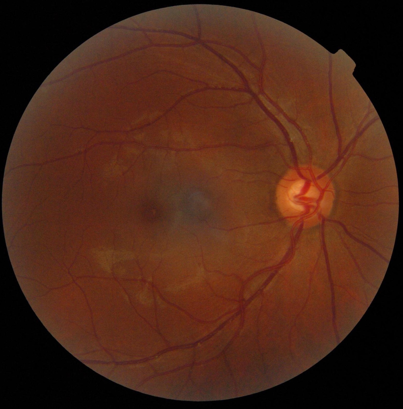
The foveola is located within a region called the macula, a yellowish, cone photoreceptor filled portion of the human retina. Approximately 0.35 mm in diameter, the foveola lies in the center of the fovea and contains only cone cells and a cone-shaped zone of Müller cells. In this region the cone receptors are found to be longer, slimmer, and more densely packed than anywhere else in the retina, thus allowing that region to have the potential to have the highest visual acuity in the eye. The center of the foveola is sometimes referred to as the umbo, a small (150-200µm) center of the floor of the foveola; features elongated cones. The umbo is the observed point corresponding to the normal light reflex but not solely responsible for this light reflex. The anatomy of the foveola was recently reinvestigated. Material was copied from this source, which is available under Creative Commons Attribution 4.0 International License Serial semithin and ultrathin sections, and focu ... [...More Info...] [...Related Items...] OR: [Wikipedia] [Google] [Baidu] |
Fovea Centralis
The fovea centralis is a small, central pit composed of closely packed cones in the eye. It is located in the center of the macula lutea of the retina. The fovea is responsible for sharp central vision (also called foveal vision), which is necessary in humans for activities for which visual detail is of primary importance, such as reading and driving. The fovea is surrounded by the ''parafovea'' belt and the ''perifovea'' outer region. The parafovea is the intermediate belt, where the ganglion cell layer is composed of more than five layers of cells, as well as the highest density of cones; the perifovea is the outermost region where the ganglion cell layer contains two to four layers of cells, and is where visual acuity is below the optimum. The perifovea contains an even more diminished density of cones, having 12 per 100 micrometres versus 50 per 100 micrometres in the most central fovea. That, in turn, is surrounded by a larger peripheral area, which delivers highly c ... [...More Info...] [...Related Items...] OR: [Wikipedia] [Google] [Baidu] |
Macula
The macula (/ˈmakjʊlə/) or macula lutea is an oval-shaped pigmented area in the center of the retina of the human eye and in other animals. The macula in humans has a diameter of around and is subdivided into the umbo, foveola, foveal avascular zone, fovea, parafovea, and perifovea areas. The anatomical macula at a size of is much larger than the clinical macula which, at a size of , corresponds to the anatomical fovea. The macula is responsible for the central, high-resolution, color vision that is possible in good light; and this kind of vision is impaired if the macula is damaged, for example in macular degeneration. The clinical macula is seen when viewed from the pupil, as in ophthalmoscopy or retinal photography. The term macula lutea comes from Latin ''macula'', "spot", and ''lutea'', "yellow". Structure The macula is an oval-shaped pigmented area in the center of the retina of the human eye and other animal eyes. Its center is shifted slightly away from t ... [...More Info...] [...Related Items...] OR: [Wikipedia] [Google] [Baidu] |
Retina
The retina (from la, rete "net") is the innermost, light-sensitive layer of tissue of the eye of most vertebrates and some molluscs. The optics of the eye create a focused two-dimensional image of the visual world on the retina, which then processes that image within the retina and sends nerve impulses along the optic nerve to the visual cortex to create visual perception. The retina serves a function which is in many ways analogous to that of the film or image sensor in a camera. The neural retina consists of several layers of neurons interconnected by synapses and is supported by an outer layer of pigmented epithelial cells. The primary light-sensing cells in the retina are the photoreceptor cells, which are of two types: rods and cones. Rods function mainly in dim light and provide monochromatic vision. Cones function in well-lit conditions and are responsible for the perception of colour through the use of a range of opsins, as well as high-acuity vision used ... [...More Info...] [...Related Items...] OR: [Wikipedia] [Google] [Baidu] |
Cone Cell
Cone cells, or cones, are photoreceptor cells in the retinas of vertebrate eyes including the human eye. They respond differently to light of different wavelengths, and the combination of their responses is responsible for color vision. Cones function best in relatively bright light, called the photopic region, as opposed to rod cells, which work better in dim light, or the scotopic region. Cone cells are densely packed in the fovea centralis, a 0.3 mm diameter rod-free area with very thin, densely packed cones which quickly reduce in number towards the periphery of the retina. Conversely, they are absent from the optic disc, contributing to the blind spot. There are about six to seven million cones in a human eye (vs ~92 million rods), with the highest concentration being towards the macula. Cones are less sensitive to light than the rod cells in the retina (which support vision at low light levels), but allow the perception of color. They are also able to perceive ... [...More Info...] [...Related Items...] OR: [Wikipedia] [Google] [Baidu] |
Müller Cell
Müller may refer to: * ''Die schöne Müllerin'' (1823) (sometimes referred to as ''Müllerlieder''; ''Müllerin'' is a female miller) is a song cycle with words by Wilhelm Müller and music by Franz Schubert * Doctor Müller, fictional character in ''The Adventures of Tintin'' by Hergé * Geiger–Müller tube, the sensing element of a Geiger counter instrument * GMD Müller, Swiss aerial lift manufacturing company * Müller (company), a German multinational dairy company * Müller (footballer, born 1966), nickname of ''Luís Antônio Corrêa da Costa'', Brazilian footballer * Muller glia, a macroglial cell in the retina * Müller Ltd. & Co. KG, a German pharmacy chain * Müller (lunar crater), impact crater on the lunar surface * Müller (Martian crater), impact crater on the Martian surface * Müller (store), a German retail store chain * Müller (surname), a common German surname * Müller-Thurgau, German wine grape * Müller Brothers, 19th-century string quartet * Müller ... [...More Info...] [...Related Items...] OR: [Wikipedia] [Google] [Baidu] |
CC-BY Icon
A Creative Commons (CC) license is one of several public copyright licenses that enable the free distribution of an otherwise copyrighted "work".A "work" is any creative material made by a person. A painting, a graphic, a book, a song/lyrics to a song, or a photograph of almost anything are all examples of "works". A CC license is used when an author wants to give other people the right to share, use, and build upon a work that the author has created. CC provides an author flexibility (for example, they might choose to allow only non-commercial uses of a given work) and protects the people who use or redistribute an author's work from concerns of copyright infringement as long as they abide by the conditions that are specified in the license by which the author distributes the work. There are several types of Creative Commons licenses. Each license differs by several combinations that condition the terms of distribution. They were initially released on December 16, 2002, by ... [...More Info...] [...Related Items...] OR: [Wikipedia] [Google] [Baidu] |
Fundus Photograph
Fundus photography involves photographing the rear of an eye, also known as the fundus. Specialized fundus cameras consisting of an intricate microscope attached to a flash enabled camera are used in fundus photography. The main structures that can be visualized on a fundus photo are the central and peripheral retina, optic disc and macula. Fundus photography can be performed with colored filters, or with specialized dyes including fluorescein and indocyanine green. The models and technology of fundus photography have advanced and evolved rapidly over the last century. Since the equipment is sophisticated and challenging to manufacture to clinical standards, only a few manufacturers/brands are available in the market: Welch Allyn, Digisight, Volk, Topcon, Zeiss, Canon, Nidek, Kowa, CSO, CenterVue, Ezer and Optos are some example of fundus camera manufacturers. History The concept of fundus photography was first introduced in the mid 19th century, after the introduction o ... [...More Info...] [...Related Items...] OR: [Wikipedia] [Google] [Baidu] |
Visual Artifact
Visual artifacts (also artefacts) are anomalies apparent during visual representation as in digital graphics and other forms of imagery, especially photography and microscopy. In digital graphics * Image quality factors, different types of visual artifacts * Compression artifacts * Digital artifacts, visual artifacts resulting from digital image processing * Noise * Screen-door effect, also known as fixed-pattern noise (FPN), a visual artifact of digital projection technology * Ghosting (television) *Screen burn-in * Distortion * Silk screen effect * Rainbow effect * Screen tearing * Moiré pattern * Color banding In video entertainment Many people who use their computers as a hobby experience artifacting due to a hardware or software malfunction. The cases can differ but the usual causes are: * Temperature issues, such as failure of cooling fan. * Unsuited video card (graphics card) drivers. * Drivers that have values that the graphics card is not suited with. * Overcloc ... [...More Info...] [...Related Items...] OR: [Wikipedia] [Google] [Baidu] |
Human Eye Anatomy
Humans (''Homo sapiens'') are the most abundant and widespread species of primate, characterized by bipedalism and exceptional cognitive skills due to a large and complex brain. This has enabled the development of advanced tools, culture, and language. Humans are highly social and tend to live in complex social structures composed of many cooperating and competing groups, from families and kinship networks to political states. Social interactions between humans have established a wide variety of values, social norms, and rituals, which bolster human society. Its intelligence and its desire to understand and influence the environment and to explain and manipulate phenomena have motivated humanity's development of science, philosophy, mythology, religion, and other fields of study. Although some scientists equate the term ''humans'' with all members of the genus ''Homo'', in common usage, it generally refers to ''Homo sapiens'', the only extant member. Anatomically modern hu ... [...More Info...] [...Related Items...] OR: [Wikipedia] [Google] [Baidu] |


