|
Fifth Metatarsal Bone
The fifth metatarsal bone is a long bone in the foot, and is palpable along the distal outer edges of the feet. It is the second smallest of the five metatarsal bones. The fifth metatarsal is analogous to the fifth metacarpal bone in the hand. As with the four other metatarsal bones it can be divided into three parts; a base, body and head. The base is the part closest to the ankle and the head is closest to the toes. The narrowed part in the middle is referred to as the body (or shaft) of the bone. The bone is somewhat flat giving it two surfaces; the plantar (towards the sole of the foot) and the dorsal side (the area facing upwards while standing). These surfaces are rough for the attachment of ligaments. The bone is curved longitudinally, so as to be concave below, slightly convex above. The base articulates behind, by a triangular surface cut obliquely in a transverse direction, with the cuboid; and medially, with the fourth metatarsal. The fifth metatarsal has a rough emine ... [...More Info...] [...Related Items...] OR: [Wikipedia] [Google] [Baidu] |
Long Bone
The long bones are those that are longer than they are wide. They are one of five types of bones: long, short, flat, irregular and sesamoid. Long bones, especially the femur and tibia, are subjected to most of the load during daily activities and they are crucial for skeletal mobility. They grow primarily by elongation of the diaphysis, with an epiphysis at each end of the growing bone. The ends of epiphyses are covered with hyaline cartilage ("articular cartilage"). The longitudinal growth of long bones is a result of endochondral ossification at the epiphyseal plate. Bone growth in length is stimulated by the production of growth hormone (GH), a secretion of the anterior lobe of the pituitary gland. The long bone category includes the femora, tibiae, and fibulae of the legs; the humeri, radii, and ulnae of the arms; metacarpals and metatarsals of the hands and feet, the phalanges of the fingers and toes, and the clavicles or collar bones. The long bones of the hum ... [...More Info...] [...Related Items...] OR: [Wikipedia] [Google] [Baidu] |
Stress Fracture
A stress fracture is a fatigue-induced bone fracture caused by repeated stress over time. Instead of resulting from a single severe impact, stress fractures are the result of accumulated injury from repeated submaximal loading, such as running or jumping. Because of this mechanism, stress fractures are common overuse injuries in athletes. Stress fractures can be described as small cracks in the bone, or hairline fractures. Stress fractures of the foot are sometimes called "march fractures" because of the injury's prevalence among heavily marching soldiers. Stress fractures most frequently occur in weight-bearing bones of the lower extremities, such as the tibia and fibula (bones of the lower leg), metatarsal and navicular bones (bones of the foot). Less common are stress fractures to the femur, pelvis, and sacrum. Treatment usually consists of rest followed by a gradual return to exercise over a period of months. Signs and symptoms Stress fractures are typically discovered afte ... [...More Info...] [...Related Items...] OR: [Wikipedia] [Google] [Baidu] |
Flexor Digiti Minimi Brevis Muscle (foot)
The Flexor digiti minimi brevis (Flexor brevis minimi digiti, Flexor digiti quinti brevis) lies under the metatarsal bone on the little toe, and resembles one of the Interossei. It arises from the base of the fifth metatarsal bone, and from the sheath of the Fibularis longus; its tendon is inserted into the lateral side of the base of the first phalanx The phalanx ( grc, φάλαγξ; plural phalanxes or phalanges, , ) was a rectangular mass military formation, usually composed entirely of heavy infantry armed with spears, pikes, sarissas, or similar pole weapons. The term is particularly ... of the fifth toe. Occasionally a few of the deeper fibers are inserted into the lateral part of the distal half of the fifth metatarsal bone; these are described by some as a distinct muscle, the opponens digiti quinti. References . Foot muscles Muscles of the lower limb {{muscle-stub ... [...More Info...] [...Related Items...] OR: [Wikipedia] [Google] [Baidu] |
Fibularis Brevis
In human anatomy, the fibularis brevis (or peroneus brevis) is a muscle that lies underneath the fibularis longus within the lateral compartment of the leg. It acts to tilt the sole of the foot away from the midline of the body (eversion) and to extend the foot downward away from the body at the ankle (plantar flexion). Structure The fibularis brevis arises from the lower two-thirds of the lateral, or outward, surface of the fibula (inward in relation to the fibularis longus) and from the connective tissue between it and the muscles on the front and back of the leg. The muscle passes downward and ends in a tendon that runs behind the lateral malleolus of the ankle in a groove that it shares with the tendon of the fibularis longus; the groove is converted into a canal by the superior fibular retinaculum, and the tendons in it are contained in a common mucous sheath. The tendon then runs forward along the lateral side of the calcaneus, above the calcaneal tubercle and the tendon ... [...More Info...] [...Related Items...] OR: [Wikipedia] [Google] [Baidu] |
Fibularis Tertius
In human anatomy, the fibularis tertius (also known as the peroneus tertius) is a muscle in the anterior compartment of the leg. It acts to tilt the sole of the foot away from the midline of the body ( eversion) and to pull the foot upward toward the body (dorsiflexion). Structure The fibularis tertius arises from the lower third of the front surface of the fibula, the lower part of the interosseous membrane, and septum, or connective tissue, between it and the fibularis brevis. The septum is sometimes called the intermuscular septum of Otto. The muscle passes downward and ends in a tendon that passes under the superior extensor retinaculum and the inferior extensor retinaculum of the foot in the same canal as the extensor digitorum longus muscle. It may be mistaken as a fifth tendon of the extensor digitorum longus. The tendon inserts into the medial part of the posterior surface of the shaft of the fifth metatarsal bone. The fibularis tertius is supplied by the deep fibular ... [...More Info...] [...Related Items...] OR: [Wikipedia] [Google] [Baidu] |
Tendon
A tendon or sinew is a tough, high-tensile-strength band of dense fibrous connective tissue that connects muscle to bone. It is able to transmit the mechanical forces of muscle contraction to the skeletal system without sacrificing its ability to withstand significant amounts of tension. Tendons are similar to ligaments; both are made of collagen. Ligaments connect one bone to another, while tendons connect muscle to bone. Structure Histologically, tendons consist of dense regular connective tissue. The main cellular component of tendons are specialized fibroblasts called tendon cells (tenocytes). Tenocytes synthesize the extracellular matrix of tendons, abundant in densely packed collagen fibers. The collagen fibers are parallel to each other and organized into tendon fascicles. Individual fascicles are bound by the endotendineum, which is a delicate loose connective tissue containing thin collagen fibrils and elastic fibres. Groups of fascicles are bounded by the epi ... [...More Info...] [...Related Items...] OR: [Wikipedia] [Google] [Baidu] |
Accessory Bone
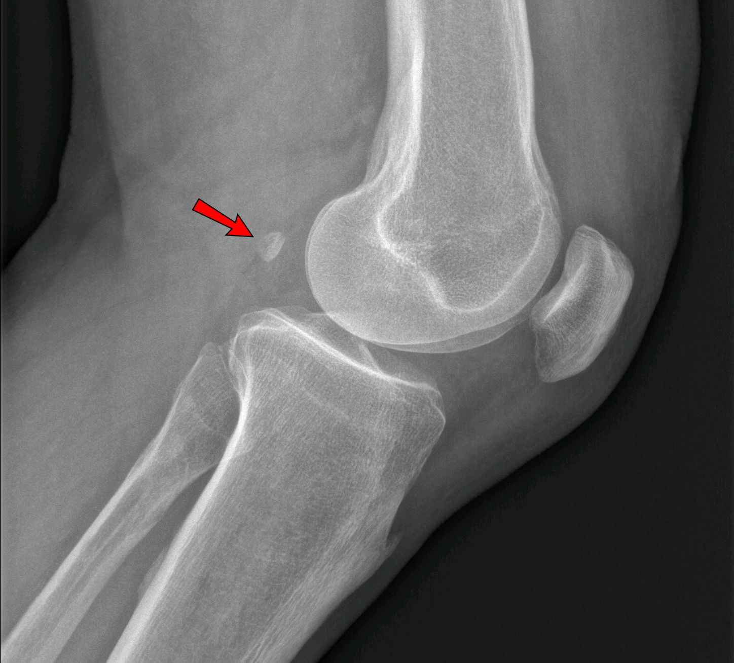
An accessory bone or supernumerary bone is a bone that is not normally present in the body, but can be found as a variant in a significant number of people. It poses a risk of being misdiagnosed as bone fractures on radiography. Wrist and hand Os ulnostyloideum The ''os ulnostyloideum'' is an ulnar styloid process that is not fused to the rest of the ulna bone.R. O'Rahilly. ''A survey of carpal and tarsal anomalies.'' J Bone Joint Surg Am. 1953; 35: 626–642 On X-rays, an ''os ulnostyloideum'' is sometimes mistaken for an avulsion fracture of the styloid process. However, the distinction between these is extremely difficult.T.E. Keats, M.W. Anderson. ''Atlas of normal roentgen variants that may simulate disease''. 7th edition, Mosby Inc. 2001 It is alleged that the os ulnostyloideum has a close relationship with or is synonymous with the os triquetrum secundarium. Os centrale The ''os carpi centrale'' (also briefly ''os centrale'') is, where present, located on the ... [...More Info...] [...Related Items...] OR: [Wikipedia] [Google] [Baidu] |
Os Vesalianum
An accessory bone or supernumerary bone is a bone that is not normally present in the body, but can be found as a variant in a significant number of people. It poses a risk of being misdiagnosed as bone fractures on radiography. Wrist and hand Os ulnostyloideum The ''os ulnostyloideum'' is an ulnar styloid process that is not fused to the rest of the ulna bone.R. O'Rahilly. ''A survey of carpal and tarsal anomalies.'' J Bone Joint Surg Am. 1953; 35: 626–642 On X-rays, an ''os ulnostyloideum'' is sometimes mistaken for an avulsion fracture of the styloid process. However, the distinction between these is extremely difficult.T.E. Keats, M.W. Anderson. ''Atlas of normal roentgen variants that may simulate disease''. 7th edition, Mosby Inc. 2001 It is alleged that the os ulnostyloideum has a close relationship with or is synonymous with the os triquetrum secundarium. Os centrale The ''os carpi centrale'' (also briefly ''os centrale'') is, where present, located on the ... [...More Info...] [...Related Items...] OR: [Wikipedia] [Google] [Baidu] |
Ossification Center
An ossification center is a point where ossification of the cartilage begins. The first step in ossification is that the cartilage cells at this point enlarge and arrange themselves in rows.Gray and Spitzka (1910), page 44. The matrix in which they are imbedded increases in quantity, so that the cells become further separated from each other. A deposit of calcareous Calcareous () is an adjective meaning "mostly or partly composed of calcium carbonate", in other words, containing lime or being chalky. The term is used in a wide variety of scientific disciplines. In zoology ''Calcareous'' is used as an ad ... material now takes place in this matrix, between the rows of cells, so that they become separated from each other by longitudinal columns of calcified matrix, presenting a granular and opaque appearance. Here and there the matrix between two cells of the same row also becomes calcified, and transverse bars of calcified substance stretch across from one calcareous colu ... [...More Info...] [...Related Items...] OR: [Wikipedia] [Google] [Baidu] |
Avulsion Fracture
An avulsion fracture is a bone fracture which occurs when a fragment of bone tears away from the main mass of bone as a result of physical trauma. This can occur at the ligament by the application of forces external to the body (such as a fall or pull) or at the tendon by a muscular contraction that is stronger than the forces holding the bone together. Generally muscular avulsion is prevented by the neurological limitations placed on muscle contractions. Highly trained athletes can overcome this neurological inhibition of strength and produce a much greater force output capable of breaking or avulsing a bone. Types Dental avulsion Traumatic complete displacement of a tooth from its socket in alveolar bone. It is a serious dental emergency in which prompt management (within 20–40 minutes of injury) affects the prognosis of the tooth. Tuberosity avulsion of the 5th metatarsal left, Proximal fractures of 5th metatarsa The tuberosity avulsion fracture (also known as ... [...More Info...] [...Related Items...] OR: [Wikipedia] [Google] [Baidu] |
Dancer's Fracture
An avulsion fracture is a bone fracture which occurs when a fragment of bone tears away from the main mass of bone as a result of physical trauma. This can occur at the ligament by the application of forces external to the body (such as a fall or pull) or at the tendon by a muscular contraction that is stronger than the forces holding the bone together. Generally muscular avulsion is prevented by the neurological limitations placed on muscle contractions. Highly trained athletes can overcome this neurological inhibition of strength and produce a much greater force output capable of breaking or avulsing a bone. Types Dental avulsion Traumatic complete displacement of a tooth from its socket in alveolar bone. It is a serious dental emergency in which prompt management (within 20–40 minutes of injury) affects the prognosis of the tooth. Tuberosity avulsion of the 5th metatarsal left, Proximal fractures of 5th metatarsa The tuberosity avulsion fracture (also known as ... [...More Info...] [...Related Items...] OR: [Wikipedia] [Google] [Baidu] |
Pseudo-Jones Fracture
An avulsion fracture is a bone fracture which occurs when a fragment of bone tears away from the main mass of bone as a result of physical trauma. This can occur at the ligament by the application of forces external to the body (such as a fall or pull) or at the tendon by a muscular contraction that is stronger than the forces holding the bone together. Generally muscular avulsion is prevented by the neurological limitations placed on muscle contractions. Highly trained athletes can overcome this neurological inhibition of strength and produce a much greater force output capable of breaking or avulsing a bone. Types Dental avulsion Traumatic complete displacement of a tooth from its socket in alveolar bone. It is a serious dental emergency in which prompt management (within 20–40 minutes of injury) affects the prognosis of the tooth. Tuberosity avulsion of the 5th metatarsal left, Proximal fractures of 5th metatarsa The tuberosity avulsion fracture (also known as ... [...More Info...] [...Related Items...] OR: [Wikipedia] [Google] [Baidu] |


