|
Falcine Sinus
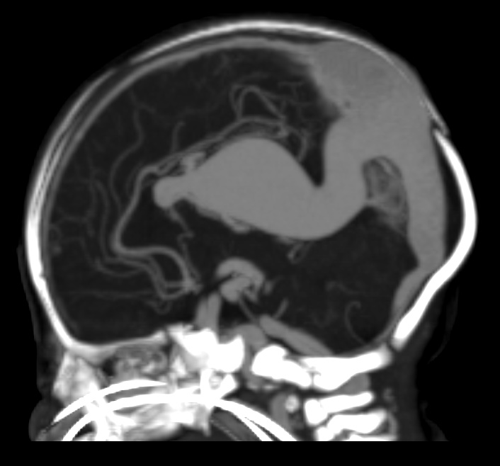
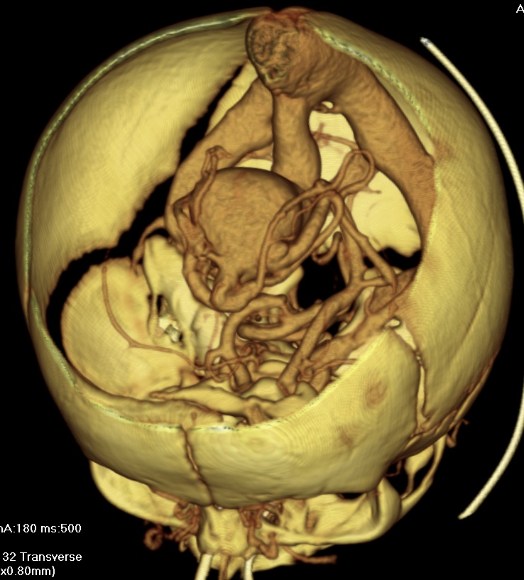
A falcine sinus is a venous channel that lies within the falx cerebri connecting the vein of Galen and the posterior part of superior sagittal sinus. It is normally present during fetal development Prenatal development () involves the development of the embryo and of the fetus during a viviparous animal's gestation. Prenatal development starts with fertilization, in the germinal stage of embryonic development, and continues in fetal deve ... and involutes after birth. The presence of a falcine sinus has been associated with a vein of Galen malformation and other vascular anomalies. The persistence of a falcine sinus after the neonatal period was previously thought to be rare, but has recently been described to be present in up to 5% of all people, appearings in approximately 2.1% of CT examinations of adult patients. Some authors have studied the plexus rather than the sinus, a rare form of the venous pathway between the layers of the cerebral falx, which connects the superi ... [...More Info...] [...Related Items...] OR: [Wikipedia] [Google] [Baidu] |
Vein Of Galen Sag Mip
Veins () are blood vessels in the circulatory system of humans and most other animals that carry blood towards the heart. Most veins carry deoxygenated blood from the tissues back to the heart; exceptions are those of the pulmonary circulation, pulmonary and fetal circulations which carry oxygenated blood to the heart. In the systemic circulation, Artery, arteries carry oxygenated blood away from the heart, and veins return deoxygenated blood to the heart, in the deep veins. There are three sizes of veins: large, medium, and small. Smaller veins are called venules, and the smallest the post-capillary venules are microscopic that make up the veins of the microcirculation. Veins are often closer to the skin than arteries. Veins have less smooth muscle and connective tissue and wider Lumen (anatomy), internal diameters than arteries. Because of their thinner walls and wider lumens they are able to expand and hold more blood. This greater venous capacitance, capacity gives them the ... [...More Info...] [...Related Items...] OR: [Wikipedia] [Google] [Baidu] |
Falx Cerebri
The falx cerebri (also known as the cerebral falx) is a large, crescent-shaped fold of dura mater that descends vertically into the longitudinal fissure to separate the cerebral hemispheres.Saladin K. "Anatomy & Physiology: The Unity of Form and Function. New York: McGraw Hill, 2014. Print. pp 512, 770-773 It supports the dural sinuses that provide venous and CSF drainage from the brain. It is attached to the crista galli anteriorly, and blends with the tentorium cerebelli posteriorly. The falx cerebri is often subject to age-related calcification, and a site of falcine meningiomas. The falx cerebri is named for its sickle-like shape. Anatomy The falx cerebri is a strong, crescent-shaped sheet of dura mater lying in the sagittal plane between the two cerebral hemispheres. It is one of four dural partitions of the brain along with the falx cerebelli, tentorium cerebelli, and diaphragma sellae; it is formed through invagination of the dura mater into the longitudina ... [...More Info...] [...Related Items...] OR: [Wikipedia] [Google] [Baidu] |
Great Cerebral Vein
The great cerebral vein is one of the large blood vessels in the skull draining the cerebrum of the brain. It is also known as the vein of Galen, named for its discoverer, the Greek physician Galen. Structure The great cerebral vein is one of the deep cerebral veins. Other deep cerebral veins are the internal cerebral veins, formed by the union of the superior thalamostriate vein and the superior choroid vein at the interventricular foramina. The internal cerebral veins can be seen on the superior surfaces of the caudate nuclei and thalami just under the corpus callosum. The veins at the anterior poles of the thalami merge posterior to the pineal gland to form the great cerebral vein. Most of the blood in the deep cerebral veins collects into the great cerebral vein. This comes from the inferior side of the posterior end of the corpus callosum and empties ie similarities, there are also differences between these two types of veins in the brain. The superficial veins at the d ... [...More Info...] [...Related Items...] OR: [Wikipedia] [Google] [Baidu] |
Superior Sagittal Sinus
The superior sagittal sinus (also known as the superior longitudinal sinus), within the human head, is an unpaired dural venous sinus lying along the attached margin of the falx cerebri. It allows blood to drain from the lateral aspects of the anterior cerebral hemispheres to the confluence of sinuses. Cerebrospinal fluid drains through arachnoid granulations into the superior sagittal sinus and is returned to the venous circulation. Structure It is triangular in section. It is narrower anteriorly, and gradually increases in size as it passes posterior-ward. It commences at the foramen cecum, through which it receives emissary veins from the nasal cavity. It passes posterior-ward along its entire course. It is accommodated within a groove which runs across the inner surface of the frontal bone, the adjacent margins of the two parietal lobes, and the superior division of the cruciate eminence of the occipital lobe. Near the internal occipital protuberance, it deviates to e ... [...More Info...] [...Related Items...] OR: [Wikipedia] [Google] [Baidu] |
Prenatal Development
Prenatal development () involves the development of the embryo and of the fetus during a viviparous animal's gestation. Prenatal development starts with fertilization, in the germinal stage of embryonic development, and continues in fetal development until birth. The term "prenate" is used to describe an unborn offspring at any stage of gestation. In human pregnancy, prenatal development is also called antenatal development. The development of the human embryo follows fertilization, and continues as fetal development. By the end of the tenth week of gestational age, the embryo has acquired its basic form and is referred to as a fetus. The next period is that of fetal development where many organs become fully developed. This fetal period is described both topically (by organ) and chronologically (by time) with major occurrences being listed by gestational age. The very early stages of embryonic development are the same in all mammals, but later stages of development, an ... [...More Info...] [...Related Items...] OR: [Wikipedia] [Google] [Baidu] |
Vein Of Galen Aneurysmal Malformations
Vein of Galen aneurysmal malformations (VGAMs) and Vein of Galen aneurysmal dilations (VGADs) are the most frequent arteriovenous malformations in infants and fetuses. A VGAM consists of a tangled mass of dilated vessels supplied by an enlarged artery. The malformation increases greatly in size with age, although the mechanism of the increase is unknown. Dilation of the great cerebral vein of Galen is a secondary result of the force of arterial blood either directly from an artery via an arteriovenous fistula or by way of a tributary vein that receives the blood directly from an artery. There is usually a venous anomaly downstream from the draining vein that, together with the high blood flow into the great cerebral vein of Galen causes its dilation. The right sided cardiac chambers and pulmonary arteries also develop mild to severe dilation. Signs and symptoms Malformations often lead to cardiac failure, cranial bruits (pattern 1), hydrocephaly, and subarachnoid hemorrhage in neon ... [...More Info...] [...Related Items...] OR: [Wikipedia] [Google] [Baidu] |
Anatomical Variations
An anatomical variation, anatomical variant, or anatomical variability is a presentation of body structure with morphological features different from those that are typically described in the majority of individuals. Anatomical variations are categorized into three types including morphometric (size or shape), consistency (present or absent), and spatial (proximal/distal or right/left). Variations are seen as normal in the sense that they are found consistently among different individuals, are mostly without symptoms, and are termed anatomical variations rather than abnormalities. Anatomical variations are mainly caused by genetics and may vary considerably between different populations. The rate of variation considerably differs between single organs, particularly in muscles. Knowledge of anatomical variations is important in order to distinguish them from pathological conditions. A very early paper published in 1898, presented anatomic variations to have a wide range and signi ... [...More Info...] [...Related Items...] OR: [Wikipedia] [Google] [Baidu] |
Neuroanatomy
Neuroanatomy is the study of the structure and organization of the nervous system. In contrast to animals with radial symmetry, whose nervous system consists of a distributed network of cells, animals with bilateral symmetry have segregated, defined nervous systems. Their neuroanatomy is therefore better understood. In vertebrates, the nervous system is segregated into the internal structure of the brain and spinal cord (together called the central nervous system, or CNS) and the series of nerves that connect the CNS to the rest of the body (known as the peripheral nervous system, or PNS). Breaking down and identifying specific parts of the nervous system has been crucial for figuring out how it operates. For example, much of what neuroscientists have learned comes from observing how damage or "lesions" to specific brain areas affects behavior or other neural functions. For information about the composition of non-human animal nervous systems, see nervous system. For information a ... [...More Info...] [...Related Items...] OR: [Wikipedia] [Google] [Baidu] |


