|
Esophageal Hiatus
In human anatomy, the esophageal hiatus is an opening in the diaphragm through which the esophagus and the vagus nerve pass. Structure The esophageal hiatus is an oval opening in (sources differ) the right crus of the diaphragm/left crus of the diaphragm, with fibres of the right crus looping around the hiatus to form a sling (upon inspiration, this sling would constrict the esophagus, forming a functional (not anatomical) sphincter that prevents gastric contents from refluxing up the esophagus when intra-abdominal pressure rises during inspiration). Fibers of the right crus decussate inferior to the hiatus. Contents The esophageal hiatus gives passage to the oesophagus as well as the anterior and the posterior vagal trunk, esophageal branches of the left gastric artery and vein, and some lymphatic vessels. The transversalis fascia lining the inferior surface of the diaphragm extends superiorly through the hiatus to blend with the endothoracic fascia and attach to the oesoph ... [...More Info...] [...Related Items...] OR: [Wikipedia] [Google] [Baidu] |
Thoracic Diaphragm
The thoracic diaphragm, or simply the diaphragm (; ), is a sheet of internal Skeletal striated muscle, skeletal muscle in humans and other mammals that extends across the bottom of the thoracic cavity. The diaphragm is the most important Muscles of respiration, muscle of respiration, and separates the thoracic cavity, containing the heart and lungs, from the abdominal cavity: as the diaphragm contracts, the volume of the thoracic cavity increases, creating a negative pressure there, which draws air into the lungs. Its high oxygen consumption is noted by the many mitochondria and capillaries present; more than in any other skeletal muscle. The term ''diaphragm'' in anatomy, created by Gerard of Cremona, can refer to other flat structures such as the urogenital diaphragm or Pelvic floor, pelvic diaphragm, but "the diaphragm" generally refers to the thoracic diaphragm. In humans, the diaphragm is slightly asymmetric—its right half is higher up (superior) to the left half, since th ... [...More Info...] [...Related Items...] OR: [Wikipedia] [Google] [Baidu] |
Transversalis Fascia
The transversalis fascia (or transverse fascia) is the fascial lining of the anterolateral abdominal wall situated between the inner surface of the transverse abdominal muscle, and the preperitoneal fascia. It is directly continuous with the iliac fascia, the internal spermatic fascia, and pelvic fascia. Structure In the inguinal region, the transversalis fascia is thick and dense; here, it is joined by fibers of the aponeurosis of the transverse abdominal muscle. It becomes thin towards to the diaphragm, blending with the fascia covering the inferior surface of the diaphragm. Posteriorly, it is lost in Below, it has the following attachments: posteriorly, to the whole length of the iliac crest, between the attachments of the transverse abdominal and Iliacus; between the anterior superior iliac spine and the femoral vessels it is connected to the posterior margin of the inguinal ligament, and is there continuous with the iliac fascia. Medial to the femoral vessels it is t ... [...More Info...] [...Related Items...] OR: [Wikipedia] [Google] [Baidu] |
Hiatal Hernia
A hiatal hernia or hiatus hernia is a type of hernia in which abdominal organs (typically the stomach) slip through the diaphragm into the middle compartment of the chest. This may result in gastroesophageal reflux disease (GERD) or laryngopharyngeal reflux (LPR) with symptoms such as a taste of acid in the back of the mouth or heartburn. Other symptoms may include trouble swallowing and chest pains. Complications may include iron deficiency anemia, volvulus, or bowel obstruction. The most common risk factors are obesity and older age. Other risk factors include major trauma, scoliosis, and certain types of surgery. There are two main types: sliding hernia, in which the body of the stomach moves up; and paraesophageal hernia, in which an abdominal organ moves beside the esophagus. The diagnosis may be confirmed with endoscopy or medical imaging. Endoscopy is typically only required when concerning symptoms are present, symptoms are resistant to treatment, or the person i ... [...More Info...] [...Related Items...] OR: [Wikipedia] [Google] [Baidu] |
Aortic Hiatus
The aortic hiatus is a midline opening in the posterior part of the diaphragm giving passage to the descending aorta as well as the thoracic duct, and variably the azygos and hemiazygos veins. It is the lowest and most posterior of the large apertures. It is located at the level of the inferior border of the twelfth thoracic vertebra (T12), posterior to the median arcuate ligament between the two crura of the diaphragm. Structure Strictly speaking, it is not an aperture in the diaphragm but an osseoaponeurotic opening between it and the vertebral column, and therefore behind the diaphragm (meaning that diaphragmatic contractions during respiration do not directly affect aortic blood flow). The hiatus is situated slightly to the left of the midline, and is bound anteriorly by the crura, and posteriorly by the body of the first lumbar vertebra The lumbar vertebrae are located between the thoracic vertebrae and pelvis. They form the lower part of the back in humans, and t ... [...More Info...] [...Related Items...] OR: [Wikipedia] [Google] [Baidu] |
Intercostal Space
The intercostal space (ICS) is the anatomic space between two ribs (Lat. costa). Since there are 12 ribs on each side, there are 11 intercostal spaces, each numbered for the rib superior to it. Structures in intercostal space * several kinds of intercostal muscle * intercostal arteries and intercostal veins * intercostal lymph nodes * intercostal nerves Order of components Muscles There are 3 muscular layers in each intercostal space, consisting of the external intercostal muscle, the internal intercostal muscle, and the thinner innermost intercostal muscle. These muscles help to move the ribs during breathing. Neurovascular bundles Neurovascular bundles are located between the internal intercostal muscle and the innermost intercostal muscle. The neurovascular bundle has a strict order of vein-artery-nerve (VAN), from top to bottom. This neurovascular bundle runs high in the intercostal space, and the smaller collateral neurovascular bundle runs just superior ... [...More Info...] [...Related Items...] OR: [Wikipedia] [Google] [Baidu] |
Costal Cartilage
Costal cartilage, also known as rib cartilage, are bars of hyaline cartilage that serve to prolong the ribs forward and contribute to the elasticity of the walls of the thorax. Costal cartilage is only found at the anterior ends of the ribs, providing medial extension. Differences from ribs 1-12 The first seven pairs are connected with the sternum; the next three are each articulated with the lower border of the cartilage of the preceding rib; the last two have pointed extremities, which end in the wall of the abdomen. Like the ribs, the costal cartilages vary in their length, breadth, and direction. They increase in length from the first to the seventh, then gradually decrease to the twelfth. Their breadth, as well as that of the intervals between them, diminishes from the first to the last. They are broad at their attachments to the ribs, and taper toward their sternal extremities, excepting the first two, which are of the same breadth throughout, and the sixth, seventh, an ... [...More Info...] [...Related Items...] OR: [Wikipedia] [Google] [Baidu] |
Thoracic Vertebrae
In vertebrates, thoracic vertebrae compose the middle segment of the vertebral column, between the cervical vertebrae and the lumbar vertebrae. In humans, there are twelve thoracic vertebra (anatomy), vertebrae of intermediate size between the cervical and lumbar vertebrae; they increase in size going towards the lumbar vertebrae. They are distinguished by the presence of Zygapophysial joint, facets on the sides of the bodies for Articulation (anatomy), articulation with the head of rib, heads of the ribs, as well as facets on the transverse processes of all, except the eleventh and twelfth, for articulation with the tubercle (rib), tubercles of the ribs. By convention, the human thoracic vertebrae are numbered T1–T12, with the first one (T1) located closest to the skull and the others going down the spine toward the lumbar region. General characteristics These are the general characteristics of the second through eighth thoracic vertebrae. The first and ninth through twelfth v ... [...More Info...] [...Related Items...] OR: [Wikipedia] [Google] [Baidu] |
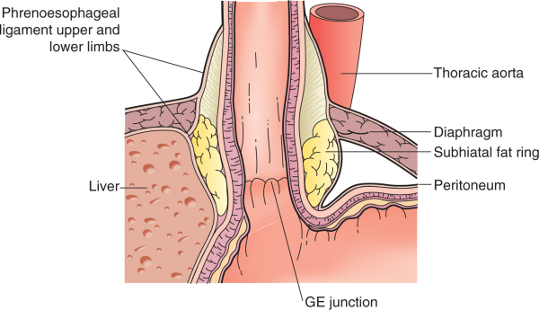
Phrenoesophageal Ligament
The phrenoesophageal ligament (phrenicoesophageal ligament, or phrenoesophageal membrane) is the ligament by which the esophagus is attached to the diaphragm. It is an extension of the inferior diaphragmatic fascia and is divided into an upper and lower limb which attach to the superior and inferior surfaces of the diaphragm respectively at the esophageal hiatus. The upper limb attaches the esophagus to the superior surface of the diaphragm and the lower limb attaches the cardia The stomach is a muscular, hollow organ in the upper gastrointestinal tract of humans and many other animals, including several invertebrates. The Ancient Greek name for the stomach is ''gaster'' which is used as ''gastric'' in medical terms re ... region of the stomach to the inferior surface of the diaphragm at the cardiac notch of stomach. The ligament allows independent movement of the diaphragm and esophagus during respiration and swallowing. References * Full text' * * Ligaments {{li ... [...More Info...] [...Related Items...] OR: [Wikipedia] [Google] [Baidu] |
Endothoracic Fascia
The endothoracic fascia is the layer of loose connective tissue deep to the intercostal spaces and ribs, separating these structures from the underlying pleura. This fascial layer is the outermost membrane of the thoracic cavity. The endothoracic fascia contains variable amounts of fat. It becomes more fibrous over the apices of the lungs as the suprapleural membrane. It separates the internal thoracic artery The internal thoracic artery (ITA), also known as the internal mammary artery, is an artery that supplies the anterior chest wall and the breasts. It is a paired artery, with one running along each side of the sternum, to continue after its bifurc ... from the parietal pleura. References Thorax (human anatomy) Fascia {{musculoskeletal-stub ... [...More Info...] [...Related Items...] OR: [Wikipedia] [Google] [Baidu] |
Left Gastric Vein
The left gastric vein (or coronary vein) is a vein that derives from tributaries draining the lesser curvature of the stomach. Structure The left gastric vein runs from right to left along the lesser curvature of the stomach. It passes to the esophageal opening of the stomach, where it receives some esophageal veins. It then turns backward and passes from left to right behind the omental bursa. It drains into the portal vein near the superior border of the pancreas. Function The left gastric vein drains deoxygenated blood from the lesser curvature of the stomach. It also acts as collaterals between the portal vein The portal vein or hepatic portal vein (HPV) is a blood vessel that carries blood from the gastrointestinal tract, gallbladder, pancreas and spleen to the liver. This blood contains nutrients and toxins extracted from digested contents. Approxima ... and the systemic venous system of the lower esophagus ( azygos vein). Clinical significance The esophageal ... [...More Info...] [...Related Items...] OR: [Wikipedia] [Google] [Baidu] |
Diaphragm (anatomy)
The thoracic diaphragm, or simply the diaphragm (; ), is a sheet of internal skeletal muscle in humans and other mammals that extends across the bottom of the thoracic cavity. The diaphragm is the most important muscle of respiration, and separates the thoracic cavity, containing the heart and lungs, from the abdominal cavity: as the diaphragm contracts, the volume of the thoracic cavity increases, creating a negative pressure there, which draws air into the lungs. Its high oxygen consumption is noted by the many mitochondria and capillaries present; more than in any other skeletal muscle. The term ''diaphragm'' in anatomy, created by Gerard of Cremona, can refer to other flat structures such as the urogenital diaphragm or Pelvic floor, pelvic diaphragm, but "the diaphragm" generally refers to the thoracic diaphragm. In humans, the diaphragm is slightly asymmetric—its right half is higher up (superior) to the left half, since the large liver rests beneath the right half of t ... [...More Info...] [...Related Items...] OR: [Wikipedia] [Google] [Baidu] |
Left Gastric Artery
In human anatomy, the left gastric artery arises from the celiac artery and runs along the superior portion of the lesser curvature of the stomach before anastomosing with the right gastric artery (which runs right to left). It also issues esophageal branches that supply lower esophagus and ascend through the esophageal hiatus to form anastomoses with the esophageal branches of thoracic part of aorta. Anatomy Origin The LGA usually arises from (the superior aspect of) the coeliac trunk - sometimes as a terminal branch of a trifurcation, and more rarely as a side branch of the splenic artery or of common hepatic artery. Sometimes it originates directly from aorta or from arteria phrenica inferior. Course From the crus of diaphragm, the LGA arches obliquely anterior-ward and to the left to reach the left curvature of the stomach just inferior to the gastric cardia (thus erecting the gastropancreatic (peritoneal) fold). Fate Upon reaching the cardia, the LGA splits ... [...More Info...] [...Related Items...] OR: [Wikipedia] [Google] [Baidu] |


