|
Electroencephalograph
Electroencephalography (EEG) is a method to record an electrogram of the spontaneous electrical activity of the brain. The bio signals detected by EEG have been shown to represent the postsynaptic potentials of pyramidal neurons in the neocortex and allocortex. It is typically non-invasive, with the EEG electrodes placed along the scalp (commonly called "scalp EEG") using the International 10–20 system, or variations of it. Electrocorticography, involving surgical placement of electrodes, is sometimes called "intracranial EEG". Clinical interpretation of EEG recordings is most often performed by visual inspection of the tracing or quantitative EEG analysis. Voltage fluctuations measured by the EEG bio amplifier and electrodes allow the evaluation of normal brain activity. As the electrical activity monitored by EEG originates in neurons in the underlying brain tissue, the recordings made by the electrodes on the surface of the scalp vary in accordance with their orie ... [...More Info...] [...Related Items...] OR: [Wikipedia] [Google] [Baidu] [Amazon] |
Alpha Wave
Alpha waves, or the alpha rhythm, are neural oscillations in the frequency range of 8–12 Hz likely originating from the synchronous and coherent ( in phase or constructive) neocortical neuronal electrical activity possibly involving thalamic pacemaker cells. Historically, they are also called "Berger's waves" after Hans Berger, who first described them when he invented the EEG in 1924. Alpha waves are one type of brain waves detected by electrophysiological methods, e.g., electroencephalography (EEG) or magnetoencephalography (MEG), and can be quantified using power spectra and time-frequency representations of power like quantitative electroencephalography (qEEG). They are predominantly recorded over parieto-occipital brain and were the earliest brain rhythm recorded in humans. Alpha waves can be observed during relaxed wakefulness, especially when there is no mental activity. During the eyes-closed condition, alpha waves are prominent at parietal locations. Atte ... [...More Info...] [...Related Items...] OR: [Wikipedia] [Google] [Baidu] [Amazon] |
Electrocorticography
Electrocorticography (ECoG), a type of intracranial electroencephalography (iEEG), is a type of electrophysiological monitoring that uses electrodes placed directly on the exposed surface of the brain to record electrical activity from the cerebral cortex. In contrast, conventional electroencephalography (EEG) electrodes monitor this activity from outside the skull. ECoG may be performed either in the operating room during surgery (intraoperative ECoG) or outside of surgery (extraoperative ECoG). Because a craniotomy (a surgical incision into the skull) is required to implant the electrode grid, ECoG is an invasive procedure. History ECoG was pioneered in the early 1950s by Wilder Penfield and Herbert Jasper, neurosurgeons at the Montreal Neurological Institute. The two developed ECoG as part of their groundbreakinMontreal procedure a surgical protocol used to treat patients with severe epilepsy. The cortical potentials recorded by ECoG were used to identify epileptogen ... [...More Info...] [...Related Items...] OR: [Wikipedia] [Google] [Baidu] [Amazon] |
Epilepsy
Epilepsy is a group of Non-communicable disease, non-communicable Neurological disorder, neurological disorders characterized by a tendency for recurrent, unprovoked Seizure, seizures. A seizure is a sudden burst of abnormal electrical activity in the brain that can cause a variety of symptoms, ranging from brief lapses of awareness or muscle jerks to prolonged convulsions. These episodes can result in physical injuries, either directly, such as broken bones, or through causing accidents. The diagnosis of epilepsy typically requires at least two unprovoked seizures occurring more than 24 hours apart. In some cases, however, it may be diagnosed after a single unprovoked seizure if clinical evidence suggests a high risk of recurrence. Isolated seizures that occur without recurrence risk or are provoked by identifiable causes are not considered indicative of epilepsy. The underlying cause is often unknown, but epilepsy can result from brain injury, stroke, infections, Brain tumor, ... [...More Info...] [...Related Items...] OR: [Wikipedia] [Google] [Baidu] [Amazon] |
Brain Activity And Meditation
Meditation and its effect on brain activity and the central nervous system became a focus of collaborative research in neuroscience, psychology and neurobiology during the latter half of the 20th century. Research on meditation sought to define and characterize various practices. The effects of meditation on the brain can be broken up into two categories: state changes and trait changes, respectively alterations in brain activities during the act of meditating and changes that are the outcome of long-term practice. Mindfulness meditation, a Buddhist meditation approach found in Zen and Vipassana, is frequently studied. Jon Kabat-Zinn describes mindfulness meditation as complete, unbiased attention to the current moment. Changes in brain state Electroencephalography Electroencephalography (EEG) has been used in many studies as a primary method for evaluating the meditating brain. Electroencephalography uses electrical leads placed all over the scalp to measure the collective e ... [...More Info...] [...Related Items...] OR: [Wikipedia] [Google] [Baidu] [Amazon] |
Quantitative EEG
Quantitative electroencephalography (qEEG or QEEG) is a field concerned with the numerical analysis of electroencephalography (EEG) data and associated behavioral correlates. Details Techniques used in digital signal analysis are extended to the analysis of electroencephalography (EEG). These include wavelet analysis and Fourier analysis, with new focus on shared activity between rhythms including phase synchrony (coherence, phase lag) and magnitude synchrony (comodulation/correlation, and asymmetry). The analog signal comprises a microvoltage time series of the EEG, sampled digitally and sampling rates adequate to over-sample the signal (using the Nyquist principle of exceeding twice the highest frequency being detected). Modern EEG amplifiers use adequate sampling to resolve the EEG across the traditional medical band from DC to 70 or 100 Hz, using sample rates of 250/256, 500/512, to over 1000 samples per second, depending on the intended application. QEEG can be p ... [...More Info...] [...Related Items...] OR: [Wikipedia] [Google] [Baidu] [Amazon] |
Human Brain
The human brain is the central organ (anatomy), organ of the nervous system, and with the spinal cord, comprises the central nervous system. It consists of the cerebrum, the brainstem and the cerebellum. The brain controls most of the activities of the human body, body, processing, integrating, and coordinating the information it receives from the sensory nervous system. The brain integrates sensory information and coordinates instructions sent to the rest of the body. The cerebrum, the largest part of the human brain, consists of two cerebral hemispheres. Each hemisphere has an inner core composed of white matter, and an outer surface – the cerebral cortex – composed of grey matter. The cortex has an outer layer, the neocortex, and an inner allocortex. The neocortex is made up of six Cerebral cortex#Layers of neocortex, neuronal layers, while the allocortex has three or four. Each hemisphere is divided into four lobes of the brain, lobes – the frontal lobe, frontal, pa ... [...More Info...] [...Related Items...] OR: [Wikipedia] [Google] [Baidu] [Amazon] |
Brain
The brain is an organ (biology), organ that serves as the center of the nervous system in all vertebrate and most invertebrate animals. It consists of nervous tissue and is typically located in the head (cephalization), usually near organs for special senses such as visual perception, vision, hearing, and olfaction. Being the most specialized organ, it is responsible for receiving information from the sensory nervous system, processing that information (thought, cognition, and intelligence) and the coordination of motor control (muscle activity and endocrine system). While invertebrate brains arise from paired segmental ganglia (each of which is only responsible for the respective segmentation (biology), body segment) of the ventral nerve cord, vertebrate brains develop axially from the midline dorsal nerve cord as a brain vesicle, vesicular enlargement at the rostral (anatomical term), rostral end of the neural tube, with centralized control over all body segments. All vertebr ... [...More Info...] [...Related Items...] OR: [Wikipedia] [Google] [Baidu] [Amazon] |
10–20 System (EEG)
The 10–20 system or International 10–20 system is an internationally recognized method to describe and apply the location of scalp electrodes in the context of an EEG exam, polysomnograph sleep study, or voluntary lab research. This method was developed to maintain standardized testing methods ensuring that a subject's study outcomes (clinical or research) could be compiled, reproduced, and effectively analyzed and compared using the scientific method. The system is based on the relationship between the location of an electrode and the underlying area of the brain, specifically the cerebral cortex. Across all phases of consciousness, brains produce different, objectively recognizable and distinguishable electrical patterns, which can be detected by electrodes on the skin. These patterns vary, and are affected by multiple extrinsic factors, including age, prescription drugs, somatic diagnoses, Medical history, history of neurologic insults/injury/trauma, and substance abuse. ... [...More Info...] [...Related Items...] OR: [Wikipedia] [Google] [Baidu] [Amazon] |
Beta Wave
Beta waves, or beta rhythm, are neural oscillations (brainwaves) in the brain with a frequency range of between 12.5 and 30 Hz (12.5 to 30 cycles per second). Several different rhythms coexist, with some being inhibitory and others excitory in function. Beta waves can be split into three sections: Low Beta Waves (12.5–16 Hz, "Beta 1"); Beta Waves (16.5–20 Hz, "Beta 2"); and High Beta Waves (20.5–28 Hz, "Beta 3"). Beta states are the states associated with normal waking consciousness. History Beta waves were discovered and named by the German psychiatrist Hans Berger, who invented electroencephalography (EEG) in 1924, as a method of recording electrical brain activity from the human scalp. Berger termed the larger amplitude, slower frequency waves that appeared over the posterior scalp when the subject's eye were closed alpha waves. The smaller amplitude, faster frequency waves that replaced alpha waves when the subject opened their eye ... [...More Info...] [...Related Items...] OR: [Wikipedia] [Google] [Baidu] [Amazon] |
Biosignal
A biosignal is any signal in living beings that can be continually measured and monitored. The term biosignal is often used to refer to bioelectrical signals, but it may refer to both electrical and non-electrical signals. The usual understanding is to refer only to time-varying signals, although spatial parameter variations (e.g. the nucleotide sequence determining the genetic code) are sometimes subsumed as well. Electrical biosignals Electrical biosignals, or bioelectrical time signals, usually refers to the change in electric current produced by the sum of an electrical potential difference across a specialized tissue, organ or cell system like the nervous system. Thus, among the best-known bioelectrical signals are: * Electroencephalogram (EEG) * Electrocardiogram (ECG) * Electromyogram (EMG) * Electrooculogram (EOG) * Electroretinogram (ERG) * Electrogastrogram (EGG) * Galvanic skin response (GSR) or electrodermal activity (EDA) EEG, ECG, EOG and EMG are measure ... [...More Info...] [...Related Items...] OR: [Wikipedia] [Google] [Baidu] [Amazon] |
Scalp
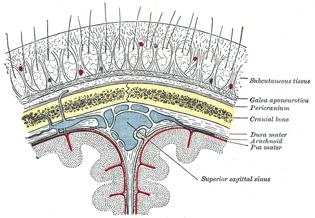
The scalp is the area of the head where head hair grows. It is made up of skin, layers of connective and fibrous tissues, and the membrane of the skull. Anatomically, the scalp is part of the epicranium, a collection of structures covering the cranium. The scalp is bordered by the face at the front, and by the neck at the sides and back. The scientific study of hair and scalp is called trichology. Structure Layers The scalp is usually described as having five layers, which can be remembered using the mnemonic 'SCALP': * S: Skin. The skin of the scalp contains numerous hair follicles and sebaceous glands. * C: Connective tissue. A dense subcutaneous layer of fat and fibrous tissue that lies beneath the skin, containing the nerves and vessels of the scalp. * A: Aponeurosis. The epicranial aponeurosis or galea aponeurotica is a tough layer of dense fibrous tissue which anchors the above layers in place. It runs from the frontalis muscle anteriorly to the occipitalis ... [...More Info...] [...Related Items...] OR: [Wikipedia] [Google] [Baidu] [Amazon] |
Hippocampus
The hippocampus (: hippocampi; via Latin from Ancient Greek, Greek , 'seahorse'), also hippocampus proper, is a major component of the brain of humans and many other vertebrates. In the human brain the hippocampus, the dentate gyrus, and the subiculum are components of the hippocampal formation located in the limbic system. The hippocampus plays important roles in the Memory consolidation, consolidation of information from short-term memory to long-term memory, and in spatial memory that enables Navigation#Navigation in spatial cognition, navigation. In humans, and other primates the hippocampus is located in the archicortex, one of the three regions of allocortex, in each cerebral hemisphere, hemisphere with direct neural projections to, and reciprocal indirect projections from the neocortex. The hippocampus, as the medial pallium, is a structure found in all vertebrates. In Alzheimer's disease (and other forms of dementia), the hippocampus is one of the first regions of th ... [...More Info...] [...Related Items...] OR: [Wikipedia] [Google] [Baidu] [Amazon] |


