|
Dorsal Root Of Spinal Nerve
The dorsal root of spinal nerve (or posterior root of spinal nerve or sensory root) is one of two "roots" which emerge from the spinal cord. It emerges directly from the spinal cord, and travels to the dorsal root ganglion. Nerve fibres with the ventral root then combine to form a spinal nerve. The dorsal root transmits sensory information, forming the afferent sensory root of a spinal nerve. Structure The root emerges from the posterior part of the spinal cord and travels to the dorsal root ganglion. The dorsal root ganglia contain the pseudo-unipolar cell bodies of the nerve fibres which travel from the ganglia through the root into the spinal cord. The lateral division of the dorsal root contains lightly myelinated and unmyelinated fibres of small diameter. These carry pain and temperature sensation. These fibers cross through the anterior white commissure to form the anterolateral system in the lateral funiculus. The medial division of the dorsal root contains myelina ... [...More Info...] [...Related Items...] OR: [Wikipedia] [Google] [Baidu] |
Spinal Cord
The spinal cord is a long, thin, tubular structure made up of nervous tissue, which extends from the medulla oblongata in the brainstem to the lumbar region of the vertebral column (backbone). The backbone encloses the central canal of the spinal cord, which contains cerebrospinal fluid. The brain and spinal cord together make up the central nervous system (CNS). In humans, the spinal cord begins at the occipital bone, passing through the foramen magnum and then enters the spinal canal at the beginning of the cervical vertebrae. The spinal cord extends down to between the first and second lumbar vertebrae, where it ends. The enclosing bony vertebral column protects the relatively shorter spinal cord. It is around long in adult men and around long in adult women. The diameter of the spinal cord ranges from in the cervical and lumbar regions to in the thoracic area. The spinal cord functions primarily in the transmission of nerve signals from the motor cortex to the ... [...More Info...] [...Related Items...] OR: [Wikipedia] [Google] [Baidu] |
Lateral Funiculus
The most lateral of the bundles of the anterior nerve roots is generally taken as a dividing line that separates the anterolateral system The spinothalamic tract is a part of the anterolateral system or the ventrolateral system, a sensory pathway to the thalamus. From the ventral posterolateral nucleus in the thalamus, sensory information is relayed upward to the somatosensory cor ... into two parts. These are the anterior funiculus, between the anterior median fissure and the most lateral of the anterior nerve roots, and the lateral funiculus (or lateral column) between the exit of these roots and the posterolateral sulcus. The lateral funiculus transmits the contralateral corticospinal and spinothalamic tracts. A lateral cutting of the spinal cord results in the transection of both ipsilateral posterior column and lateral funiculus and this produces Brown-Séquard syndrome.Kaplan Qbook - USMLE Step 1 - 5th edition - page References Central nervous system { ... [...More Info...] [...Related Items...] OR: [Wikipedia] [Google] [Baidu] |
Vertebral Foramen
In a typical vertebra, the vertebral foramen is the foramen (opening) formed by the anterior segment (the body), and the posterior part, the vertebral arch. The vertebral foramen begins at cervical vertebra #1 (C1 or atlas) and continues inferior to lumbar vertebra #5 (L5). The vertebral foramen houses the spinal cord and its meninges In anatomy, the meninges (, ''singular:'' meninx ( or ), ) are the three membranes that envelop the brain and spinal cord. In mammals, the meninges are the dura mater, the arachnoid mater, and the pia mater. Cerebrospinal fluid is located in .... This large tunnel running up and down inside all of the vertebrae contains the spinal cord and is typically called the spinal canal, not the vertebral foramen. See also * Atlas (anatomy)#Vertebral foramen References * External links * - "Superior and lateral views of typical vertebrae"Vertebral foramen- BlueLink Anatomy - University of Michigan Medical School * - "Typical Lumbar Vertebra, Superi ... [...More Info...] [...Related Items...] OR: [Wikipedia] [Google] [Baidu] |
Ventral Root
In anatomy and neurology, the ventral root of spinal nerve, anterior root, or motor root is the efferent motor root of a spinal nerve. At its distal end, the ventral root joins with the dorsal root The dorsal root of spinal nerve (or posterior root of spinal nerve or sensory root) is one of two "roots" which emerge from the spinal cord. It emerges directly from the spinal cord, and travels to the dorsal root ganglion. Nerve fibres with the ve ... to form a mixed spinal nerve. Additional images Image:Cervical vertebra english.png, Cervical vertebra Image:Medulla spinalis - Section - English.svg, Medulla spinalis Image:Gray675.png, A spinal nerve with its anterior and posterior. Image:Gray764.png, The motor tract. Image:Gray770-en.svg, Diagrammatic transverse section of the medulla spinalis and its membranes. Image:Gray796.png, A portion of the spinal cord, showing its right lateral surface. The dura is opened and arranged to show the nerve roots. Image:Gray799.svg, Sc ... [...More Info...] [...Related Items...] OR: [Wikipedia] [Google] [Baidu] |
Posterior Column–medial Lemniscus Pathway
Posterior may refer to: * Posterior (anatomy), the end of an organism opposite to its head ** Buttocks, as a euphemism * Posterior horn (other) * Posterior probability The posterior probability is a type of conditional probability that results from updating the prior probability with information summarized by the likelihood via an application of Bayes' rule. From an epistemological perspective, the posterior ..., the conditional probability that is assigned when the relevant evidence is taken into account * Posterior tense, a relative future tense {{disambiguation ... [...More Info...] [...Related Items...] OR: [Wikipedia] [Google] [Baidu] |
Cuneate Fasciculus , a tract from the spinal cord into the brainstem
{{disambiguation ...
Cuneate means "wedge-shaped", and can apply to: * Cuneate leaf, a leaf shape * Cuneate nucleus, a part of the brainstem * Cuneate fasciculus Cuneate means "wedge-shaped", and can apply to: * Cuneate leaf The following is a list of terms which are used to describe leaf morphology in the description and taxonomy of plants. Leaves may be simple (a single leaf blade or lamina) or compou ... [...More Info...] [...Related Items...] OR: [Wikipedia] [Google] [Baidu] |
Gracile Fasciculus
Gracility is slenderness, the condition of being gracile, which means slender. It derives from the Latin adjective ''gracilis'' ( masculine or feminine), or ''gracile'' ( neuter), which in either form means slender, and when transferred for example to discourse takes the sense of "without ornament", "simple" or various similar connotations. In ''Glossary of Botanic Terms'', B. D. Jackson speaks dismissively of an entry in earlier dictionary of A. A. Crozier as follows: ''Gracilis (Lat.), slender. Crozier has the needless word "gracile"''. However, his objection would be hard to sustain in current usage; apart from the fact that ''gracile'' is a natural and convenient term, it is hardly a neologism. The ''Shorter Oxford English Dictionary'' gives the source date for that usage as 1623 and indicates the word is misused (through association with ''grace'') for Gracefully slender." This misuse is unfortunate at least, because the terms ''gracile'' and ''grace'' are unrelated: the et ... [...More Info...] [...Related Items...] OR: [Wikipedia] [Google] [Baidu] |
Sacral Spinal Nerve 5
Sacral may refer to: *Sacred, associated with divinity and considered worthy of spiritual respect or devotion *Of the sacrum The sacrum (plural: ''sacra'' or ''sacrums''), in human anatomy, is a large, triangular bone at the base of the spine that forms by the fusing of the sacral vertebrae (S1S5) between ages 18 and 30. The sacrum situates at the upper, back part o ..., a large, triangular bone at the base of the spine See also * {{disambiguation ... [...More Info...] [...Related Items...] OR: [Wikipedia] [Google] [Baidu] |
Cervical Spinal Nerve 2
The cervical spinal nerve 2 (C2) is a spinal nerve of the cervical segment. Nervous System -- Groups of Nerves It is a part of the along with the C1 and C3 nerves sometimes forming part of . it also connects into the with the C3. [...More Info...] [...Related Items...] OR: [Wikipedia] [Google] [Baidu] |
Proprioception
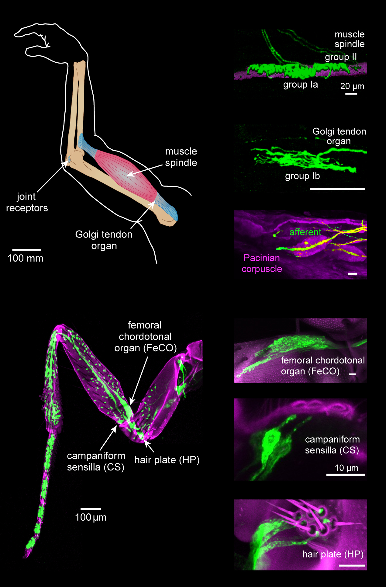
Proprioception ( ), also referred to as kinaesthesia (or kinesthesia), is the sense of self-movement, force, and body position. It is sometimes described as the "sixth sense". Proprioception is mediated by proprioceptors, mechanosensory neurons located within muscles, tendons, and joints. Most animals possess multiple subtypes of proprioceptors, which detect distinct kinematic parameters, such as joint position, movement, and load. Although all mobile animals possess proprioceptors, the structure of the sensory organs can vary across species. Proprioceptive signals are transmitted to the central nervous system, where they are integrated with information from other sensory systems, such as the visual system and the vestibular system, to create an overall representation of body position, movement, and acceleration. In many animals, sensory feedback from proprioceptors is essential for stabilizing body posture and coordinating body movement. System overview In vertebrates, limb v ... [...More Info...] [...Related Items...] OR: [Wikipedia] [Google] [Baidu] |
Myelinated
Myelin is a lipid-rich material that surrounds nerve cell axons (the nervous system's "wires") to insulate them and increase the rate at which electrical impulses (called action potentials) are passed along the axon. The myelinated axon can be likened to an electrical wire (the axon) with insulating material (myelin) around it. However, unlike the plastic covering on an electrical wire, myelin does not form a single long sheath over the entire length of the axon. Rather, myelin sheaths the nerve in segments: in general, each axon is encased with multiple long myelinated sections with short gaps in between called nodes of Ranvier. Myelin is formed in the central nervous system (CNS; brain, spinal cord and optic nerve) by glial cells called oligodendrocytes and in the peripheral nervous system (PNS) by glial cells called Schwann cells. In the CNS, axons carry electrical signals from one nerve cell body to another. In the PNS, axons carry signals to muscles and glands or from sens ... [...More Info...] [...Related Items...] OR: [Wikipedia] [Google] [Baidu] |
Anterolateral System
The spinothalamic tract is a part of the anterolateral system or the ventrolateral system, a sensory pathway to the thalamus. From the ventral posterolateral nucleus in the thalamus, sensory information is relayed upward to the somatosensory cortex of the postcentral gyrus. The spinothalamic tract consists of two adjacent pathways: anterior and lateral. The anterior spinothalamic tract carries information about crude touch. The lateral spinothalamic tract conveys pain and temperature. In the spinal cord, the spinothalamic tract has somatotopic organization. This is the segmental organization of its cervical, thoracic, lumbar, and sacral components, which is arranged from most medial to most lateral respectively. The pathway crosses over (decussates) at the level of the spinal cord, rather than in the brainstem like the dorsal column-medial lemniscus pathway and lateral corticospinal tract. It is one of the three tracts which make up the anterolateral system. Structure Th ... [...More Info...] [...Related Items...] OR: [Wikipedia] [Google] [Baidu] |
