|
Dorsal Mesentery
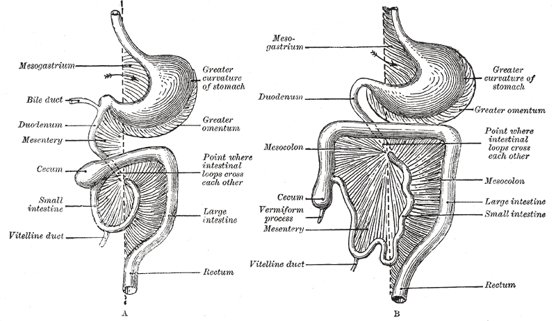
The mesentery is an organ that attaches the intestines to the posterior abdominal wall in humans and is formed by the double fold of peritoneum. It helps in storing fat and allowing blood vessels, lymphatics, and nerves to supply the intestines, among other functions. The mesocolon was thought to be a fragmented structure, with all named parts—the ascending, transverse, descending, and sigmoid mesocolons, the mesoappendix, and the mesorectum—separately terminating their insertion into the posterior abdominal wall. However, in 2012, new microscopic and electron microscopic examinations showed the mesocolon to be a single structure derived from the duodenojejunal flexure and extending to the distal mesorectal layer. Thus, the mesentery is an internal organ. Structure The mesentery of the small intestine arises from the root of the mesentery (or mesenteric root) and is the part connected with the structures in front of the vertebral column. The root is narrow, about 15&n ... [...More Info...] [...Related Items...] OR: [Wikipedia] [Google] [Baidu] |
Duodenojejunal Flexure
The duodenojejunal flexure or duodenojejunal junction is the border between the duodenum and the jejunum. Structure The ascending portion of the duodenum ascends on the left side of the aorta, as far as the level of the upper border of the second lumbar vertebra. At this point, it turns abruptly forward to merge with the jejunum, forming the duodenojejunal flexure. This forms the beginning of the jejunum. The duodenojejunal flexure is surrounded by the suspensory muscle of the duodenum. It is retroperitoneal, so is less mobile than the jejunum that comes after it, helping to stabilise the jejunum. The duodenojejunal flexure lies in front of the left psoas major muscle, the left renal artery, and the left renal vein. It is covered in front, and partly at the sides, by peritoneum continuous with the left portion of the mesentery. Clinical significance The ligament of Treitz, a peritoneal fold, from the right crus of diaphragm, is an identification point for the duodenojejunal flex ... [...More Info...] [...Related Items...] OR: [Wikipedia] [Google] [Baidu] |
Sacroiliac Joint
The sacroiliac joint or SI joint (SIJ) is the joint between the sacrum and the ilium bones of the pelvis, which are connected by strong ligaments. In humans, the sacrum supports the spine and is supported in turn by an ilium on each side. The joint is strong, supporting the entire weight of the upper body. It is a synovial plane joint with irregular elevations and depressions that produce interlocking of the two bones. The human body has two sacroiliac joints, one on the left and one on the right, that often match each other but are highly variable from person to person. Structure Sacroiliac joints are paired C-shaped or L-shaped joints capable of a small amount of movement (2–18 degrees, which is debatable at this time) that are formed between the auricular surfaces of the sacrum and the ilium bones. However mostBogduk, Nicolai "Clinical and Radiological Anatomy of the Lumbar Spine" Elsevier Health Sciences, 2022, p. 172. agree that there are only slight movements o ... [...More Info...] [...Related Items...] OR: [Wikipedia] [Google] [Baidu] |
Vermiform Appendix
The appendix (or vermiform appendix; also cecal r caecalappendix; vermix; or vermiform process) is a finger-like, blind-ended tube connected to the cecum, from which it develops in the embryo. The cecum is a pouch-like structure of the large intestine, located at the junction of the small and the large intestines. The term " vermiform" comes from Latin and means "worm-shaped". The appendix was once considered a vestigial organ, but this view has changed since the early 2000s. Research suggests that the appendix may serve an important purpose. In particular, it may serve as a reservoir for beneficial gut bacteria. Structure The human appendix averages in length but can range from . The diameter of the appendix is , and more than is considered a thickened or inflamed appendix. The longest appendix ever removed was long. The appendix is usually located in the lower right quadrant of the abdomen, near the right hip bone. The base of the appendix is located beneath the ... [...More Info...] [...Related Items...] OR: [Wikipedia] [Google] [Baidu] |
Ileum
The ileum () is the final section of the small intestine in most higher vertebrates, including mammals, reptiles, and birds. In fish, the divisions of the small intestine are not as clear and the terms posterior intestine or distal intestine may be used instead of ileum. Its main function is to absorb vitamin B12, bile salts, and whatever products of digestion that were not absorbed by the jejunum. The ileum follows the duodenum and jejunum and is separated from the cecum by the ileocecal valve (ICV). In humans, the ileum is about 2–4 m long, and the pH is usually between 7 and 8 (neutral or slightly basic). ''Ileum ''is derived from the Greek word ''eilein'', meaning "to twist up tightly". Structure The ileum is the third and final part of the small intestine. It follows the jejunum and ends at the ileocecal junction, where the terminal ileum communicates with the cecum of the large intestine through the ileocecal valve. The ileum, along with the jejunum, is susp ... [...More Info...] [...Related Items...] OR: [Wikipedia] [Google] [Baidu] |
Sigmoid Colon
The sigmoid colon (or pelvic colon) is the part of the large intestine that is closest to the rectum and anus. It forms a loop that averages about in length. The loop is typically shaped like a Greek letter sigma (ς) or Latin letter S (thus '' sigma'' + '' -oid''). This part of the colon normally lies within the pelvis, but due to its freedom of movement it is liable to be displaced into the abdominal cavity. Structure The sigmoid colon begins at the superior aperture of the lesser pelvis, where it is continuous with the iliac colon, and passes transversely across the front of the sacrum to the right side of the pelvis. It then curves on itself and turns toward the left to reach the middle line at the level of the third piece of the sacrum, where it bends downward and ends in the rectum. Its function is to expel solid and gaseous waste from the gastrointestinal tract. The curving path it takes toward the anus allows it to store gas in the superior arched portion, enab ... [...More Info...] [...Related Items...] OR: [Wikipedia] [Google] [Baidu] |
Transverse Colon
In human anatomy, the transverse colon is the longest and most movable part of the colon. Anatomical position It crosses the abdomen from the ascending colon at the right colic flexure (hepatic flexure) with a downward convexity to the descending colon where it curves sharply on itself beneath the lower end of the spleen forming the left colic flexure (splenic flexure). In its course, it describes an arch, the concavity of which is directed backward and a little upward. Toward its splenic end there is often an abrupt U-shaped curve which may descend lower than the main curve. It is almost completely invested by the peritoneum, and is connected to the inferior border of the pancreas by a large and wide duplicature of that membrane, the transverse mesocolon. It is in relation, by its upper surface, with the liver and gall-bladder, the greater curvature of the stomach, and the lower end of the spleen; by its under surface, with the small intestine; by its anterior surface, with ... [...More Info...] [...Related Items...] OR: [Wikipedia] [Google] [Baidu] |
Colic Flexures
In the anatomy of the human digestive tract, there are two colic flexures, or curvatures in the transverse colon. The right colic flexure is also known as the hepatic flexure, and the left colic flexure is also known as the splenic flexure. Note that "right" refers to the patient's anatomical right, which may be depicted on the left of a diagram. Structure Right colic flexure The right colic flexure or hepatic flexure (as it is next to the liver) is the sharp bend between the ascending colon and the transverse colon. The hepatic flexure lies in the right upper quadrant of the human abdomen. It receives blood supply from the superior mesenteric artery. Left colic flexure The left colic flexure or splenic flexure (as it is close to the spleen) is the sharp bend between the transverse colon and the descending colon. The splenic flexure receives dual blood supply from the terminal branches of the superior mesenteric artery and the inferior mesenteric artery. Clinical significan ... [...More Info...] [...Related Items...] OR: [Wikipedia] [Google] [Baidu] |
Presacral Fascia
The presacral fascia lines the anterior aspect of the sacrum, enclosing the sacral vessels and nerves. It continues anteriorly as the pelvic parietal fascia, covering the entire pelvic cavity. The presacral fascia is limited postero-inferiorly, as it fuses with the mesorectal fascia, lying above the levator ani muscle, at the level of the anorectal junction. These two fascias have been erroneously confused, though they are in fact, separate anatomical entities. The colloquial term, among colo-rectal surgeons, for this inter-fascial plane, is known as the holy plane of dissection first coined by Bill Heald. During rectal surgery and mesorectum excision, dissection along the avascular alveolar plane between these two fascias, facilitates a straightforward dissection and preserves the sacral vessels and hypogastric nerves. Waldeyer's fascia (a.k.a. rectosacral fascia) originates from the presacral parietal fascia at the S2 to S4 level fusing with the rectal visceral fascia at th ... [...More Info...] [...Related Items...] OR: [Wikipedia] [Google] [Baidu] |
Pelvis
The pelvis (plural pelves or pelvises) is the lower part of the trunk, between the abdomen and the thighs (sometimes also called pelvic region), together with its embedded skeleton (sometimes also called bony pelvis, or pelvic skeleton). The pelvic region of the trunk includes the bony pelvis, the pelvic cavity (the space enclosed by the bony pelvis), the pelvic floor, below the pelvic cavity, and the perineum, below the pelvic floor. The pelvic skeleton is formed in the area of the back, by the sacrum and the coccyx and anteriorly and to the left and right sides, by a pair of hip bones. The two hip bones connect the spine with the lower limbs. They are attached to the sacrum posteriorly, connected to each other anteriorly, and joined with the two femurs at the hip joints. The gap enclosed by the bony pelvis, called the pelvic cavity, is the section of the body underneath the abdomen and mainly consists of the reproductive organs (sex organs) and the rectum, while the ... [...More Info...] [...Related Items...] OR: [Wikipedia] [Google] [Baidu] |
Retroperitoneum
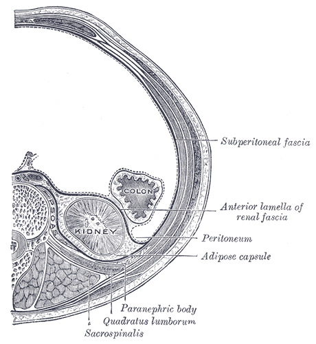
The retroperitoneal space (retroperitoneum) is the anatomical space (sometimes a potential space) behind (''retro'') the peritoneum. It has no specific delineating anatomical structures. Organs are retroperitoneal if they have peritoneum on their anterior side only. Structures that are not suspended by mesentery in the abdominal cavity and that lie between the parietal peritoneum and abdominal wall are classified as retroperitoneal. This is different from organs that are not retroperitoneal, which have peritoneum on their posterior side and are suspended by mesentery in the abdominal cavity. The retroperitoneum can be further subdivided into the following: *Perirenal (or perinephric) space *Anterior pararenal (or paranephric) space *Posterior pararenal (or paranephric) space Retroperitoneal structures Structures that lie behind the peritoneum are termed "retroperitoneal". Organs that were once suspended within the abdominal cavity by mesentery but migrated posterior to the p ... [...More Info...] [...Related Items...] OR: [Wikipedia] [Google] [Baidu] |
Colic Flexures
In the anatomy of the human digestive tract, there are two colic flexures, or curvatures in the transverse colon. The right colic flexure is also known as the hepatic flexure, and the left colic flexure is also known as the splenic flexure. Note that "right" refers to the patient's anatomical right, which may be depicted on the left of a diagram. Structure Right colic flexure The right colic flexure or hepatic flexure (as it is next to the liver) is the sharp bend between the ascending colon and the transverse colon. The hepatic flexure lies in the right upper quadrant of the human abdomen. It receives blood supply from the superior mesenteric artery. Left colic flexure The left colic flexure or splenic flexure (as it is close to the spleen) is the sharp bend between the transverse colon and the descending colon. The splenic flexure receives dual blood supply from the terminal branches of the superior mesenteric artery and the inferior mesenteric artery. Clinical significan ... [...More Info...] [...Related Items...] OR: [Wikipedia] [Google] [Baidu] |
Colectomy
Colectomy ('' col-'' + '' -ectomy'') is bowel resection of the large bowel ( colon). It consists of the surgical removal of any extent of the colon, usually segmental resection (partial colectomy). In extreme cases where the entire large intestine is removed, it is called total colectomy, and proctocolectomy ('' procto-'' + ''colectomy'') denotes that the rectum is included. Indications Some of the most common indications for colectomy are: * Colon cancer * Diverticulitis and diverticular disease of the large intestine * Trauma * Inflammatory bowel disease such as ulcerative colitis or Crohn's disease. Colectomy neither cures nor eliminates Crohn's disease, instead only removing part of the entire diseased large intestine. A colectomy is considered a "cure" for ulcerative colitis because the disease attacks only the large intestine and therefore will not be able to flare up again if the entire large intestine (cecum, ascending colon, transverse colon, descending colon and ... [...More Info...] [...Related Items...] OR: [Wikipedia] [Google] [Baidu] |


