|
Dorsal Rami
The dorsal ramus of spinal nerve, posterior ramus of spinal nerve, or posterior primary division is the posterior division of a spinal nerve. The dorsal rami provide motor innervation to the deep (a.k.a. intrinsic or true) muscles of the back, and sensory innervation to the skin of the posterior portion of the head, neck and back. A spinal nerve splits within the intervertebral foramen to form a dorsal ramus and a ventral ramus. The dorsal ramus then turns to course posterior-ward before splitting into a medial branch and a lateral branch. Both these branches provide motor innervation to deep back muscles. In the neck and upper back, the medial branch is also responsible for providing sensory innervation of the skin; in the lower back, the lateral branch does so. All medial branches additionally also provide sensory innervation to the zygapophyseal joints and periosteum of the vertebral column. Structure Ventral root axons join with dorsal root ganglia to form mixed spinal nerves ... [...More Info...] [...Related Items...] OR: [Wikipedia] [Google] [Baidu] |
Spinal Nerve
A spinal nerve is a mixed nerve, which carries Motor neuron, motor, Sensory neuron, sensory, and Autonomic nervous system, autonomic signals between the spinal cord and the body. In the human body there are 31 pairs of spinal nerves, one on each side of the vertebral column. These are grouped into the corresponding cervical vertebrae, cervical, thoracic vertebrae, thoracic, lumbar vertebrae, lumbar, sacral vertebrae, sacral and coccygeal vertebrae, coccygeal regions of the spine. There are eight pairs of cervical nerves, twelve pairs of thoracic nerves, five pairs of lumbar nerves, five pairs of sacral nerves, and one pair of coccygeal nerves. The spinal nerves are part of the peripheral nervous system. Structure Each spinal nerve is a mixed nerve, formed from the combination of nerve root axon, fibers from its Dorsal root of spinal nerve, dorsal and Ventral root of spinal nerve, ventral roots. The dorsal root is the afferent nerve fiber, afferent sensory root and carries sen ... [...More Info...] [...Related Items...] OR: [Wikipedia] [Google] [Baidu] |
Sympathetic Ganglion
The sympathetic ganglia, or paravertebral ganglia, are autonomic ganglia of the sympathetic nervous system. Ganglia are 20,000 to 30,000 afferent and efferent nerve cell bodies that run along on either side of the spinal cord. Afferent nerve cell bodies bring information from the body to the brain and spinal cord, while efferent nerve cell bodies bring information from the brain and spinal cord to the rest of the body. The cell bodies create long sympathetic chains that are on either side of the spinal cord. They also form para- or pre-vertebral ganglia of gross anatomy. The efferent nerve cell bodies bring information from the body to the brain regarding perceptions of danger. This perception of danger can instigate the fight-or-flight response associated with the sympathetic nervous system. The fight-or-flight response is adaptive when there is a real and present danger which can be avoided or diminished through increased sympathetic activity. Sympathetic activity could be incr ... [...More Info...] [...Related Items...] OR: [Wikipedia] [Google] [Baidu] |
Zygapophyseal Joint
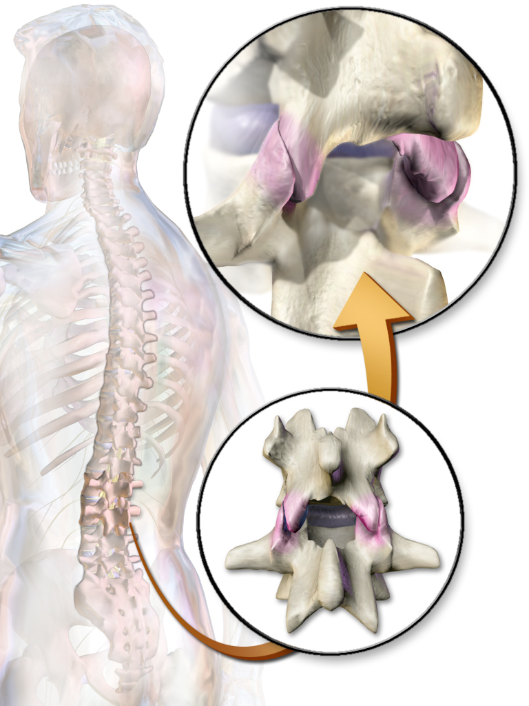
The facet joints (also zygapophysial joints, zygapophyseal, apophyseal, or Z-joints) are a set of synovial, plane joints between the articular processes of two adjacent vertebrae. There are two facet joints in each spinal motion segment and each facet joint is innervated by the recurrent meningeal nerves. Innervation Innervation to the facet joints vary between segments of the spinal, but they are generally innervated by medial branch nerves that come off the dorsal rami. It is thought that these nerves are for primary sensory input, though there is some evidence that they have some motor input local musculature. Within the cervical spine, most joints are innervated by the medial branch nerve (a branch of the dorsal rami) from the same levels. In other words, the facet joint between C4 and C5 vertebral segments is innervated by the C4 and C5 medial branch nerves. However, there are two exceptions: # The facet joint between C2 and C3 is innervated by the third occipital ne ... [...More Info...] [...Related Items...] OR: [Wikipedia] [Google] [Baidu] |
Transversospinales
The transversospinales are a group of muscles of the human back. Their combined action is rotation and extension of the vertebral column. These muscles are small and have a poor mechanical advantage for contributing to motion. They include: the three semispinalis muscles, the multifidus muscle, and the rotatores muscles. Location The three semispinalis muscles, span 4-6 vertebral segments: ** semispinalis thoracis ** semispinalis cervicis ** semispinalis capitis The multifidus muscle The multifidus (multifidus spinae; : ''multifidi'') muscle consists of a number of fleshy and tendinous fasciculi, which fill up the groove on either side of the spinous processes of the vertebrae, from the sacrum to the axis. While very thin, ..., and spans 2-4 vertebral segments The rotatores muscles, lie beneath the multifidus, and spans 1-2 vertebral segments ** rotatores cervicis ** rotatores thoracis ** rotatores lumborum External links * Musculoskeletal Interventions: Techniques fo ... [...More Info...] [...Related Items...] OR: [Wikipedia] [Google] [Baidu] |
Iliocostalis
Iliocostalis muscle is the muscle immediately lateral to the longissimus that is the nearest to the furrow that separates the epaxial muscles from the hypaxial. It lies very deep to the fleshy portion of the serratus posterior muscle. It laterally flexes the vertebral column to the same side. Structure Iliocostalis muscle has a common origin from the iliac crest, the sacrum, the thoracolumbar fascia, and the spinous processes of the vertebrae from T11 to L5. Iliocostalis cervicis (cervicalis ascendens) arises from the angles of the third, fourth, fifth, and sixth ribs, and is inserted into the posterior tubercles of the transverse processes of the fourth, fifth, and sixth cervical vertebrae. Iliocostalis thoracis (musculus accessorius; iliocostalis thoracis) arises by flattened tendons from the upper borders of the angles of the lower six ribs medial to the tendons of insertion of the iliocostalis lumborum; these become muscular, and are inserted into the upper bord ... [...More Info...] [...Related Items...] OR: [Wikipedia] [Google] [Baidu] |
Anatomical Terms Of Location
Standard anatomical terms of location are used to describe unambiguously the anatomy of humans and other animals. The terms, typically derived from Latin or Greek roots, describe something in its standard anatomical position. This position provides a definition of what is at the front ("anterior"), behind ("posterior") and so on. As part of defining and describing terms, the body is described through the use of anatomical planes and axes. The meaning of terms that are used can change depending on whether a vertebrate is a biped or a quadruped, due to the difference in the neuraxis, or if an invertebrate is a non-bilaterian. A non-bilaterian has no anterior or posterior surface for example but can still have a descriptor used such as proximal or distal in relation to a body part that is nearest to, or furthest from its middle. International organisations have determined vocabularies that are often used as standards for subdisciplines of anatomy. For example, '' Termi ... [...More Info...] [...Related Items...] OR: [Wikipedia] [Google] [Baidu] |
Coccygeal
The coccyx (: coccyges or coccyxes), commonly referred to as the tailbone, is the final segment of the vertebral column in all apes, and analogous structures in certain other mammals such as horses. In tailless primates (e.g. humans and other great apes) since '' Nacholapithecus'' (a Miocene hominoid),Nakatsukasa 2004, ''Acquisition of bipedalism'' (SeFig. 5entitled ''First coccygeal/caudal vertebra in short-tailed or tailless primates.''.) the coccyx is the remnant of a vestigial tail. In animals with bony tails, it is known as ''tailhead'' or ''dock'', in bird anatomy as ''tailfan''. It comprises three to five separate or fused coccygeal vertebrae below the sacrum, attached to the sacrum by a fibrocartilaginous joint, the sacrococcygeal symphysis, which permits limited movement between the sacrum and the coccyx. Structure The coccyx is formed of three, four or five rudimentary vertebrae. It articulates superiorly with the sacrum. In each of the first three segments may be ... [...More Info...] [...Related Items...] OR: [Wikipedia] [Google] [Baidu] |
Sacrum
The sacrum (: sacra or sacrums), in human anatomy, is a triangular bone at the base of the spine that forms by the fusing of the sacral vertebrae (S1S5) between ages 18 and 30. The sacrum situates at the upper, back part of the pelvic cavity, between the two wings of the pelvis. It forms joints with four other bones. The two projections at the sides of the sacrum are called the alae (wings), and articulate with the ilium at the L-shaped sacroiliac joints. The upper part of the sacrum connects with the last lumbar vertebra (L5), and its lower part with the coccyx (tailbone) via the sacral and coccygeal cornua. The sacrum has three different surfaces which are shaped to accommodate surrounding pelvic structures. Overall, it is concave (curved upon itself). The base of the sacrum, the broadest and uppermost part, is tilted forward as the sacral promontory internally. The central part is curved outward toward the posterior, allowing greater room for the pelvic cavity. In a ... [...More Info...] [...Related Items...] OR: [Wikipedia] [Google] [Baidu] |
Neck
The neck is the part of the body in many vertebrates that connects the head to the torso. It supports the weight of the head and protects the nerves that transmit sensory and motor information between the brain and the rest of the body. Additionally, the neck is highly flexible, allowing the head to turn and move in all directions. Anatomically, the human neck is divided into four compartments: vertebral, visceral, and two vascular compartments. Within these compartments, the neck houses the cervical vertebrae, the cervical portion of the spinal cord, upper parts of the respiratory and digestive tracts, endocrine glands, nerves, arteries and veins. The muscles of the neck, which are separate from the compartments, form the boundaries of the neck triangles. In anatomy, the neck is also referred to as the or . However, when the term ''cervix'' is used alone, it often refers to the uterine cervix, the neck of the uterus. Therefore, the adjective ''cervical'' ... [...More Info...] [...Related Items...] OR: [Wikipedia] [Google] [Baidu] |
Ramus Communicans
Ramus communicans (: rami communicantes) is the Latin term used for a nerve which connects two other nerves, and can be translated as "communicating branch". Structure When used without further definition, it almost always refers to a communicating branch between a spinal nerve and the sympathetic trunk. More specifically, it usually refers to one of the following : * Gray ramus communicans * White ramus communicans The grey and white rami communicantes are responsible for conveying autonomic signals, specifically for the sympathetic nervous system. Their difference in colouration is caused by differences in myelination of the nerve fibres contained within, i.e. there are more myelinated than unmyelinated fibres in the white rami communicantes while the converse is true for the grey rami communicantes. Gray ramus communicans The grey rami communicantes exist at every level of the spinal cord and are responsible for carrying postganglionic nerve fibres from the paravertebral ... [...More Info...] [...Related Items...] OR: [Wikipedia] [Google] [Baidu] |
Intervertebral Foramen
The intervertebral foramen (also neural foramen) (often abbreviated as IV foramen or IVF) is an opening between (the intervertebral notches of) two pedicles (one above and one below) of adjacent vertebra in the articulated spine. Each intervertebral foramen gives passage to a spinal nerve and spinal blood vessels, and lodges a posterior (dorsal) root ganglion. Cervical, thoracic, and lumbar vertebrae all have intervertebral foramina. Anatomy Structure In the thoracic region and lumbar region, each vertebral foramen is additionally bounded anteriorly by (the inferior portion of) the body of vertebra (particularly in the thoracic region) and adjacent intervertebral disc (particularly in the lumbar region). In the cervical region, a small part of the body of vertebra inferior to the intervertebral disc also forms the anterior boundary of the IVF (due to the fact that the junction of the pedicle with the body of vertebra is situated somewhat more inferiorly on the body). ... [...More Info...] [...Related Items...] OR: [Wikipedia] [Google] [Baidu] |
Motor System
The motor system is the set of central nervous system, central and peripheral nervous system, peripheral structures in the nervous system that support motor functions, i.e. movement. Peripheral structures may include skeletal muscles and Efferent nerve fiber#Motor nerve, neural connections with muscle tissues. Central structures include cerebral cortex, brainstem, spinal cord, pyramidal system including the upper motor neurons, extrapyramidal system, cerebellum, and the lower motor neurons in the brainstem and the spinal cord. The motor system is a biological system with close ties to the muscular system and the circulatory system. To achieve motor skill, the motor system must accommodate the working state of the muscles, whether hot or cold, stiff or loose, as well as physiological fatigue. Pyramidal motor system The pyramidal tracts, pyramidal motor system, also called the pyramidal tract or the corticospinal tract, start in the motor center of the cerebral cortex.Rizzolatti ... [...More Info...] [...Related Items...] OR: [Wikipedia] [Google] [Baidu] |


