|
Diagonal Band Of Broca
The diagonal band of Broca interconnects the amygdala and the septal area. It is one of the olfactory structures. It is situated upon the inferior aspect of the brain. It forms the medial margin of the anterior perforated substance. It was described by the French neuroanatomist Paul Broca. Structure It consists of fibers that are said to arise in the parolfactory area, the gyrus subcallosus and the anterior perforated substance, and course backward in the longitudinal striae to the dentate gyrus and the hippocampal region. This is a cholinergic bundle of nerve fibers posterior to the anterior perforated substance. It interconnects the subcallosal gyrus in the septal area with the hippocampus and lateral olfactory area. Nuclei Two structures are often described in this brain regions, namely the nuclei of the vertical and horizontal limbs of the diagonal band of Broca (nvlDBB and nhlDBB, respectively). nvlDBB projects to the hippocampal formation through the fornix and ... [...More Info...] [...Related Items...] OR: [Wikipedia] [Google] [Baidu] [Amazon] |
Amygdala
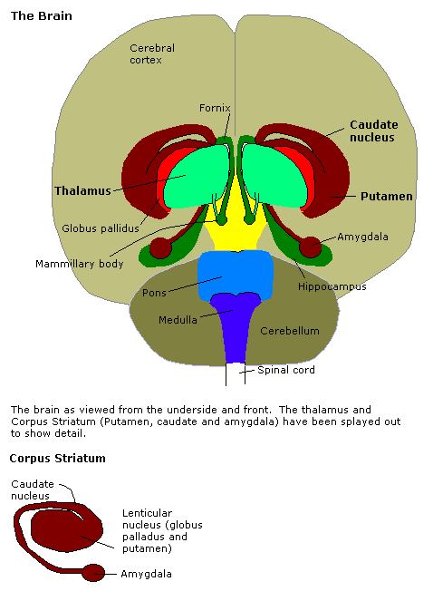
The amygdala (; : amygdalae or amygdalas; also '; Latin from Greek language, Greek, , ', 'almond', 'tonsil') is a paired nucleus (neuroanatomy), nuclear complex present in the Cerebral hemisphere, cerebral hemispheres of vertebrates. It is considered part of the limbic system. In Primate, primates, it is located lateral and medial, medially within the temporal lobes. It consists of many nuclei, each made up of further subnuclei. The subdivision most commonly made is into the Basolateral amygdala, basolateral, Central nucleus of the amygdala, central, cortical, and medial nuclei together with the intercalated cells of the amygdala, intercalated cell clusters. The amygdala has a primary role in the processing of memory, decision making, decision-making, and emotions, emotional responses (including fear, anxiety, and aggression). The amygdala was first identified and named by Karl Friedrich Burdach in 1822. Structure Thirteen Nucleus (neuroanatomy), nuclei have been identif ... [...More Info...] [...Related Items...] OR: [Wikipedia] [Google] [Baidu] [Amazon] |
Hippocampal Formation
The hippocampal formation is a compound structure in the medial temporal lobe of the brain. It forms a c-shaped bulge on the floor of the inferior horn of the lateral ventricle. Typically, the hippocampal formation is said to included the dentate gyrus, the hippocampus, and the subiculum. The presubiculum, parasubiculum, and the entorhinal cortex may also be included. The hippocampal formation is thought to play a role in memory, spatial navigation and control of attention. The neural layout and pathways within the hippocampal formation are very similar in all mammals. History and function During the nineteenth and early twentieth centuries, based largely on the observation that, between species, the size of the olfactory bulb varies with the size of the parahippocampal gyrus, the hippocampal formation was thought to be part of the olfactory system. In 1937, Papez theorized that a circuit (the Papez circuit) including the hippocampal formation constitutes the neural subst ... [...More Info...] [...Related Items...] OR: [Wikipedia] [Google] [Baidu] [Amazon] |
Nerve Growth Factor
Nerve growth factor (NGF) is a neurotrophic factor and neuropeptide primarily involved in the regulation of growth, maintenance, proliferation, and survival of certain target neurons. It is perhaps the prototypical growth factor, in that it was one of the first to be described. Since it was first isolated by Nobel laureates Rita Levi-Montalcini and Stanley Cohen in 1954, numerous biological processes involving NGF have been identified, two of them being the survival of pancreatic beta cells and the regulation of the immune system. Structure NGF is initially in a 7S, 130- kDa complex of 3 proteins – Alpha-NGF, Beta-NGF, and Gamma-NGF (2:1:2 ratio) when expressed. This form of NGF is also referred to as proNGF (NGF precursor). The gamma subunit of this complex acts as a serine protease, and cleaves the N-terminal of the beta subunit, thereby activating the protein into functional NGF. The term ''nerve growth factor'' usually refers to the 2.5S, 26-kDa beta subunit of the p ... [...More Info...] [...Related Items...] OR: [Wikipedia] [Google] [Baidu] [Amazon] |
Magnocellular Neurosecretory Cell
Magnocellular neurosecretory cells are large neuroendocrine cells within the supraoptic nucleus and paraventricular nucleus of the hypothalamus. They are also found in smaller numbers in accessory cell groups between these two nuclei, the largest one being the circular nucleus. There are two types of magnocellular neurosecretory cells, oxytocin-producing cells and vasopressin-producing cells, but a small number can produce both hormones. These cells are neuroendocrine neurons, are electrically excitable, and generate action potentials in response to afferent stimulation. Vasopressin is produced from the vasopressin-producing cells via the AVP gene, a molecular output of circadian pathways. Magnocellular neurosecretory cells in rats (where these neurons have been most extensively studied) in general have a single long varicose axon, which projects to the posterior pituitary. Each axon gives rise to about 10,000 neurosecretory terminals and many axon swellings that store very l ... [...More Info...] [...Related Items...] OR: [Wikipedia] [Google] [Baidu] [Amazon] |
Theta Wave
Theta waves generate the theta rhythm, a neural oscillation in the brain that underlies various aspects of cognition and behavior, including learning, memory, and spatial navigation in many animals. It can be recorded using various electrophysiological methods, such as electroencephalogram (EEG), recorded either from inside the brain or from electrodes attached to the scalp. At least two types of theta rhythm have been described. The hippocampal theta rhythm is a strong oscillation that can be observed in the hippocampus and other brain structures in numerous species of mammals including rodents, rabbits, dogs, cats, and marsupials. ''"Cortical theta rhythms"'' are low-frequency components of scalp EEG, usually recorded from humans. Theta rhythms can be quantified using quantitative electroencephalography (qEEG) using freely available toolboxes, such as, EEGLAB or the Neurophysiological Biomarker Toolbox (NBT). In rats, theta wave rhythmicity is easily observed in the hipp ... [...More Info...] [...Related Items...] OR: [Wikipedia] [Google] [Baidu] [Amazon] |
Medial Septal Nucleus
The medial septal nucleus (MS) is one of the septal nuclei. Neurons in this nucleus give rise to the bulk of efferents from the septal nuclei. A major projection from the medial septal nucleus terminates in the hippocampal formation. It plays a role in the generation of theta waves in the hippocampus. Specifically, the GABAergic cells of the medial septum that act as theta pacemakers target dentate gyrus, CA3, and CA1 interneurons. Pacemaking MS interneurons express hyperpolarization-activated cyclic nucleotide-gated (HCN) channels which likely, at least partially, mediate their pacemaker properties. It is composed of GABAergic cells, glutamatergic cells, and cholinergic cells. Each cell-type carries out different functions. In addition to the theta wave generation, it has recently been discovered that medial septum also serves as an important node for sensory valence processing. For example, Vglut2 neurons in medial septum respond strongly to noxious sensory stimulation. Re ... [...More Info...] [...Related Items...] OR: [Wikipedia] [Google] [Baidu] [Amazon] |
Septum Pellucidum
The septum pellucidum (Latin for "translucent wall") is a thin, triangular, vertical double membrane separating the anterior horns of the left and right lateral ventricles of the brain. It runs as a sheet from the corpus callosum down to the fornix. The septum is not present in the syndrome septo-optic dysplasia. Structure The septum pellucidum is located in the septal area in the midline of the brain between the two cerebral hemispheres. The septal area is also the location of the septal nuclei. It is attached to the lower part of the corpus callosum, the large collection of nerve fibers that connect the two cerebral hemispheres. It is attached to the front forward part of the fornix. The lateral ventricles sit on either side of the septum. The septum pellucidum consists of two layers or ''laminae'' of both white and gray matter. During fetal development, there is a space between the two laminae called the cave of septum pellucidum that, in ninety percent of cases, dis ... [...More Info...] [...Related Items...] OR: [Wikipedia] [Google] [Baidu] [Amazon] |
Cerebrum
The cerebrum (: cerebra), telencephalon or endbrain is the largest part of the brain, containing the cerebral cortex (of the two cerebral hemispheres) as well as several subcortical structures, including the hippocampus, basal ganglia, and olfactory bulb. In the human brain, the cerebrum is the uppermost region of the central nervous system. The cerebrum prenatal development, develops prenatally from the forebrain (prosencephalon). In mammals, the Dorsum (biology), dorsal telencephalon, or Pallium (neuroanatomy), pallium, develops into the cerebral cortex, and the ventral telencephalon, or Pallium (neuroanatomy), subpallium, becomes the basal ganglia. The cerebrum is also divided into approximately symmetric Lateralization of brain function, left and right cerebral hemispheres. With the assistance of the cerebellum, the cerebrum controls all voluntary actions in the human body. Structure The cerebrum is the largest part of the brain. Depending upon the position of the animal, ... [...More Info...] [...Related Items...] OR: [Wikipedia] [Google] [Baidu] [Amazon] |
Basal Forebrain
Part of the human brain, the basal forebrain structures are located in the forebrain to the front of and below the striatum. They include the ventral basal ganglia (including nucleus accumbens and ventral pallidum), nucleus basalis, diagonal band of Broca, substantia innominata, and the medial septal nucleus. These structures are important in the production of acetylcholine, which is then distributed widely throughout the brain. The basal forebrain is considered to be the major cholinergic output of the central nervous system (CNS) centred on the output of the nucleus basalis. The presence of non-cholinergic neurons projecting to the cortex have been found to act with the cholinergic neurons to dynamically modulate activity in the cortex. Function Acetylcholine is known to promote wakefulness in the basal forebrain. Stimulating the basal forebrain gives rise to acetylcholine release, which induces wakefulness and REM sleep, whereas inhibition of acetylcholine release in the ba ... [...More Info...] [...Related Items...] OR: [Wikipedia] [Google] [Baidu] [Amazon] |
Olfactory Bulb
The olfactory bulb (Latin: ''bulbus olfactorius'') is a neural structure of the vertebrate forebrain involved in olfaction, the sense of smell. It sends olfactory information to be further processed in the amygdala, the orbitofrontal cortex (OFC) and the hippocampus where it plays a role in emotion, memory and learning. The bulb is divided into two distinct structures: the main olfactory bulb and the accessory olfactory bulb. The main olfactory bulb connects to the amygdala via the piriform cortex of the primary olfactory cortex and directly projects from the main olfactory bulb to specific amygdala areas. The accessory olfactory bulb resides on the dorsal-posterior region of the main olfactory bulb and forms a parallel pathway. Destruction of the olfactory bulb results in ipsilateral anosmia, while irritative lesions of the uncus can result in olfactory and gustatory hallucinations. Structure In most vertebrates, the olfactory bulb is the most rostral (forward) part ... [...More Info...] [...Related Items...] OR: [Wikipedia] [Google] [Baidu] [Amazon] |
Fornix Of The Brain
The fornix (from ; : fornices) is a C-shaped bundle of nerve fibers in the brain that acts as the major output tract of the hippocampus. The fornix also carries some afferent fibers to the hippocampus from structures in the diencephalon and basal forebrain. The fornix is part of the limbic system. While its exact function and importance in the physiology of the brain are still not entirely clear, it has been demonstrated in humans that surgical transection—the cutting of the fornix along its body—can cause memory loss. There is some debate over what type of memory is affected by this damage, but it has been found to most closely correlate with recall memory rather than recognition memory. This means that damage to the fornix can cause difficulty in recalling long-term information such as details of past events, but it has little effect on the ability to recognize objects or familiar situations. Structure The fibers begin in the hippocampus on each side of the brain ... [...More Info...] [...Related Items...] OR: [Wikipedia] [Google] [Baidu] [Amazon] |
Lateral Olfactory Area
Lateral is a geometric term of location which may also refer to: Biology and healthcare * Lateral (anatomy), a term of location meaning "towards the side" * Lateral cricoarytenoid muscle, an intrinsic muscle of the larynx * Lateral release (surgery), a surgical procedure to release tight capsular structures Other uses * ''Lateral'', a digital journal and production of the Cultural Studies Association * ''Lateral'', a podcast by English YouTuber and web developer Tom Scott * Lateral canal, a canal built along the same right-of-way as an existing stream * Lateral consonant, a consonant in which the airstream proceeds along one or both of the sides of the tongue * Lateral mark, a sea mark used in maritime pilotage to indicate the edge of a channel * Lateral modes, an aspect of dynamic stability and control in the field of aircraft flight dynamics * Lateral pass, a non-advancing move in gridiron football * Lateral release (phonetics), the release of a plosive consonant into a lat ... [...More Info...] [...Related Items...] OR: [Wikipedia] [Google] [Baidu] [Amazon] |


