|
Deltoid Tuberosity
In human anatomy, the deltoid tuberosity is a rough, triangular area on the anterolateral (front-side) surface of the middle of the humerus. It is a site of attachment of deltoid muscle. Structure Variation The deltoid tuberosity has been reported as very prominent in less than 10% of people. Development The deltoid tuberosity develops through endochondral ossification in a two-phase process. The initiating signal is tendon-dependent, whilst the growth phase is muscle-dependent. Clinical significance The deltoid tuberosity is at risk of avulsion fracture. These fractures may be managed conservatively with rest. Other animals In mammals, the humerus displays a wide morphological variation. The size and orientation of its functionally important features, including the deltoid tubercle, greater tubercle, and medial epicondyle, are pivotal to an animal's style of locomotion and habitat. In cursorial (running) animals such as the pronghorn, the deltoid tubercle is locate ... [...More Info...] [...Related Items...] OR: [Wikipedia] [Google] [Baidu] |
Human Anatomy
Human anatomy (gr. ἀνατομία, "dissection", from ἀνά, "up", and τέμνειν, "cut") is primarily the scientific study of the morphology of the human body. Anatomy is subdivided into gross anatomy and microscopic anatomy. Gross anatomy (also called macroscopic anatomy, topographical anatomy, regional anatomy, or anthropotomy) is the study of anatomical structures that can be seen by the naked eye. Microscopic anatomy is the study of minute anatomical structures assisted with microscopes, which includes histology (the study of the organization of tissues), and cytology (the study of cells). Anatomy, human physiology (the study of function), and biochemistry (the study of the chemistry of living structures) are complementary basic medical sciences that are generally together (or in tandem) to students studying medical sciences. In some of its facets human anatomy is closely related to embryology, comparative anatomy and comparative embryology, through common ... [...More Info...] [...Related Items...] OR: [Wikipedia] [Google] [Baidu] |
Medial Epicondyle Of The Humerus
The medial epicondyle of the humerus is an epicondyle of the humerus bone of the upper arm in humans. It is larger and more prominent than the Lateral epicondyle of the humerus, lateral epicondyle and is directed slightly more posteriorly in the Anatomical position#Medical (human) anatomy, anatomical position. In birds, where the arm is somewhat rotated compared to other tetrapods, it is called the ventral epicondyle of the humerus. In comparative anatomy, the more neutral term entepicondyle is used. The medial epicondyle gives attachment to the ulnar collateral ligament of elbow joint, to the pronator teres, and to a common tendon of origin (the common flexor tendon) of some of the flexor muscles of the forearm: the flexor carpi radialis, the flexor carpi ulnaris, the flexor digitorum superficialis, and the palmaris longus. The medial epicondyle is located on the distal end of the humerus. Additionally, the medial epicondyle is inferior to the medial supracondylar ridge. It is ... [...More Info...] [...Related Items...] OR: [Wikipedia] [Google] [Baidu] |
Deltoid Tubercle Of Spine Of Scapula
The deltoid tubercle of spine of scapula is a prominence on the spine of scapula. The spine, at lateral to the root of the spine, curves down and laterally to form a lip.R.M.H. McMin"Lasts Anatomy Regional and Applied"Elsevier Australia, 2003. p.129 This lip is called the deltoid tubercle. Muscles Middle and inferior fibres of trapezius muscle, and deltoid muscle, attached to the deltoid tubercle.Alison Middleditch, Jean Oliver"Functional Anatomy of the Spine,"Butterworth-Heinemann (2002) p.113 The deltoid tubercle marks the beginning of attachment of deltoid muscle. Additional images File:Deltoid tubercle of spine - left scapula - animation01.gif, Left scapula. Animation. Deltoid tubercle is shown in red. File:Deltoid tubercle of spine of scapula - animation01.gif, Position of deltoid tubercle (shown in red). Animation. File:Deltoid tubercle of spine - left scapula02.png, Medial view of left scapula. Deltoid tubercle shown in red. File:Gray203.png, Posterior surface of sc ... [...More Info...] [...Related Items...] OR: [Wikipedia] [Google] [Baidu] |
Saunders (imprint)
Saunders is an American academic publisher based in the United States. It is currently an imprint of Elsevier. Formerly independent, the W. B. Saunders company was acquired by CBS in 1968, who added it to their publishing division Holt, Rinehart & Winston. When CBS left the publishing field in 1986, it sold the academic publishing units to Harcourt Brace Jovanovich. Harcourt was acquired by Reed Elsevier in 2001. . Northern Illinois University Libraries. Retrieved May 2, 2015. W. B. Saunders published the Kinsey Reports
[...More Info...] [...Related Items...] OR: [Wikipedia] [Google] [Baidu] |
Horse
The horse (''Equus ferus caballus'') is a domesticated, one-toed, hoofed mammal. It belongs to the taxonomic family Equidae and is one of two extant subspecies of ''Equus ferus''. The horse has evolved over the past 45 to 55 million years from a small multi-toed creature, '' Eohippus'', into the large, single-toed animal of today. Humans began domesticating horses around 4000 BCE in Central Asia, and their domestication is believed to have been widespread by 3000 BCE. Horses in the subspecies ''caballus'' are domesticated, although some domesticated populations live in the wild as feral horses. These feral populations are not true wild horses, which are horses that have never been domesticated. There is an extensive, specialized vocabulary used to describe equine-related concepts, covering everything from anatomy to life stages, size, colors, markings, breeds, locomotion, and behavior. Horses are adapted to run, allowing them to quickly escape predator ... [...More Info...] [...Related Items...] OR: [Wikipedia] [Google] [Baidu] |
Mountain Beaver
The mountain beaver (''Aplodontia rufa'')Other names include boomer, mountain boomer, ground bear, giant mole, gehalis, sewellel, suwellel, showhurll, showtl, and showte, as well as a number of other Native American terms. "Mountain beaver" is a misnomer as the animal is not a true beaver (''Castor'') and it is not restricted to mountains. "Boomer" refers to the loud vocalizations that these usually-solitary animals make when in social situations, but this has not been recorded nor verified. Lewis and Clark originally called the animal "sewellel", a misunderstanding of the Chinook word "she-wal-lal", the name for garments made from the skin of the creature. See Borrecco and Anderson, 1980. is a North American rodent. It is the only living member of its genus, ''Aplodontia'', and Family (biology), family, Aplodontiidae. It should not be confused with true North American beaver, North American and Eurasian beavers, to which it is not closely related; the mountain beaver is instead m ... [...More Info...] [...Related Items...] OR: [Wikipedia] [Google] [Baidu] |
Fossorial
A fossorial animal () is one that is adapted to digging and which lives primarily (but not solely) underground. Examples of fossorial vertebrates are Mole (animal), moles, badgers, naked mole-rats, meerkats, armadillos, wombats, and mole salamanders. Among invertebrates, many molluscs (e.g., clams), insects (e.g., beetles, wasps, bees), and arachnids (e.g. spiders) are fossorial. Prehistoric evidence The physical adaptation of fossoriality is widely accepted as being widespread among many Prehistory, prehistoric Phylum, phyla and Taxon, taxa, such as bacteria and early eukaryotes. Furthermore, fossoriality has evolved independently multiple times, even within a single Family (biology), family. Fossorial animals appeared simultaneously with the colonization of land by arthropods in the late Ordovician period (over 440 million years ago). Other notable early burrowers include ''Eocaecilia'' and possibly ''Dinilysia''. The oldest example of burrowing in synapsids, the lineag ... [...More Info...] [...Related Items...] OR: [Wikipedia] [Google] [Baidu] |
North American River Otter
The North American river otter (''Lontra canadensis''), also known as the northern river otter and river otter, is a semiaquatic mammal that endemism, lives only on the North American continent throughout most of Canada, along the coasts of the United States and its inland waterways. An adult North American river otter can weigh between . The river otter is protected and insulated by a thick, water-repellent coat of fur. The North American river otter, a member of the subfamily Lutrinae in the weasel family (Mustelidae), is equally versatile in the water and on land. It establishes a burrow close to the water's edge in river, lake, swamp, coastal shoreline, tidal flat, or estuary ecosystems. The den typically has many tunnel openings, one of which generally allows the otter to enter and exit the body of water. Female North American river otters give birth in these burrows, producing litters of one to six young. North American river otters, like most predators, prey upon the most ... [...More Info...] [...Related Items...] OR: [Wikipedia] [Google] [Baidu] |
Pronghorn
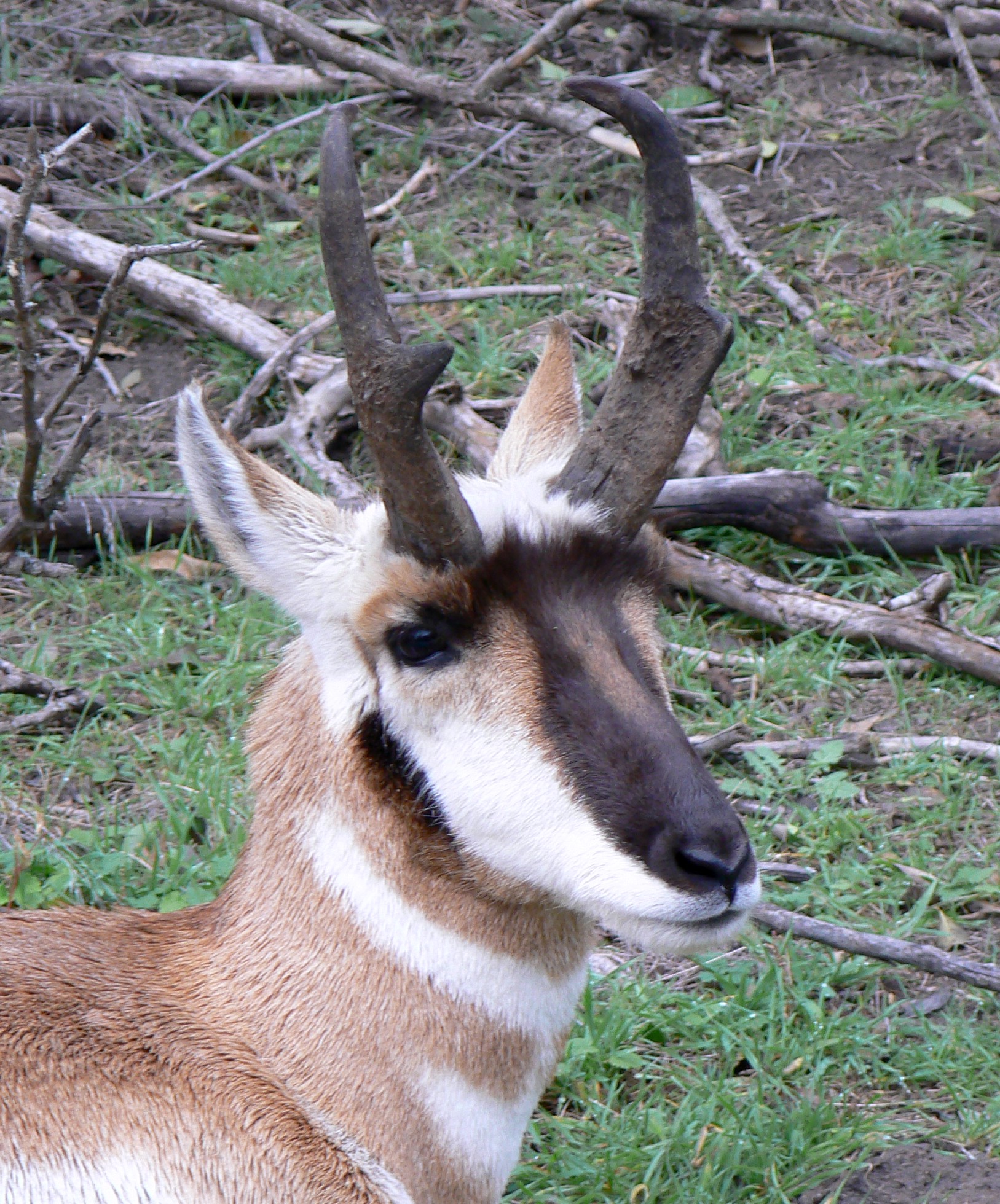
The pronghorn (, ) (''Antilocapra americana'') is a species of artiodactyl (even-toed, hoofed) mammal indigenous to interior western and central North America. Though not an antelope, it is known colloquially in North America as the American antelope, prong buck, pronghorn antelope, and prairie antelope, because it closely resembles the antelopes of the Old World and fills a similar ecological niche due to parallel evolution. It is the only surviving member of the family Antilocapridae. During the Pleistocene epoch, about 11 other antilocaprid species existed in North America, many with long or spectacularly twisted horns.Smithsonian Institution. North American MammalsPronghorn ''Antilocapra americana'' Three other genera ('' Capromeryx'', '' Stockoceros'' and '' Tetrameryx'') existed when humans entered North America but are now extinct. The pronghorn's closest living relatives are the giraffe and okapi. See Fig. S10 in Supplementary Information. The antilocaprids are part of ... [...More Info...] [...Related Items...] OR: [Wikipedia] [Google] [Baidu] |
Cursorial
A cursorial organism is one that is adapted specifically to run. An animal can be considered cursorial if it has the ability to run fast (e.g. cheetah) or if it can keep a constant speed for a long distance (high endurance). "Cursorial" is often used to categorize a certain locomotor mode, which is helpful for biologists who examine behaviors of different animals and the way they move in their environment. Cursorial adaptations can be identified by morphological characteristics (e.g. loss of lateral digits as in ungulate species), physiological characteristics, maximum speed, and how often running is used in life. Much debate exists over how to define a cursorial animal specifically. The most accepted definitions include that a cursorial organism could be considered adapted to long-distance running at high speeds or has the ability to accelerate quickly over short distances. Among vertebrates, animals under 1 kg of mass are rarely considered cursorial, and cursorial behaviors ... [...More Info...] [...Related Items...] OR: [Wikipedia] [Google] [Baidu] |
Greater Tubercle
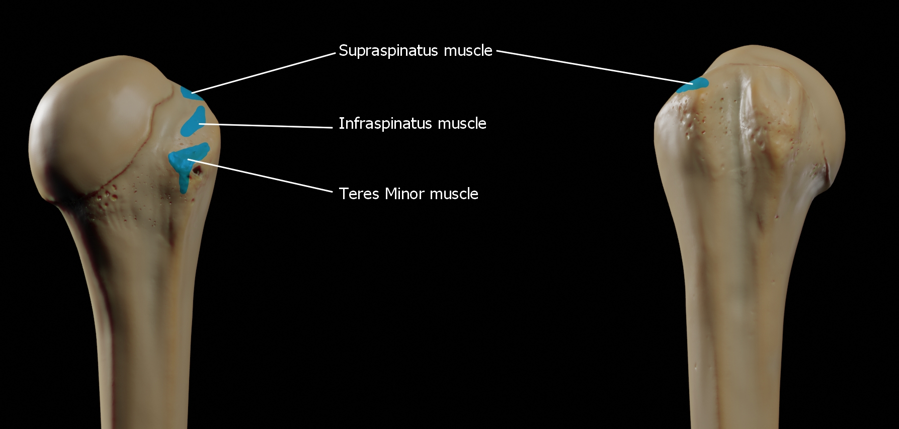
The greater tubercle of the humerus is the outward part the upper end of that bone, adjacent to the large rounded prominence of the humerus head. It provides attachment points for the supraspinatus, infraspinatus, and teres minor muscles, three of the four muscles of the rotator cuff, a muscle group that stabilizes the shoulder joint. In doing so the tubercle acts as a location for the transfer of forces from the rotator cuff muscles to the humerus. Structure The upper surface of the greater tubercle is rounded, and marked by three flat impressions: * the highest ("superior facet") gives insertion to the supraspinatus muscle. * the middle ("middle facet") gives insertion to the infraspinatus muscle. * the lowest ("inferior facet"), and the body of the bone for about 2.5 cm, gives insertion to the teres minor muscle. The lateral surface of the greater tubercle is convex, rough, and continuous with the lateral surface of the body of the humerus. It can be describ ... [...More Info...] [...Related Items...] OR: [Wikipedia] [Google] [Baidu] |


