|
Coronaviridae
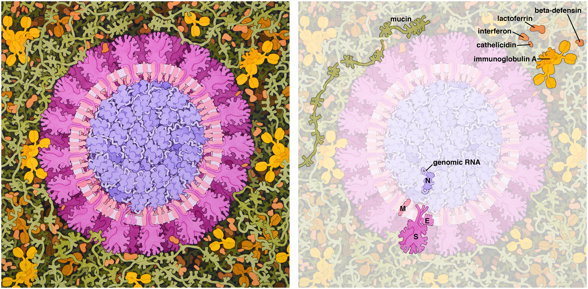
''Coronaviridae'' is a family of enveloped, positive-strand RNA viruses which infect amphibians, birds, and mammals. The group includes the subfamilies '' Letovirinae'' and ''Orthocoronavirinae;'' the members of the latter are known as coronaviruses. The viral genome is 26–32 kilobases in length. The particles are typically decorated with large (~20 nm), club- or petal-shaped surface projections (the " peplomers" or "spikes"), which in electron micrographs of spherical particles create an image reminiscent of the solar corona. Virology The 5' and 3' ends of the genome have a cap and poly(A) tract, respectively. The viral envelope, obtained by budding through membranes of the endoplasmic reticulum (ER) or Golgi apparatus, invariably contains two virus-specified glycoprotein species, known as the spike (S) and membrane (M) proteins. The spike protein makes up the large surface projections (sometimes known as peplomers), while the membrane protein is a triple-spanning ... [...More Info...] [...Related Items...] OR: [Wikipedia] [Google] [Baidu] |
Electron Micrograph
A micrograph or photomicrograph is a photograph or digital image taken through a microscope or similar device to show a magnified image of an object. This is opposed to a macrograph or photomacrograph, an image which is also taken on a microscope but is only slightly magnified, usually less than 10 times. Micrography is the practice or art of using microscopes to make photographs. A micrograph contains extensive details of microstructure. A wealth of information can be obtained from a simple micrograph like behavior of the material under different conditions, the phases found in the system, failure analysis, grain size estimation, elemental analysis and so on. Micrographs are widely used in all fields of microscopy. Types Photomicrograph A light micrograph or photomicrograph is a micrograph prepared using an optical microscope, a process referred to as ''photomicroscopy''. At a basic level, photomicroscopy may be performed simply by connecting a camera to a microscope, t ... [...More Info...] [...Related Items...] OR: [Wikipedia] [Google] [Baidu] |
Peplomer
In virology, a spike protein or peplomer protein is a protein that forms a large structure known as a spike or peplomer projecting from the surface of an enveloped virus. as cited in The proteins are usually glycoproteins that form dimers or trimers. History and etymology The term "peplomer" refers to an individual spike from the viral surface; collectively the layer of material at the outer surface of the virion has been referred to as the "peplos". The term is derived from the Greek peplos, "a loose outer garment", "robe or cloak", or "woman smantle". Early systems of viral taxonomy, such as the Lwoff- Horne- Tournier system proposed in the 1960s, used the appearance and morphology of the "peplos" and peplomers as important characteristics for classification. More recently, the term "peplos" is considered a synonym for viral envelope. Properties Spikes or peplomers are usually rod- or club-shaped projections from the viral surface. Spike proteins are membrane prot ... [...More Info...] [...Related Items...] OR: [Wikipedia] [Google] [Baidu] |
Torovirus
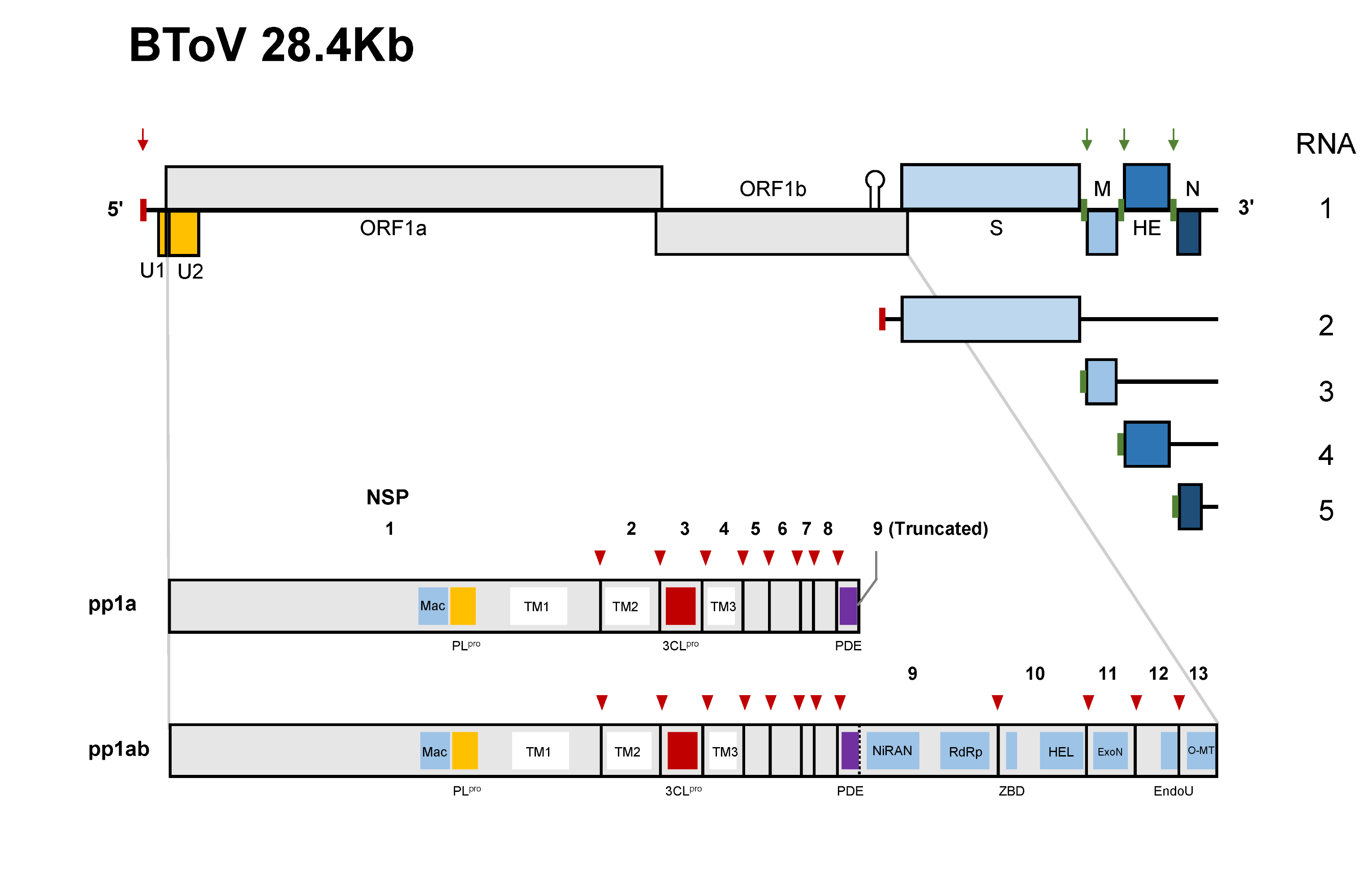
''Torovirus'' is a genus of enveloped, positive-strand RNA viruses in the order '' Nidovirales'' and family '' Tobaniviridae''. They primarily infect vertebrates, especially cattle, pigs, and horses. Diseases associated with this genus include gastroenteritis, which commonly presents in mammals. ''Torovirus'' is the only genus in the monotypic subfamily ''Torovirinae''. ''Torovirus'' is also a monotypic taxon, containing only one subgenus, ''Renitovirus''. The discovery of the first torovirus can be traced back to 1970s. Equine torovirus (EToV) was accidentally found in the rectal sample from a horse who was experiencing severe diarrhea. The 'Breda' bovine torovirus was later found in 1979 while investigation in a dairy farm in Breda. They had several calves experiencing severe diarrhea for months. In 1984, torovirus-like particles were detected with electron microscope (EM) technique in the human patients with gastroenteritis.Cho, K. O., & Hoet, A. E. (2014). Toroviruses (Coro ... [...More Info...] [...Related Items...] OR: [Wikipedia] [Google] [Baidu] |
Transmembrane Protein
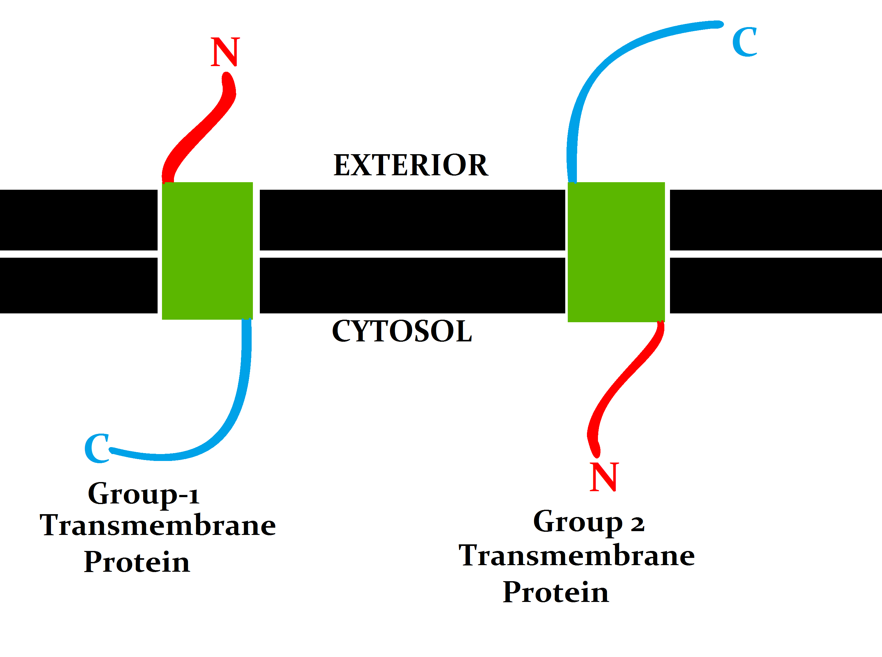
A transmembrane protein (TP) is a type of integral membrane protein that spans the entirety of the cell membrane. Many transmembrane proteins function as gateways to permit the transport of specific substances across the membrane. They frequently undergo significant conformational changes to move a substance through the membrane. They are usually highly hydrophobic and aggregate and precipitate in water. They require detergents or nonpolar solvents for extraction, although some of them ( beta-barrels) can be also extracted using denaturing agents. The peptide sequence that spans the membrane, or the transmembrane segment, is largely hydrophobic and can be visualized using the hydropathy plot. Depending on the number of transmembrane segments, transmembrane proteins can be classified as single-span (or bitopic) or multi-span (polytopic). Some other integral membrane proteins are called monotopic, meaning that they are also permanently attached to the membrane, but do not ... [...More Info...] [...Related Items...] OR: [Wikipedia] [Google] [Baidu] |
Coronavirus Membrane Protein
The membrane (M) protein (previously called E1, sometimes also matrix protein) is an integral membrane protein that is the most abundant of the four major structural proteins found in coronaviruses. The M protein organizes the assembly of coronavirus virions through protein-protein interactions with other M protein molecules as well as with the other three structural proteins, the envelope (E), spike (S), and nucleocapsid (N) proteins. Structure The M protein is a transmembrane protein with three transmembrane domains and is around 230 amino acid residues long. In SARS-CoV-2, the causative agent of Covid-19, the M protein is 222 residues long. Its membrane topology orients the C-terminus toward the cytosolic face of the membrane and thus into the interior of the virion. It has a short N-terminal segment and a larger C-terminal domain. Although the protein sequence is not well conserved across all coronavirus groups, there is a conserved amphipathic region near the C-terminal ... [...More Info...] [...Related Items...] OR: [Wikipedia] [Google] [Baidu] |
Coronavirus Spike Protein
Spike (S) glycoprotein (sometimes also called spike protein, formerly known as E2) is the largest of the four major structural proteins found in coronaviruses. The spike protein assembles into trimers that form large structures, called spikes or peplomers, that project from the surface of the virion. The distinctive appearance of these spikes when visualized using negative stain transmission electron microscopy, "recalling the solar corona", gives the virus family its name. The function of the spike glycoprotein is to mediate viral entry into the host cell by first interacting with molecules on the exterior cell surface and then fusing the viral and cellular membranes. Spike glycoprotein is a class I fusion protein that contains two regions, known as S1 and S2, responsible for these two functions. The S1 region contains the receptor-binding domain that binds to receptors on the cell surface. Coronaviruses use a very diverse range of receptors; SARS-CoV (which causes ... [...More Info...] [...Related Items...] OR: [Wikipedia] [Google] [Baidu] |
Glycoprotein
Glycoproteins are proteins which contain oligosaccharide chains covalently attached to amino acid side-chains. The carbohydrate is attached to the protein in a cotranslational or posttranslational modification. This process is known as glycosylation. Secreted extracellular proteins are often glycosylated. In proteins that have segments extending extracellularly, the extracellular segments are also often glycosylated. Glycoproteins are also often important integral membrane proteins, where they play a role in cell–cell interactions. It is important to distinguish endoplasmic reticulum-based glycosylation of the secretory system from reversible cytosolic-nuclear glycosylation. Glycoproteins of the cytosol and nucleus can be modified through the reversible addition of a single GlcNAc residue that is considered reciprocal to phosphorylation and the functions of these are likely to be an additional regulatory mechanism that controls phosphorylation-based signalling. In contra ... [...More Info...] [...Related Items...] OR: [Wikipedia] [Google] [Baidu] |
Golgi Apparatus
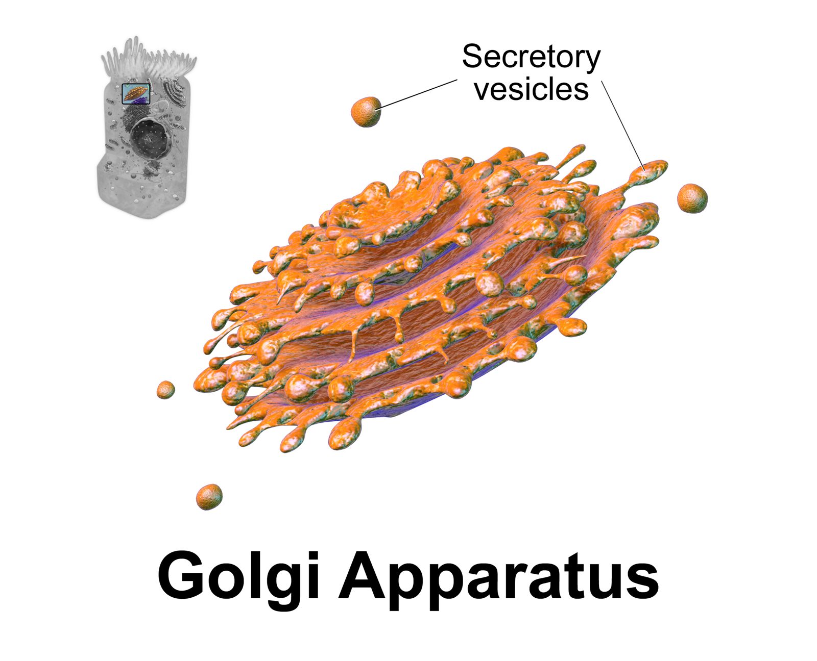
The Golgi apparatus (), also known as the Golgi complex, Golgi body, or simply the Golgi, is an organelle found in most eukaryotic cells. Part of the endomembrane system in the cytoplasm, it packages proteins into membrane-bound vesicles inside the cell before the vesicles are sent to their destination. It resides at the intersection of the secretory, lysosomal, and endocytic pathways. It is of particular importance in processing proteins for secretion, containing a set of glycosylation enzymes that attach various sugar monomers to proteins as the proteins move through the apparatus. It was identified in 1897 by the Italian scientist Camillo Golgi and was named after him in 1898. Discovery Owing to its large size and distinctive structure, the Golgi apparatus was one of the first organelles to be discovered and observed in detail. It was discovered in 1898 by Italian physician Camillo Golgi during an investigation of the nervous system. After first observing it unde ... [...More Info...] [...Related Items...] OR: [Wikipedia] [Google] [Baidu] |
Endoplasmic Reticulum
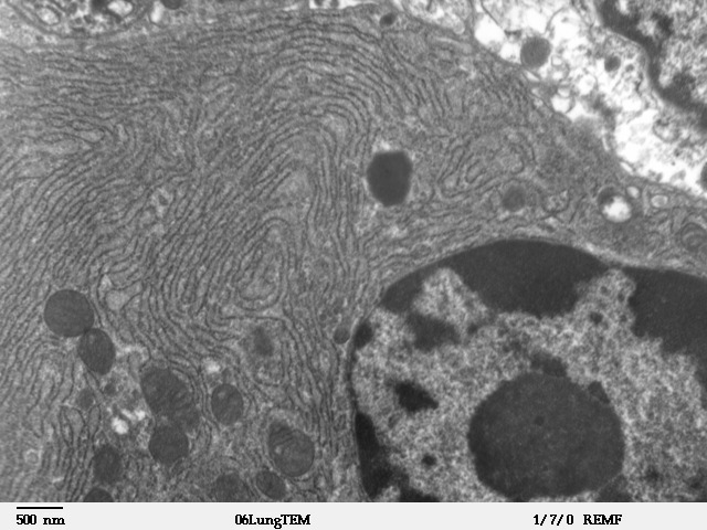
The endoplasmic reticulum (ER) is, in essence, the transportation system of the eukaryotic cell, and has many other important functions such as protein folding. It is a type of organelle made up of two subunits – rough endoplasmic reticulum (RER), and smooth endoplasmic reticulum (SER). The endoplasmic reticulum is found in most eukaryotic cells and forms an interconnected network of flattened, membrane-enclosed sacs known as cisternae (in the RER), and tubular structures in the SER. The membranes of the ER are continuous with the outer nuclear membrane. The endoplasmic reticulum is not found in red blood cells, or spermatozoa. The two types of ER share many of the same proteins and engage in certain common activities such as the synthesis of certain lipids and cholesterol. Different types of cells contain different ratios of the two types of ER depending on the activities of the cell. RER is found mainly toward the nucleus of cell and SER towards the cell membrane or plasma ... [...More Info...] [...Related Items...] OR: [Wikipedia] [Google] [Baidu] |
Viral Envelope
A viral envelope is the outermost layer of many types of viruses. It protects the genetic material in their life cycle when traveling between host cells. Not all viruses have envelopes. Numerous human pathogenic viruses in circulation are encased in lipid bilayers, and they infect their target cells by causing the viral envelope and cell membrane to fuse. Although there are effective vaccines against some of these viruses, there is no preventative or curative medicine for the majority of them. In most cases, the known vaccines operate by inducing antibodies that prevent the pathogen from entering cells. This happens in the case of enveloped viruses when the antibodies bind to the viral envelope proteins. The membrane fusion event that triggers viral entrance is caused by the viral fusion protein. Many enveloped viruses only have one protein visible on the surface of the particle, which is required for both mediating adhesion to the cell surface and for the subsequent membrane fusi ... [...More Info...] [...Related Items...] OR: [Wikipedia] [Google] [Baidu] |
Poly(A) Tract
Polyadenylation is the addition of a poly(A) tail to an RNA transcript, typically a messenger RNA (mRNA). The poly(A) tail consists of multiple adenosine monophosphates; in other words, it is a stretch of RNA that has only adenine bases. In eukaryotes, polyadenylation is part of the process that produces mature mRNA for translation. In many bacteria, the poly(A) tail promotes degradation of the mRNA. It, therefore, forms part of the larger process of gene expression. The process of polyadenylation begins as the transcription of a gene terminates. The 3′-most segment of the newly made pre-mRNA is first cleaved off by a set of proteins; these proteins then synthesize the poly(A) tail at the RNA's 3′ end. In some genes these proteins add a poly(A) tail at one of several possible sites. Therefore, polyadenylation can produce more than one transcript from a single gene (alternative polyadenylation), similar to alternative splicing. The poly(A) tail is important for the nuc ... [...More Info...] [...Related Items...] OR: [Wikipedia] [Google] [Baidu] |
Five-prime Cap
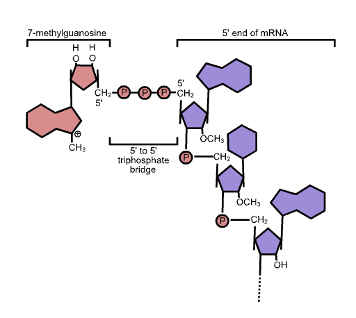
In molecular biology, the five-prime cap (5′ cap) is a specially altered nucleotide on the 5′ end of some primary transcripts such as precursor messenger RNA. This process, known as mRNA capping, is highly regulated and vital in the creation of stable and mature messenger RNA able to undergo translation during protein synthesis. Mitochondrial mRNA and chloroplastic mRNA are not capped. Structure In eukaryotes, the 5′ cap (cap-0), found on the 5′ end of an mRNA molecule, consists of a guanine nucleotide connected to mRNA via an unusual 5′ to 5′ triphosphate linkage. This guanosine is methylated on the 7 position directly after capping ''in vivo'' by a methyltransferase. It is referred to as a 7-methylguanylate cap, abbreviated m7G. In multicellular eukaryotes and some viruses, further modifications exist, including the methylation of the 2′ hydroxy-groups of the first 2 ribose sugars of the 5′ end of the mRNA. cap-1 has a methylated 2′-hydroxy group ... [...More Info...] [...Related Items...] OR: [Wikipedia] [Google] [Baidu] |


