|
Cervical Spinal Nerve 4
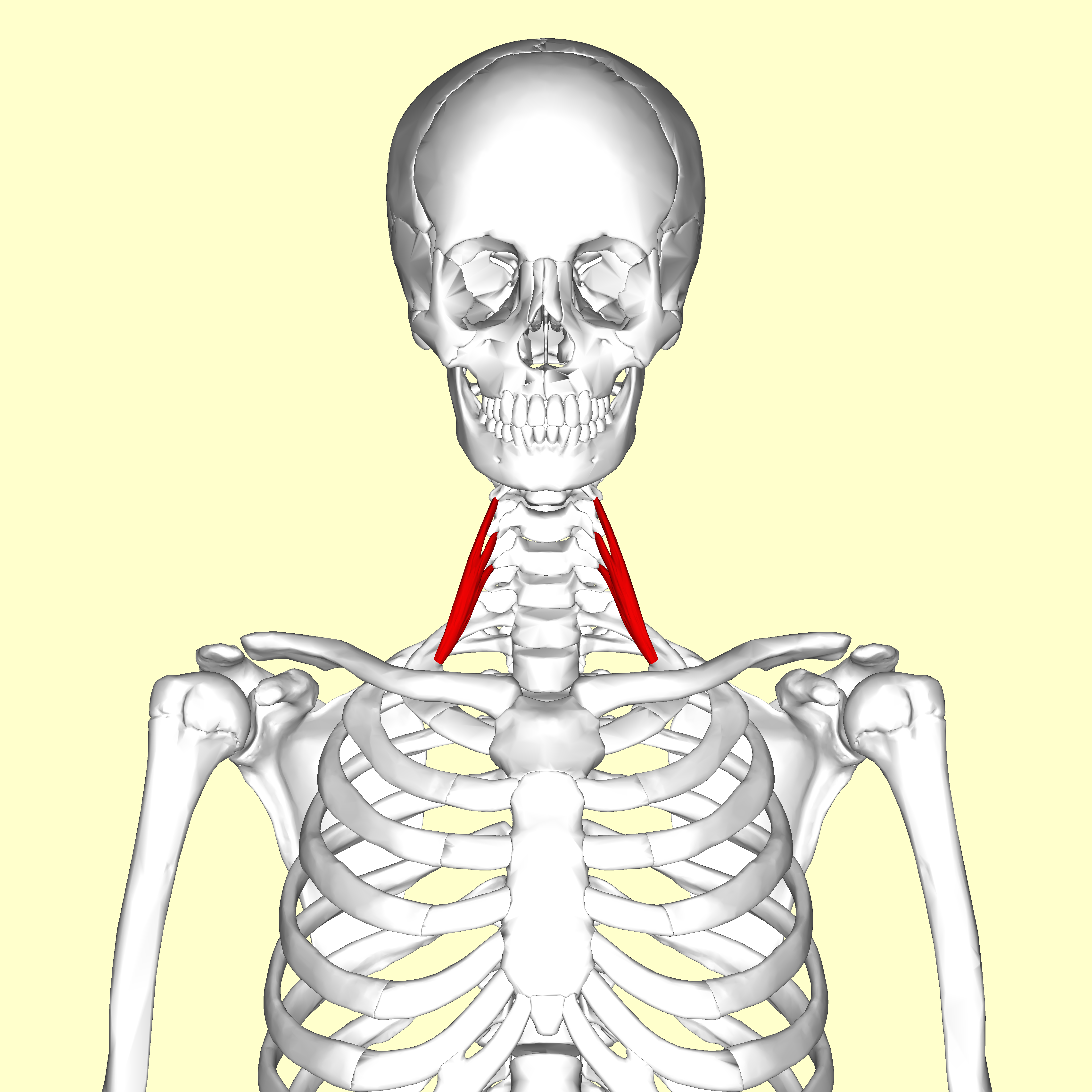
Cervical spinal nerve 4, also called C4, is a spinal nerve of the cervical segment. It originates from the spinal cord above the 4th cervical vertebra (C4). It contributes nerve fibers to the phrenic nerve, the motor nerve to the thoracoabdominal diaphragm. It also provides motor nerves for the longus capitis, longus colli, anterior scalene, middle scalene, and levator scapulae muscles. C4 contributes some sensory fibers to the supraclavicular nerves, responsible for sensation from the skin above the clavicle. Additional Images File:Slide1y.JPG, Cervical spinal nerve 4 File:Projectional radiograph of cervical foraminal stenosis, annotated.jpg, Projectional radiograph of a man presenting with pain by the nape and left shoulder, showing a stenosis in the intervertebral foramen The intervertebral foramen (also called neural foramen, and often abbreviated as IV foramen or IVF) is a foramen between two spinal vertebrae. Cervical, thoracic, and lumbar vertebrae all have in ... [...More Info...] [...Related Items...] OR: [Wikipedia] [Google] [Baidu] |
Spinal Nerve
A spinal nerve is a mixed nerve, which carries motor, sensory, and autonomic signals between the spinal cord and the body. In the human body there are 31 pairs of spinal nerves, one on each side of the vertebral column. These are grouped into the corresponding cervical, thoracic, lumbar, sacral and coccygeal regions of the spine. There are eight pairs of cervical nerves, twelve pairs of thoracic nerves, five pairs of lumbar nerves, five pairs of sacral nerves, and one pair of coccygeal nerves. The spinal nerves are part of the peripheral nervous system. Structure Each spinal nerve is a mixed nerve, formed from the combination of nerve fibers from its dorsal and ventral roots. The dorsal root is the afferent sensory root and carries sensory information to the brain. The ventral root is the efferent motor root and carries motor information from the brain. The spinal nerve emerges from the spinal column through an opening ( intervertebral foramen) between adjacent verteb ... [...More Info...] [...Related Items...] OR: [Wikipedia] [Google] [Baidu] |
Scalene Muscles
The scalene muscles are a group of three pairs of muscles in the lateral neck, namely the anterior scalene, middle scalene, and posterior scalene. They are innervated by the third to the eight cervical spinal nerves (C3-C8). The anterior and middle scalene muscles lift the first rib and bend the neck to the same side; the posterior scalene lifts the second rib and tilts the neck to the same side. The muscles are named . Structure The scalene muscles originate from the transverse processes from the cervical vertebrae of C2 to C7 and insert onto the first and second ribs. Anterior scalene The anterior scalene muscle ( la, scalenus anterior), lies deeply at the side of the neck, behind the sternocleidomastoid muscle. It arises from the anterior tubercles of the transverse processes of the third, fourth, fifth, and sixth cervical vertebrae, and descending, almost vertically, is inserted by a narrow, flat tendon into the scalene tubercle on the inner border of the first rib ... [...More Info...] [...Related Items...] OR: [Wikipedia] [Google] [Baidu] |
Intervertebral Foramen
The intervertebral foramen (also called neural foramen, and often abbreviated as IV foramen or IVF) is a foramen between two spinal vertebrae. Cervical, thoracic, and lumbar vertebrae all have intervertebral foramina. The foramina, or openings, are present between every pair of vertebrae in these areas. A number of structures pass through the foramen. These are the root of each spinal nerve, the spinal artery of the segmental artery, communicating veins between the internal and external plexuses, recurrent meningeal (sinu-vertebral) nerves, and transforaminal ligaments. When the spinal vertebrae are articulated with each other, the bodies form a strong pillar that supports the head and trunk, and the vertebral foramen constitutes a canal for the protection of the medulla spinalis ( spinal cord). The size of the foramina is variable due to placement, pathology, spinal loading, and posture. Foramina can be occluded by arthritic degenerative changes and space-occupying lesi ... [...More Info...] [...Related Items...] OR: [Wikipedia] [Google] [Baidu] |
Nape
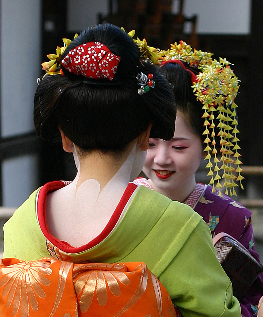
The nape is the back of the neck. In technical anatomical/medical terminology, the nape is also called the nucha (from the Medieval Latin rendering of the Arabic , "spinal marrow"). The corresponding adjective is ''nuchal'', as in the term ''nuchal rigidity'' for neck stiffness. In many mammals the nape bears a loose, non-sensitive area of skin, known as the scruff, by which a mother carries her young by her teeth, temporarily immobilizing it during transport. In the mating of cats the male will grip the female's scruff with his teeth to help immobilize her during the act, a form of pinch-induced behavioral inhibition Pinch-induced behavioural inhibition (PIBI), also called dorsal immobility, transport immobility or clipnosis, is a partially inert state which results from a gentle squeeze of the skin behind the neck. It is mostly observed among cats and allows .... Cultural connotations In traditional Japanese culture, the was one of the few areas of the body (other than f ... [...More Info...] [...Related Items...] OR: [Wikipedia] [Google] [Baidu] |
Projectional Radiograph
Projectional radiography, also known as conventional radiography, is a form of radiography and medical imaging that produces two-dimensional images by x-ray radiation. The image acquisition is generally performed by radiographers, and the images are often examined by radiologists. Both the procedure and any resultant images are often simply called "X-ray". Plain radiography or roentgenography generally refers to projectional radiography (without the use of more advanced techniques such as computed tomography that can generate 3D-images). ''Plain radiography'' can also refer to radiography without a radiocontrast agent or radiography that generates single static images, as contrasted to fluoroscopy, which are technically also projectional. Equipment X-ray generator Projectional radiographs generally use X-rays created by X-ray generators, which generate X-rays from X-ray tubes. Grid An anti-scatter grid may be placed between the patient and the detector to reduce the quantity ... [...More Info...] [...Related Items...] OR: [Wikipedia] [Google] [Baidu] |
Clavicle
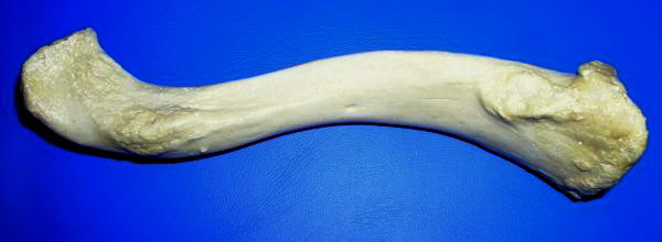
The clavicle, or collarbone, is a slender, S-shaped long bone approximately 6 inches (15 cm) long that serves as a strut between the shoulder blade and the sternum (breastbone). There are two clavicles, one on the left and one on the right. The clavicle is the only long bone in the body that lies horizontally. Together with the shoulder blade, it makes up the shoulder girdle. It is a palpable bone and, in people who have less fat in this region, the location of the bone is clearly visible. It receives its name from the Latin ''clavicula'' ("little key"), because the bone rotates along its axis like a key when the shoulder is abducted. The clavicle is the most commonly fractured bone. It can easily be fractured by impacts to the shoulder from the force of falling on outstretched arms or by a direct hit. Structure The collarbone is a thin doubly curved long bone that connects the arm to the trunk of the body. Located directly above the first rib, it acts as a strut t ... [...More Info...] [...Related Items...] OR: [Wikipedia] [Google] [Baidu] |
Supraclavicular Nerves
The supraclavicular nerves (descending branches) arise from the third and fourth cervical nerves. They emerge beneath the posterior border of the sternocleidomastoideus (sternocleidomastoid muscle), and descend in the posterior triangle of the neck beneath the platysma muscle and the deep cervical fascia. Together, they innervate skin over the shoulder. The supraclavicular nerve can be blocked during shoulder surgery. Branches The supraclavicular nerves arise from C3 and C4 spinal nerve roots. Near the clavicle, the supraclavicular nerves perforate the fascia and the platysma muscle to become cutaneous. They are arranged, according to their position, into three groups—anterior, middle, and posterior. Medial supraclavicular nerve The medial supraclavicular nerves or ''anterior supraclavicular nerves'' (nn. supraclaviculares anteriores; suprasternal nerves) cross obliquely over the external jugular vein and the clavicular and sternal heads of the sternocleidomastoideus, and supp ... [...More Info...] [...Related Items...] OR: [Wikipedia] [Google] [Baidu] |
Levator Scapulae Muscle
The levator scapulae is a slender skeletal muscle situated at the back and side of the neck. As the Latin name suggests, its main function is to lift the scapula. Anatomy Attachments The muscle descends diagonally from its origin to its insertion. Origin The levator scapulae originates from the posterior tubercles of the transverse processes of cervical vertebrae C1-4. At its origin, it attaches via tendinous slips. Insertion It inserts onto the medial border of the scapula (with its site of insertion extending between the superior angle of the scapula superiorly, and the junction of spine of scapula and medial border of scapula inferiorly). Relations One of the muscles within the floor of the posterior triangle of the neck, the superior part of levator scapulae is covered by sternocleidomastoid and its inferior part by the trapezius. It is bounded in front by the scalenus medius and behind by splenius cervicis. The spinal accessory nerve crosses laterally in the midd ... [...More Info...] [...Related Items...] OR: [Wikipedia] [Google] [Baidu] |
Longus Colli Muscle
The longus colli muscle (Latin for ''long muscle of the neck'') is a muscle of the human body. The longus colli is situated on the anterior surface of the vertebral column, between the atlas and the third thoracic vertebra. It is broad in the middle, narrow and pointed at either end, and consists of three portions, a superior oblique, an inferior oblique, and a vertical. * The ''superior oblique portion'' arises from the anterior tubercles of the transverse processes of the third, fourth, and fifth cervical vertebrae and, ascending obliquely with a medial inclination, is inserted by a narrow tendon into the tubercle on the anterior arch of the atlas. * The ''inferior oblique portion'', the smallest part of the muscle, arises from the front of the bodies of the first two or three thoracic vertebrae; and, ascending obliquely in a lateral direction, is inserted into the anterior tubercles of the transverse processes of the fifth and sixth cervical vertebrae. * The ''vertical porti ... [...More Info...] [...Related Items...] OR: [Wikipedia] [Google] [Baidu] |
Cervical Vertebrae
In tetrapods, cervical vertebrae (singular: vertebra) are the vertebrae of the neck, immediately below the skull. Truncal vertebrae (divided into thoracic and lumbar vertebrae in mammals) lie caudal (toward the tail) of cervical vertebrae. In sauropsid species, the cervical vertebrae bear cervical ribs. In lizards and saurischian dinosaurs, the cervical ribs are large; in birds, they are small and completely fused to the vertebrae. The vertebral transverse processes of mammals are homologous to the cervical ribs of other amniotes. Most mammals have seven cervical vertebrae, with the only three known exceptions being the manatee with six, the two-toed sloth with five or six, and the three-toed sloth with nine. In humans, cervical vertebrae are the smallest of the true vertebrae and can be readily distinguished from those of the thoracic or lumbar regions by the presence of a foramen (hole) in each transverse process, through which the vertebral artery, vertebral veins, and ... [...More Info...] [...Related Items...] OR: [Wikipedia] [Google] [Baidu] |
Longus Capitis Muscle
The longus capitis muscle (Latin for ''long muscle of the head'', alternatively rectus capitis anticus major), is broad and thick above, narrow below, and arises by four tendinous slips, from the anterior tubercles of the transverse processes of the third, fourth, fifth, and sixth cervical vertebræ, and ascends, converging toward its fellow of the opposite side, to be inserted into the inferior surface of the basilar part of the occipital bone. It is innervated by a branch of cervical plexus. Longus capitis has several actions: acting unilaterally, to: *flex the head and neck laterally *rotate the head ipsilaterally acting bilaterally: *flex the head and neck Additional images File:Gray129.png, Occipital bone. Outer surface. File:Gray187.png, Base of skull. Inferior surface. File:Slide4ccc.JPG, Longus capitis muscle File:Slide6jjj.JPG, Longus capitis muscle References External links * PTCentral Muscles of the head and neck {{muscle-stub ... [...More Info...] [...Related Items...] OR: [Wikipedia] [Google] [Baidu] |
Thoracic Diaphragm
The thoracic diaphragm, or simply the diaphragm ( grc, διάφραγμα, diáphragma, partition), is a sheet of internal skeletal muscle in humans and other mammals that extends across the bottom of the thoracic cavity. The diaphragm is the most important muscle of respiration, and separates the thoracic cavity, containing the heart and lungs, from the abdominal cavity: as the diaphragm contracts, the volume of the thoracic cavity increases, creating a negative pressure there, which draws air into the lungs. Its high oxygen consumption is noted by the many mitochondria and capillaries present; more than in any other skeletal muscle. The term ''diaphragm'' in anatomy, created by Gerard of Cremona, can refer to other flat structures such as the urogenital diaphragm or pelvic diaphragm, but "the diaphragm" generally refers to the thoracic diaphragm. In humans, the diaphragm is slightly asymmetric—its right half is higher up (superior) to the left half, since the large li ... [...More Info...] [...Related Items...] OR: [Wikipedia] [Google] [Baidu] |


