|
Cruciate Ligament Of Atlas
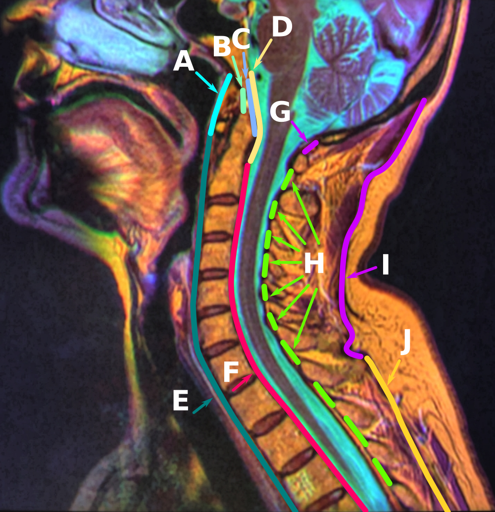
The cruciate ligament of the atlas (cruciform ligament) is a cross-shaped (thus the name) ligament in the neck forming part of the atlanto-axial joint. It consists of the transverse ligament of atlas, a superior longitudinal band, and an inferior longitudinal band. The cruciate ligament of the atlas prevents abnormal movement of the atlanto-axial joint. It may be torn, such as by fractures of the atlas bone. Structure The cruciate ligament of the atlas consists of the transverse ligament of the atlas, a superior longitudinal band, and an inferior longitudinal band. The superior longitudinal band connects the transverse ligament to the anterior side of the foramen magnum (near the basilar part) in the occipital bone of the skull. The inferior longitudinal band connects the transverse ligament to the body of the axis bone (C2). Variation The inferior longitudinal band may be absent in some people; the rest of the ligament is invariably present. Gerber's ligament In about h ... [...More Info...] [...Related Items...] OR: [Wikipedia] [Google] [Baidu] |
Atlas Bone
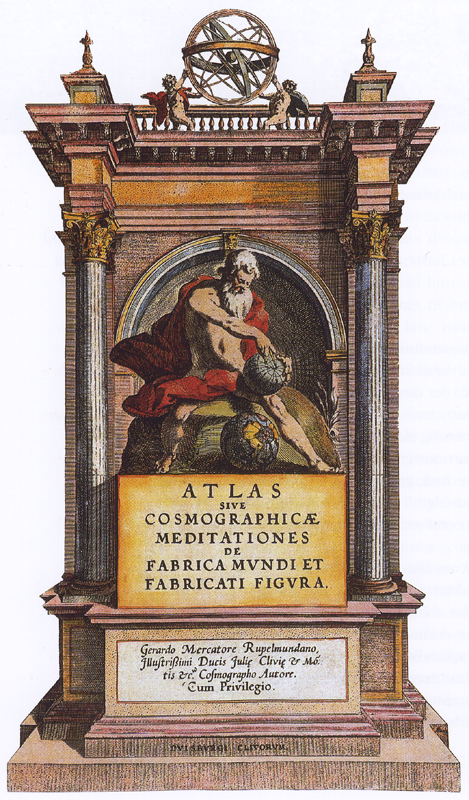
An atlas is a collection of maps; it is typically a bundle of maps of Earth or of a continent or region of Earth. Advances in astronomy have also resulted in atlases of the celestial sphere or of other planets. Atlases have traditionally been bound into book form, but today, many atlases are in multimedia formats. In addition to presenting geographical features and political boundaries, many atlases often feature geopolitical, social, religious, and economic statistics. They also have information about the map and places in it. Etymology The use of the word "atlas" in a geographical context dates from 1595 when the German-Flemish geographer Gerardus Mercator published ("Atlas or cosmographical meditations upon the creation of the universe and the universe as created"). This title provides Mercator's definition of the word as a description of the creation and form of the whole universe, not simply as a collection of maps. The volume that was published posthumously one year af ... [...More Info...] [...Related Items...] OR: [Wikipedia] [Google] [Baidu] |
Axis (anatomy)
In anatomy, the axis (from Latin ''axis'', "axle") is the second cervical vertebra (C2) of the spine, immediately inferior to the atlas, upon which the head rests. The spinal cord passes through the axis. The defining feature of the axis is its strong bony protrusion known as the dens, which rises from the superior aspect of the bone. Structure The body is deeper in front or in the back and is prolonged downward anteriorly to overlap the upper and front part of the third vertebra. It presents a median longitudinal ridge in front, separating two lateral depressions for the attachment of the longus colli muscles. Dens The dens, also called the odontoid process, or the peg, is the most pronounced projecting feature of the axis. The dens exhibits a slight constriction where it joins the main body of the vertebra. The condition where the dens is separated from the body of the axis is called ''os odontoideum'' and may cause nerve and circulation compression syndrome. On its an ... [...More Info...] [...Related Items...] OR: [Wikipedia] [Google] [Baidu] |
Cross
A cross is a religious symbol consisting of two Intersection (set theory), intersecting Line (geometry), lines, usually perpendicular to each other. The lines usually run vertically and horizontally. A cross of oblique lines, in the shape of the Latin letter X, is termed a saltire in heraldic terminology. The cross shape has been widely officially recognized as an absolute and exclusive religious symbol of Christianity from an early period in that religion's history.''Christianity: an introduction'' by Alister E. McGrath 2006 pages 321-323 Before then, it was used as a religious or cultural symbol throughout Europe, in West Asia, west and south Asia (the latter, in the form of the original Swastika); and in Ancient Egypt, where the Ankh was a hieroglyph that represented "life" and was used in the wo ... [...More Info...] [...Related Items...] OR: [Wikipedia] [Google] [Baidu] |
Spinal Cord
The spinal cord is a long, thin, tubular structure made up of nervous tissue that extends from the medulla oblongata in the lower brainstem to the lumbar region of the vertebral column (backbone) of vertebrate animals. The center of the spinal cord is hollow and contains a structure called the central canal, which contains cerebrospinal fluid. The spinal cord is also covered by meninges and enclosed by the neural arches. Together, the brain and spinal cord make up the central nervous system. In humans, the spinal cord is a continuation of the brainstem and anatomically begins at the occipital bone, passing out of the foramen magnum and then enters the spinal canal at the beginning of the cervical vertebrae. The spinal cord extends down to between the first and second lumbar vertebrae, where it tapers to become the cauda equina. The enclosing bony vertebral column protects the relatively shorter spinal cord. It is around long in adult men and around long in adult women. The diam ... [...More Info...] [...Related Items...] OR: [Wikipedia] [Google] [Baidu] |
Cervical Myelopathy
Myelopathy describes any neurologic deficit related to the spinal cord. When due to Physical trauma, trauma, myelopathy is known as (acute) spinal cord injury. When inflammatory, it is known as myelitis. Disease that is vascular in nature is known as vascular myelopathy. The most common form of myelopathy in humans, cervical spondylotic myelopathy, cervical spondylotic myelopathy (CSM) also called ''degenerative cervical myelopathy'', results from narrowing of the spinal canal (spinal stenosis) ultimately causing compression of the spinal cord. In Asian populations, spinal cord compression often occurs due to a different, inflammatory process affecting the posterior longitudinal ligament. Presentation Clinical signs and symptoms depend on which spinal cord level (cervical, thoracic, or lumbar) is affected and the extent (anterior, posterior, or lateral) of the pathology, and may include: * Upper motor neuron syndrome, Upper motor neuron signs—weakness, spasticity, clumsiness, ... [...More Info...] [...Related Items...] OR: [Wikipedia] [Google] [Baidu] |
Ossification
Ossification (also called osteogenesis or bone mineralization) in bone remodeling is the process of laying down new bone material by cells named osteoblasts. It is synonymous with bone tissue formation. There are two processes resulting in the formation of normal, healthy bone tissue: Intramembranous ossification is the direct laying down of bone into the primitive connective tissue ( mesenchyme), while endochondral ossification involves cartilage as a precursor. In fracture healing, endochondral osteogenesis is the most commonly occurring process, for example in fractures of long bones treated by plaster of Paris, whereas fractures treated by open reduction and internal fixation with metal plates, screws, pins, rods and nails may heal by intramembranous osteogenesis. Heterotopic ossification is a process resulting in the formation of bone tissue that is often atypical, at an extraskeletal location. Calcification is often confused with ossification. Calcificatio ... [...More Info...] [...Related Items...] OR: [Wikipedia] [Google] [Baidu] |
Magnetic Resonance Imaging
Magnetic resonance imaging (MRI) is a medical imaging technique used in radiology to generate pictures of the anatomy and the physiological processes inside the body. MRI scanners use strong magnetic fields, magnetic field gradients, and radio waves to form images of the organs in the body. MRI does not involve X-rays or the use of ionizing radiation, which distinguishes it from computed tomography (CT) and positron emission tomography (PET) scans. MRI is a medical application of nuclear magnetic resonance (NMR) which can also be used for imaging in other NMR applications, such as NMR spectroscopy. MRI is widely used in hospitals and clinics for medical diagnosis, staging and follow-up of disease. Compared to CT, MRI provides better contrast in images of soft tissues, e.g. in the brain or abdomen. However, it may be perceived as less comfortable by patients, due to the usually longer and louder measurements with the subject in a long, confining tube, although ... [...More Info...] [...Related Items...] OR: [Wikipedia] [Google] [Baidu] |
CT Scan
A computed tomography scan (CT scan), formerly called computed axial tomography scan (CAT scan), is a medical imaging technique used to obtain detailed internal images of the body. The personnel that perform CT scans are called radiographers or radiology technologists. CT scanners use a rotating X-ray tube and a row of detectors placed in a gantry (medical), gantry to measure X-ray Attenuation#Radiography, attenuations by different tissues inside the body. The multiple X-ray measurements taken from different angles are then processed on a computer using tomographic reconstruction algorithms to produce Tomography, tomographic (cross-sectional) images (virtual "slices") of a body. CT scans can be used in patients with metallic implants or pacemakers, for whom magnetic resonance imaging (MRI) is Contraindication, contraindicated. Since its development in the 1970s, CT scanning has proven to be a versatile imaging technique. While CT is most prominently used in medical diagnosis, i ... [...More Info...] [...Related Items...] OR: [Wikipedia] [Google] [Baidu] |
Radiography
Radiography is an imaging technology, imaging technique using X-rays, gamma rays, or similar ionizing radiation and non-ionizing radiation to view the internal form of an object. Applications of radiography include medical ("diagnostic" radiography and "therapeutic radiography") and industrial radiography. Similar techniques are used in airport security, (where "body scanners" generally use backscatter X-ray). To create an image in conventional radiography, a beam of X-rays is produced by an X-ray generator and it is projected towards the object. A certain amount of the X-rays or other radiation are absorbed by the object, dependent on the object's density and structural composition. The X-rays that pass through the object are captured behind the object by a X-ray detector, detector (either photographic film or a digital detector). The generation of flat two-dimensional images by this technique is called Projection radiography, projectional radiography. In computed tomography (C ... [...More Info...] [...Related Items...] OR: [Wikipedia] [Google] [Baidu] |
Sprain
A sprain is a soft tissue injury of the ligaments within a joint, often caused by a sudden movement abruptly forcing the joint to exceed its functional range of motion. Ligaments are tough, inelastic fibers made of collagen that connect two or more bones to form a joint and are important for joint stability and proprioception, which is the body's sense of limb position and movement. Sprains may be mild (first degree), moderate (second degree), or severe (third degree), with the latter two classes involving some degree of tearing of the ligament. Sprains can occur at any joint but most commonly occur in the ankle, knee, or wrist. An equivalent injury to a muscle or tendon is known as a strain. The majority of sprains are mild, causing minor swelling and bruising that can be resolved with conservative treatment, typically summarized as RICE: rest, ice, compression, elevation. However, severe sprains involve complete tears, ruptures, or avulsion fractures, often leading to j ... [...More Info...] [...Related Items...] OR: [Wikipedia] [Google] [Baidu] |
Transverse Ligament Of Atlas
In anatomy, the transverse ligament of the atlas is a broad, tough ligament which arches across the ring of the atlas (first cervical vertebra) posterior to the dens to keep the dens (odontoid process) in contact with the atlas. It forms the transverse component of the cruciform ligament of atlas. Structure The length of the ligament is variable; its mean length is 2 cm. The ligament broadens and thickensGray's anatomy, 1918 medially. The anterior medial aspect of the ligament is lined by a thin layer of articular cartilage. The neck of the odontoid process is constricted where it is embraced posteriorly by the transverse ligament so it retains the dens in position even after all other ligaments have been sectioned. Attachments The ligament attaches on either side onto a small yet prominent tubercle upon the medial aspect of either lateral mass of atlas. Cruciate ligament A strong median band (''crus superius'') extends superiorly from the superior margin of the ligamen ... [...More Info...] [...Related Items...] OR: [Wikipedia] [Google] [Baidu] |
Hyperextension
Motion, the process of movement, is described using specific anatomical terms. Motion includes movement of organs, joints, limbs, and specific sections of the body. The terminology used describes this motion according to its direction relative to the anatomical position of the body parts involved. Anatomists and others use a unified set of terms to describe most of the movements, although other, more specialized terms are necessary for describing unique movements such as those of the hands, feet, and eyes. In general, motion is classified according to the anatomical plane it occurs in. ''Flexion'' and ''extension'' are examples of ''angular'' motions, in which two axes of a joint are brought closer together or moved further apart. ''Rotational'' motion may occur at other joints, for example the shoulder, and are described as ''internal'' or ''external''. Other terms, such as ''elevation'' and ''depression'', describe movement above or below the horizontal plane. Many anatomica ... [...More Info...] [...Related Items...] OR: [Wikipedia] [Google] [Baidu] |


