|
Condenser Lens
A condenser is an optical lens that renders a divergent light beam from a point light source into a parallel or converging beam to illuminate an object to be imaged. Condensers are an essential part of any imaging device, such as microscopes, enlargers, slide projectors, and telescopes. The concept is applicable to all kinds of radiation undergoing optical transformation, such as electrons in electron microscopy, neutron radiation, and synchrotron radiation optics. Microscope condenser Condensers are located above the light source and under the sample in an upright microscope, and above the stage and below the light source in an inverted microscope. They act to gather light from the microscope's light source and concentrate it into a cone of light that illuminates the specimen. The aperture and angle of the light cone must be adjusted (via the size of the diaphragm) for each different objective lens with different numerical apertures. Condensers typically consist of a variabl ... [...More Info...] [...Related Items...] OR: [Wikipedia] [Google] [Baidu] |
Aplanatic
In optics, spherical aberration (SA) is a type of aberration found in optical systems that have elements with spherical surfaces. This phenomenon commonly affects lenses and curved mirrors, as these components are often shaped in a spherical manner for ease of manufacturing. Light rays that strike a spherical surface off-centre are refracted or reflected more or less than those that strike close to the centre. This deviation reduces the quality of images produced by optical systems. The effect of spherical aberration was first identified in the 11th century by Ibn al-Haytham who discussed it in his work Kitāb al-Manāẓir. Overview A spherical lens has an aplanatic point (i.e., no spherical aberration) only at a lateral distance from the optical axis that equals the radius of the spherical surface divided by the index of refraction of the lens material. Spherical aberration makes the focus of telescopes and other instruments less than ideal. This is an important effect, b ... [...More Info...] [...Related Items...] OR: [Wikipedia] [Google] [Baidu] |
Köhler Illumination
Köhler illumination is a method of specimen illumination used for transmitted and reflected light (trans- and epi-illuminated) optical microscopy. Köhler illumination acts to generate an even illumination of the sample and ensures that an image of the illumination source (for example a halogen lamp filament) is not visible in the resulting image. Köhler illumination is the predominant technique for sample illumination in modern scientific light microscopy. It requires additional optical elements which are more expensive and may not be present in more basic light microscopes. History and motivation Prior to Köhler illumination critical illumination was the predominant technique for sample illumination. Critical illumination has the major limitation that the image of the light source (typically a light bulb) falls in the same plane as the image of the specimen, i.e., the bulb filament is visible in the final image. The image of the light source is often referred to as the fil ... [...More Info...] [...Related Items...] OR: [Wikipedia] [Google] [Baidu] |
Aperture
In optics, the aperture of an optical system (including a system consisting of a single lens) is the hole or opening that primarily limits light propagated through the system. More specifically, the entrance pupil as the front side image of the aperture and focal length of an optical system determine the cone angle of a bundle of rays that comes to a focus in the image plane. An optical system typically has many structures that limit ray bundles (ray bundles are also known as ''pencils'' of light). These structures may be the edge of a lens or mirror, or a ring or other fixture that holds an optical element in place or may be a special element such as a diaphragm placed in the optical path to limit the light admitted by the system. In general, these structures are called stops, and the aperture stop is the stop that primarily determines the cone of rays that an optical system accepts (see entrance pupil). As a result, it also determines the ray cone angle and brightne ... [...More Info...] [...Related Items...] OR: [Wikipedia] [Google] [Baidu] |
Angular Resolution
Angular resolution describes the ability of any image-forming device such as an Optical telescope, optical or radio telescope, a microscope, a camera, or an Human eye, eye, to distinguish small details of an object, thereby making it a major determinant of image resolution. It is used in optics applied to light waves, in antenna (radio), antenna theory applied to radio waves, and in acoustics applied to sound waves. The colloquial use of the term "resolution" sometimes causes confusion; when an optical system is said to have a high resolution or high angular resolution, it means that the perceived distance, or actual angular distance, between resolved neighboring objects is small. The value that quantifies this property, ''θ,'' which is given by the Rayleigh criterion, is low for a system with a high resolution. The closely related term spatial resolution refers to the precision of a measurement with respect to space, which is directly connected to angular resolution in imaging ... [...More Info...] [...Related Items...] OR: [Wikipedia] [Google] [Baidu] |
Numerical Aperture
In optics, the numerical aperture (NA) of an optical system is a dimensionless number that characterizes the range of angles over which the system can accept or emit light. By incorporating index of refraction in its definition, has the property that it is constant for a beam as it goes from one material to another, provided there is no refractive power at the interface (e.g., a flat interface). The exact definition of the term varies slightly between different areas of optics. Numerical aperture is commonly used in microscopy to describe the acceptance cone of an Objective (optics), objective (and hence its light-gathering ability and Optical resolution, resolution), and in fiber optics, in which it describes the range of angles within which light that is incident on the fiber will be transmitted along it. General optics In most areas of optics, and especially in microscopy, the numerical aperture of an optical system such as an objective lens is defined by \mathrm = n \sin \t ... [...More Info...] [...Related Items...] OR: [Wikipedia] [Google] [Baidu] |
Incident Light
In optics, a ray is an idealized geometrical model of light or other electromagnetic radiation, obtained by choosing a curve that is perpendicular to the ''wavefronts'' of the actual light, and that points in the direction of energy flow. Rays are used to model the propagation of light through an optical system, by dividing the real light field up into discrete rays that can be computationally propagated through the system by the techniques of '' ray tracing''. This allows even very complex optical systems to be analyzed mathematically or simulated by computer. Ray tracing uses approximate solutions to Maxwell's equations that are valid as long as the light waves propagate through and around objects whose dimensions are much greater than the light's wavelength. '' Ray optics'' or ''geometrical optics'' does not describe phenomena such as diffraction, which require wave optics theory. Some wave phenomena such as interference can be modeled in limited circumstances by adding ... [...More Info...] [...Related Items...] OR: [Wikipedia] [Google] [Baidu] |
Fluorescing
Fluorescence is one of two kinds of photoluminescence, the emission of light by a substance that has absorbed light or other electromagnetic radiation. When exposed to ultraviolet radiation, many substances will glow (fluoresce) with colored visible light. The color of the light emitted depends on the chemical composition of the substance. Fluorescent materials generally cease to glow nearly immediately when the radiation source stops. This distinguishes them from the other type of light emission, phosphorescence. Phosphorescent materials continue to emit light for some time after the radiation stops. This difference in duration is a result of quantum spin effects. Fluorescence occurs when a photon from incoming radiation is absorbed by a molecule, exciting it to a higher energy level, followed by the emission of light as the molecule returns to a lower energy state. The emitted light may have a longer wavelength and, therefore, a lower photon energy than the absorbed radi ... [...More Info...] [...Related Items...] OR: [Wikipedia] [Google] [Baidu] |
Objective Lens
In optical engineering, an objective is an optical element that gathers light from an object being observed and focuses the light rays from it to produce a real image of the object. Objectives can be a single lens or mirror, or combinations of several optical elements. They are used in microscopes, binoculars, telescopes, cameras, slide projectors, CD players and many other optical instruments. Objectives are also called object lenses, object glasses, or objective glasses. Microscope objectives The objective lens of a microscope is the one at the bottom near the sample. At its simplest, it is a very high-powered magnifying glass, with very short focal length. This is brought very close to the specimen being examined so that the light from the specimen comes to a focus inside the microscope tube. The objective itself is usually a cylinder containing one or more lenses that are typically made of glass; its function is to collect light from the sample. Magnification One of the ... [...More Info...] [...Related Items...] OR: [Wikipedia] [Google] [Baidu] |
Epifluorescence Microscopy
A fluorescence microscope is an optical microscope that uses fluorescence instead of, or in addition to, scattering, reflection (physics), reflection, and attenuation or absorption (electromagnetic radiation), absorption, to study the properties of organic or inorganic substances. A fluorescence microscope is any microscope that uses fluorescence to generate an image, whether it is a simple setup like an epifluorescence microscope or a more complicated design such as a confocal microscope, which uses optical sectioning to get better resolution of the fluorescence image. Principle The specimen is illuminated with light of a specific wavelength (or wavelengths) which is absorbed by the fluorophores, causing them to emit light of longer wavelengths (i.e., of a different color than the absorbed light). The illumination light is separated from the much weaker emitted fluorescence through the use of a spectral emission filter. Typical components of a fluorescence microscope are a lig ... [...More Info...] [...Related Items...] OR: [Wikipedia] [Google] [Baidu] |
Hoffman Modulation Contrast
Hoffman modulation contrast microscopy (HMC microscopy) is an optical microscopy technique for enhancing the contrast in unstained biological specimens. The technique was invented by Robert Hoffman in 1975. Like differential interference contrast microscopy (DIC microscopy), contrast is increased by using components in the light path which convert phase gradients in the specimen into differences in light intensity that are rendered in an image that appears three-dimensional. The 3D appearance may be misleading, as a feature which appears to cast a shadow may not necessarily have a distinct physical geometry corresponding to the shadow. The technique is particularly suitable for optical sectioning at lower magnifications.Douglas B. Murphy (2001): ''Fundamentals of light microscopy and electronic imaging'', Wiley-Liss, Inc., New York, An example of the use of HMC illumination is in in-vitro fertilisation, where under brightfield illumination the near-transparent oocyte is hard to s ... [...More Info...] [...Related Items...] OR: [Wikipedia] [Google] [Baidu] |
Differential Interference Contrast Microscopy
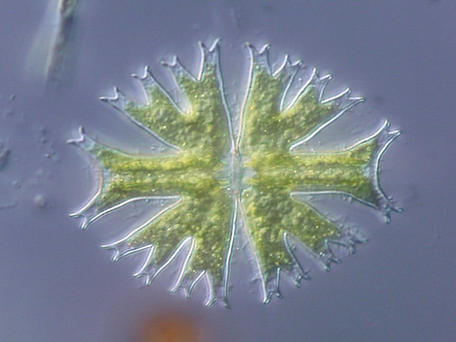
Differential interference contrast (DIC) microscopy, also known as Nomarski interference contrast (NIC) or Nomarski microscopy, is an optical microscopy technique used to enhance the contrast in unstained, transparent samples. DIC works on the principle of interferometry to gain information about the optical path length of the sample, to see otherwise invisible features. A relatively complex optical system produces an image with the object appearing black to white on a grey background. This image is similar to that obtained by phase contrast microscopy but without the bright diffraction halo. The technique was invented by Francis Hughes Smith. The "Smith DIK" was produced by Ernst Leitz Wetzlar in Germany and was difficult to manufacture. DIC was then developed further by Polish physicist Georges Nomarski in 1952. DIC works by separating a polarized light source into two orthogonally polarized mutually coherent parts which are spatially displaced (sheared) at the sample plane ... [...More Info...] [...Related Items...] OR: [Wikipedia] [Google] [Baidu] |


