|
Collateral Circulation
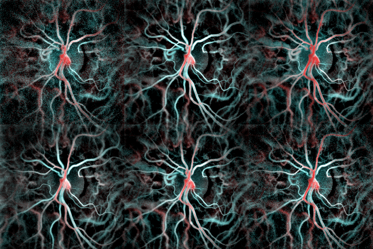
Collateral circulation is the alternate Circulatory system, circulation around a blocked blood vessel, artery or vein via another path, such as nearby minor vessels. It may occur via preexisting vascular redundancy (analogous to redundancy (engineering), engineered redundancy), as in the circle of Willis in the brain, or it may occur via new branches formed between adjacent blood vessels (neovascularization), as in the eye after a retinal embolism or in the brain when an instance of arterial constriction occurs due to Moyamoya disease. Its formation may be related by pathological conditions such as high vascular resistance or ischaemia. It is occasionally also known as accessory circulation, auxiliary circulation, or secondary circulation. It has surgery, surgically created analogues in which shunt (medical), shunts or circulatory anastomosis, anastomoses are constructed to bypass circulatory problems. An example of the usefulness of collateral circulation is a systemic thromboem ... [...More Info...] [...Related Items...] OR: [Wikipedia] [Google] [Baidu] [Amazon] |
Circulatory System
In vertebrates, the circulatory system is a system of organs that includes the heart, blood vessels, and blood which is circulated throughout the body. It includes the cardiovascular system, or vascular system, that consists of the heart and blood vessels (from Greek meaning ''heart'', and Latin meaning ''vessels''). The circulatory system has two divisions, a systemic circulation or circuit, and a pulmonary circulation or circuit. Some sources use the terms ''cardiovascular system'' and ''vascular system'' interchangeably with ''circulatory system''. The network of blood vessels are the great vessels of the heart including large elastic arteries, and large veins; other arteries, smaller arterioles, capillaries that join with venules (small veins), and other veins. The circulatory system is closed in vertebrates, which means that the blood never leaves the network of blood vessels. Many invertebrates such as arthropods have an open circulatory system with a he ... [...More Info...] [...Related Items...] OR: [Wikipedia] [Google] [Baidu] [Amazon] |
Communicating Artery (other)
Communication is commonly defined as the transmission of information. Its precise definition is disputed and there are disagreements about whether Intention, unintentional or failed transmissions are included and whether communication not only transmits semantics, meaning but also creates it. Models of communication are simplified overviews of its main components and their interactions. Many models include the idea that a source uses a code, coding system to express information in the form of a message. The message is sent through a Communication channel, channel to a receiver who has to decode it to understand it. The main field of inquiry investigating communication is called communication studies. A common way to classify communication is by whether information is exchanged between humans, members of other species, or non-living entities such as computers. For human communication, a central contrast is between Verbal communication, verbal and non-verbal communication. Verba ... [...More Info...] [...Related Items...] OR: [Wikipedia] [Google] [Baidu] [Amazon] |
Hepatic Cirrhosis
Cirrhosis, also known as liver cirrhosis or hepatic cirrhosis, chronic liver failure or chronic hepatic failure and end-stage liver disease, is a chronic condition of the liver in which the normal functioning tissue, or parenchyma, is replaced with scar tissue (fibrosis) and regenerative nodules as a result of chronic liver disease. Damage to the liver leads to repair of liver tissue and subsequent formation of scar tissue. Over time, scar tissue and nodules of regenerating hepatocytes can replace the parenchyma, causing increased resistance to blood flow in the liver's capillaries—the hepatic sinusoids—and consequently portal hypertension, as well as impairment in other aspects of liver function. The disease typically develops slowly over months or years. Stages include compensated cirrhosis and decompensated cirrhosis. Early symptoms may include tiredness, weakness, loss of appetite, unexplained weight loss, nausea and vomiting, and discomfort in the right upper quadrant ... [...More Info...] [...Related Items...] OR: [Wikipedia] [Google] [Baidu] [Amazon] |
Princeps Pollicis Artery
The princeps pollicis artery, or principal artery of the thumb, arises from the radial artery just as it turns medially towards the deep part of the hand; it descends between the first dorsal interosseous muscle and the oblique head of the adductor pollicis, along the medial side of the first metacarpal bone to the base of the proximal phalanx, where it lies beneath the tendon of the flexor pollicis longus muscle and divides into two branches. These make their appearance between the medial and lateral insertions of the adductor pollicis, and run along the sides of the thumb, forming an arch on the palmar surface of the distal phalanx, from which branches are distributed to the integument and subcutaneous tissue of the thumb. The princeps pollicis has a particularly strong pulse and can be used for ascertaining one's heart rate. For this reason, the pulse of the thumb is substantially stronger than that of the other digit, and thus it should not be used to read the pulses of othe ... [...More Info...] [...Related Items...] OR: [Wikipedia] [Google] [Baidu] [Amazon] |
Thumb
The thumb is the first digit of the hand, next to the index finger. When a person is standing in the medical anatomical position (where the palm is facing to the front), the thumb is the outermost digit. The Medical Latin English noun for thumb is ''pollex'' (compare ''hallux'' for big toe), and the corresponding adjective for thumb is ''pollical''. Definition Thumb and fingers The English word ''finger'' has two senses, even in the context of appendages of a single typical human hand: 1) Any of the five terminal members of the hand. 2) Any of the four terminal members of the hand, other than the thumb. Linguistically, it appears that the original sense was the first of these two: (also rendered as ) was, in the inferred Proto-Indo-European language, a suffixed form of (or ), which has given rise to many Indo-European-family words (tens of them defined in English dictionaries) that involve, or stem from, concepts of fiveness. The thumb shares the following with each of ... [...More Info...] [...Related Items...] OR: [Wikipedia] [Google] [Baidu] [Amazon] |
Proper Palmar Digital Arteries
The proper palmar digital arteries travel along the sides of the phalanges (along the contiguous sides of the index, middle, ring, and little fingers), each artery lying just below (dorsal (anatomy), dorsal to) its corresponding Dorsal digital nerves of ulnar nerve, digital nerve. Alternative names for these arteries are: proper volar digital arteries, collateral digital arteries, arteriae digitales palmares propriae, or aa. digitales volares propriae. Proper palmar digital arteries anastomose freely in the subcutaneous tissue of the finger tips and by smaller branches near the Interphalangeal articulations of hand, interphalangeal joints. Dorsal branches supplied by the arteries anastomose with the dorsal digital arteries of hand, dorsal digital arteries, and supply the soft parts on the back of the second and third phalanges, including the matrix of the fingernail. The proper palmar digital artery for the medial side of the little finger arises directly from the ulnar artery dee ... [...More Info...] [...Related Items...] OR: [Wikipedia] [Google] [Baidu] [Amazon] |
Superficial Palmar Arch
The superficial palmar arch is formed predominantly by the ulnar artery, with a contribution from the superficial palmar branch of the radial artery. However, in some individuals the contribution from the radial artery might be absent, and instead anastomoses with either the princeps pollicis artery, the radialis indicis artery, or the median artery, the former two of which are branches from the radial artery. Alternative names for this arterial arch are: superficial volar arch, superficial ulnar arch, arcus palmaris superficialis, or arcus volaris superficialis.Again, ''palmar'' and ''volar'' may be used synonymously, but ''arcus volaris superficialis'' does not occur in the TA, and can therefore be considered deprecated. The arch passes across the palm in a curve (Boeckel's line) with its convexity downward, With the thumb fully extended, the superficial palmar arch would lie approximately 1 cm from a line drawn between the first web space to the hook of hamate (Kapla ... [...More Info...] [...Related Items...] OR: [Wikipedia] [Google] [Baidu] [Amazon] |
Deep Palmar Arch
The deep palmar arch (deep volar arch) is an arterial network found in the palm. It is usually primarily formed from the terminal part of the radial artery. The ulnar artery also contributes through an anastomosis. This is in contrast to the superficial palmar arch, which is formed predominantly by the ulnar artery. Structure The deep palmar arch is usually primarily formed from the radial artery. The ulnar artery also contributes through an anastomosis. The deep palmar arch lies upon the bases of the metacarpal bones and on the interossei of the hand. It is deep to the oblique head of the adductor pollicis muscle, the flexor tendons of the fingers, and the lumbricals of the hand. Alongside of it, but running in the opposite direction—toward the radial side of the hand—is the deep branch of the ulnar nerve. The superficial palmar arch is more distally located than the deep palmar arch. If one were to fully extend the thumb and draw a line from the distal border of the thu ... [...More Info...] [...Related Items...] OR: [Wikipedia] [Google] [Baidu] [Amazon] |
Choroid
The choroid, also known as the choroidea or choroid coat, is a part of the uvea, the vascular layer of the eye. It contains connective tissues, and lies between the retina and the sclera. The human choroid is thickest at the far extreme rear of the eye (at 0.2 mm), while in the outlying areas it narrows to 0.1 mm. The choroid provides oxygen and nourishment to the outer layers of the retina. Along with the ciliary body and iris, the choroid forms the uveal tract. The structure of the choroid is generally divided into four layers (classified in order of furthest away from the retina to closest): *Haller's layer – outermost layer of the choroid consisting of larger diameter blood vessels; * Sattler's layer – layer of medium diameter blood vessels; * Choriocapillaris – layer of capillaries; and * Bruch's membrane (synonyms: Lamina basalis, Complexus basalis, Lamina vitra) – innermost layer of the choroid. Blood supply There are two circulations of the eye: ... [...More Info...] [...Related Items...] OR: [Wikipedia] [Google] [Baidu] [Amazon] |
Aqueous Humour
The aqueous humour is a transparent water-like fluid similar to blood plasma, but containing low protein concentrations. It is secreted from the ciliary body, a structure supporting the lens of the eyeball. It fills both the anterior and the posterior chambers of the eye, and is not to be confused with the vitreous humour, which is located in the space between the lens and the retina, also known as the posterior cavity or vitreous chamber. Blood cannot normally enter the eyeball. Structure Composition * Amino acids: transported by ciliary muscles * 98% water * Electrolytes ( pH = 7.4 -one source gives 7.1) ** Sodium = 142.09 ** Potassium = 2.2 - 4.0 ** Calcium = 1.8 ** Magnesium = 1.1 ** Chloride = 131.6 ** HCO3− = 20.15 ** Phosphate = 0.62 ** Osm = 304 * Ascorbic acid * Glutathione * Immunoglobulins Function * Maintains the intraocular pressure and inflates the globe of the eye. It is this hydrostatic pressure that keeps the eyeball in a roughly spherical shape and keeps ... [...More Info...] [...Related Items...] OR: [Wikipedia] [Google] [Baidu] [Amazon] |
Glaucoma
Glaucoma is a group of eye diseases that can lead to damage of the optic nerve. The optic nerve transmits visual information from the eye to the brain. Glaucoma may cause vision loss if left untreated. It has been called the "silent thief of sight" because the loss of vision usually occurs slowly over a long period of time. A major risk factor for glaucoma is increased pressure within the eye, known as Intraocular pressure, intraocular pressure (IOP). It is associated with old age, a family history of glaucoma, and certain medical conditions or the use of some medications. The word ''glaucoma'' comes from the Ancient Greek word (), meaning 'gleaming, blue-green, gray'. Of the different types of glaucoma, the most common are called open-angle glaucoma and closed-angle glaucoma. Inside the eye, a liquid called Aqueous humour, aqueous humor helps to maintain shape and provides nutrients. The aqueous humor normally drains through the trabecular meshwork. In open-angle glaucoma, ... [...More Info...] [...Related Items...] OR: [Wikipedia] [Google] [Baidu] [Amazon] |
Central Retinal Vein Occlusion
Central retinal vein occlusion, also CRVO, is when the central retinal vein becomes occluded, usually through thrombosis. The central retinal vein is the venous equivalent of the central retinal artery and both may become occluded. Since the central retinal artery and vein are the sole source of blood supply and drainage for the retina, such occlusion can lead to severe damage to the retina and blindness, due to ischemia (restriction in blood supply) and edema (swelling). CRVO can cause ocular ischemic syndrome. Nonischemic CRVO is the milder form of the disease. It may progress to the more severe ischemic type. CRVO can also cause glaucoma. Diagnosis Despite the role of thrombosis in the development of CRVO, a systematic review found no increased prevalence of thrombophilia (an inherent propensity to thrombosis) in patients with retinal vascular occlusion. Treatment Treatment consists of Anti-VEGF drugs like Lucentis or intravitreal steroid implant (Ozurdex) and Pan-Retinal L ... [...More Info...] [...Related Items...] OR: [Wikipedia] [Google] [Baidu] [Amazon] |


