|
Ciliary Process
In the anatomy of the eye, the ciliary processes are formed by the inward folding of the various layers of the choroid, viz. the choroid proper and the lamina basalis, and are received between corresponding foldings of the suspensory ligament of the lens. Anatomy They are arranged in a circle, and form a sort of frill behind the iris, around the margin of the lens. They vary from sixty to eighty in number, lie side by side, and may be divided into large and small; the former are about 2.5 mm. in length, and the latter, consisting of about one-third of the entire number, are situated in spaces between them, but without regular arrangement. They are attached by their periphery to three or four of the ridges of the orbiculus ciliaris, and are continuous with the layers of the choroid: their opposite extremities are free and rounded, and are directed toward the posterior chamber of the eyeball and circumference of the lens. In front, they are continuous with the periph ... [...More Info...] [...Related Items...] OR: [Wikipedia] [Google] [Baidu] [Amazon] |
Bulb Of Eye
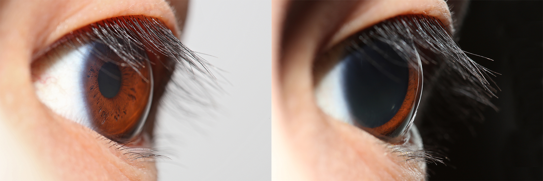
The human eye is a sensory organ in the visual system that reacts to visible light allowing eyesight. Other functions include maintaining the circadian rhythm, and keeping balance. The eye can be considered as a living optical device. It is approximately spherical in shape, with its outer layers, such as the outermost, white part of the eye (the sclera) and one of its inner layers (the pigmented choroid) keeping the eye essentially light tight except on the eye's optic axis. In order, along the optic axis, the optical components consist of a first lens (the cornea—the clear part of the eye) that accounts for most of the optical power of the eye and accomplishes most of the focusing of light from the outside world; then an aperture (the pupil) in a diaphragm (the iris—the coloured part of the eye) that controls the amount of light entering the interior of the eye; then another lens (the crystalline lens) that accomplishes the remaining focusing of light into images; and f ... [...More Info...] [...Related Items...] OR: [Wikipedia] [Google] [Baidu] [Amazon] |
Sagittal
The sagittal plane (; also known as the longitudinal plane) is an anatomical plane that divides the body into right and left sections. It is perpendicular to the transverse plane, transverse and coronal plane, coronal planes. The plane may be in the center of the body and divide it into two equal parts (mid-sagittal plane, mid-sagittal), or away from the midline and divide it into unequal parts (para-sagittal). The term ''sagittal'' was coined by Gerard of Cremona. Variations in terminology Examples of sagittal planes include: * The terms ''median plane'' or ''mid-sagittal plane'' are sometimes used to describe the wikt:sagittal plane, sagittal plane running through the midline. This plane cuts the body into halves (assuming bilateral symmetry), passing through midline structures such as the navel and Vertebral column, spine. It is one of the planes which, combined with the umbilical plane, defines the Quadrant (abdomen), four quadrants of the human abdomen. * The term ''parasagi ... [...More Info...] [...Related Items...] OR: [Wikipedia] [Google] [Baidu] [Amazon] |
Lens (vertebrate Anatomy)
The lens, or crystalline lens, is a transparent biconvex structure in most land vertebrate eyes. Relatively long, thin fiber cells make up the majority of the lens. These cells vary in architecture and are arranged in concentric layers. New layers of cells are recruited from a thin epithelium at the front of the lens, just below the basement membrane surrounding the lens. As a result the vertebrate lens grows throughout life. The surrounding lens membrane referred to as the lens capsule also grows in a systematic way, ensuring the lens maintains an optically suitable shape in concert with the underlying fiber cells. Thousands of suspensory ligaments are embedded into the capsule at its largest diameter which suspend the lens within the eye. Most of these lens structures are derived from the epithelium of the embryo before birth. Along with the cornea, aqueous, and vitreous humours, the lens refracts light, focusing it onto the retina. In many land animals the shape of the lens c ... [...More Info...] [...Related Items...] OR: [Wikipedia] [Google] [Baidu] [Amazon] |
Zonule Of Zinn
The zonule of Zinn () (Zinn's membrane, ciliary zonule) (after Johann Gottfried Zinn) is a ring of fibrous strands forming a zonule (little band) that connects the ciliary body with the crystalline lens of the eye. The Zonular fibers are viscoelastic cables, although their component microfibrils are stiff structures. These fibers are sometimes collectively referred to as the suspensory ligaments of the lens, as they act like suspensory ligaments. Development The non-pigmented ciliary epithelial cells of the eye synthesize portions of the zonules. Anatomy The zonule of Zinn is split into two layers: a thin layer, which lies near the hyaloid fossa, and a thicker layer, which is a collection of zonular fibers. Together, the fibers are known as the suspensory ligament of the lens. The zonules are about 1–2 μm in diameter. The zonules attach to the lens capsule 2 mm anterior and 1 mm posterior to the equator, and arise of the ciliary epithelium from the pars plana regi ... [...More Info...] [...Related Items...] OR: [Wikipedia] [Google] [Baidu] [Amazon] |
Short Posterior Ciliary Arteries
The short posterior ciliary arteries are a number of branches of the ophthalmic artery. They pass forward with the optic nerve to reach the eyeball, piercing the sclera around the entry of the optic nerve into the eyeball. Anatomy The number of short posterior ciliary arteries varies between individuals; one or more short posterior ciliary arteries initially branch off the ophthalmic artery, subsequently dividing to form up to 20 short posterior ciliary arteries. Origin The short posterior ciliary arteries branch off the ophthalmic artery as it crosses the optic nerve medially. Course and relations About 7 short posterior ciliary arteries accompany the optic nerve, passing anterior-ward to reach the posterior part of the eyeball, where they divide into 15-20 branches and pierce the sclera around the entrance of the optic nerve. Distribution The short posterior ciliary arteries contribute arterial supply to the choroid, ciliary processes, optic disc, the outer retina, and ... [...More Info...] [...Related Items...] OR: [Wikipedia] [Google] [Baidu] [Amazon] |
Anatomy
Anatomy () is the branch of morphology concerned with the study of the internal structure of organisms and their parts. Anatomy is a branch of natural science that deals with the structural organization of living things. It is an old science, having its beginnings in prehistoric times. Anatomy is inherently tied to developmental biology, embryology, comparative anatomy, evolutionary biology, and phylogeny, as these are the processes by which anatomy is generated, both over immediate and long-term timescales. Anatomy and physiology, which study the structure and function of organisms and their parts respectively, make a natural pair of related disciplines, and are often studied together. Human anatomy is one of the essential basic sciences that are applied in medicine, and is often studied alongside physiology. Anatomy is a complex and dynamic field that is constantly evolving as discoveries are made. In recent years, there has been a significant increase in the use of ... [...More Info...] [...Related Items...] OR: [Wikipedia] [Google] [Baidu] [Amazon] |
Choroid
The choroid, also known as the choroidea or choroid coat, is a part of the uvea, the vascular layer of the eye. It contains connective tissues, and lies between the retina and the sclera. The human choroid is thickest at the far extreme rear of the eye (at 0.2 mm), while in the outlying areas it narrows to 0.1 mm. The choroid provides oxygen and nourishment to the outer layers of the retina. Along with the ciliary body and iris, the choroid forms the uveal tract. The structure of the choroid is generally divided into four layers (classified in order of furthest away from the retina to closest): *Haller's layer – outermost layer of the choroid consisting of larger diameter blood vessels; * Sattler's layer – layer of medium diameter blood vessels; * Choriocapillaris – layer of capillaries; and * Bruch's membrane (synonyms: Lamina basalis, Complexus basalis, Lamina vitra) – innermost layer of the choroid. Blood supply There are two circulations of the eye: ... [...More Info...] [...Related Items...] OR: [Wikipedia] [Google] [Baidu] [Amazon] |
Choroid Proper
The choroid, also known as the choroidea or choroid coat, is a part of the uvea, the vascular layer of the eye. It contains connective tissues, and lies between the retina and the sclera. The human choroid is thickest at the far extreme rear of the eye (at 0.2 mm), while in the outlying areas it narrows to 0.1 mm. The choroid provides oxygen and nourishment to the outer layers of the retina. Along with the ciliary body and iris, the choroid forms the uveal tract. The structure of the choroid is generally divided into four layers (classified in order of furthest away from the retina to closest): *Haller's layer – outermost layer of the choroid consisting of larger diameter blood vessels; *Sattler's layer – layer of medium diameter blood vessels; *Choriocapillaris – layer of capillaries; and *Bruch's membrane (synonyms: Lamina basalis, Complexus basalis, Lamina vitra) – innermost layer of the choroid. Blood supply There are two circulations of the eye: the re ... [...More Info...] [...Related Items...] OR: [Wikipedia] [Google] [Baidu] [Amazon] |
Lamina Basalis Choroideae
Lamina may refer to: People * Saa Emerson Lamina, Sierra Leonean politician * Tamba Lamina, Sierra Leonean politician and diplomat Science and technology * Planar lamina, a two-dimensional planar closed surface with mass and density, in mathematics * Laminar flow, (or streamline flow) occurs when a fluid flows in parallel layers, with no disruption between the layers * Lamina (algae), a structure in seaweeds * Lamina (anatomy), with several meanings * Lamina (leaf), the flat part of a leaf, an organ of a plant * Lamina, the largest petal of a floret in an aster family flowerhead: see * ''Lamina'' (spider), a genus in the family Toxopidae * Lamina (neuropil), the most peripheral neuropil of the insect visual system *Nuclear lamina, another structure of a living cell *Basal lamina, a structure of a living cell *Lamina propria, the connective part of the mucous *Lamina of the vertebral arch *Lamination (geology), a layering structure in sedimentary rocks usually less than 1 cm in t ... [...More Info...] [...Related Items...] OR: [Wikipedia] [Google] [Baidu] [Amazon] |
Suspensory Ligament Of The Lens
The zonule of Zinn () (Zinn's membrane, ciliary zonule) (after Johann Gottfried Zinn) is a ring of fibrous strands forming a zonule (little band) that connects the ciliary body with the crystalline lens of the eye. The Zonular fibers are viscoelastic cables, although their component microfibrils are stiff structures. These fibers are sometimes collectively referred to as the suspensory ligaments of the lens, as they act like suspensory ligaments. Development The non-pigmented ciliary epithelial cells of the eye synthesize portions of the zonules. Anatomy The zonule of Zinn is split into two layers: a thin layer, which lies near the hyaloid fossa, and a thicker layer, which is a collection of zonular fibers. Together, the fibers are known as the suspensory ligament of the lens. The zonules are about 1–2 μm in diameter. The zonules attach to the lens capsule 2 mm anterior and 1 mm posterior to the equator, and arise of the ciliary epithelium from the pars plana region ... [...More Info...] [...Related Items...] OR: [Wikipedia] [Google] [Baidu] [Amazon] |
Iris (anatomy)
The iris (: irides or irises) is a thin, annular structure in the eye in most mammals and birds that is responsible for controlling the diameter and size of the pupil, and thus the amount of light reaching the retina. In optical terms, the pupil is the eye's aperture, while the iris is the diaphragm (optics), diaphragm. Eye color is defined by the iris. Etymology The word "iris" is derived from the Greek word for "rainbow", also Iris (mythology), its goddess plus messenger of the gods in the ''Iliad'', because of the many eye color, colours of this eye part. Structure The iris consists of two layers: the front pigmented Wikt:fibrovascular, fibrovascular layer known as a stroma of iris, stroma and, behind the stroma, pigmented epithelial cells. The stroma is connected to a sphincter muscle (sphincter pupillae), which contracts the pupil in a circular motion, and a set of dilator muscles (dilator pupillae), which pull the iris radially to enlarge the pupil, pulling it in folds. ... [...More Info...] [...Related Items...] OR: [Wikipedia] [Google] [Baidu] [Amazon] |
Lens (anatomy)
The lens, or crystalline lens, is a Transparency and translucency, transparent Biconvex lens, biconvex structure in most land vertebrate eyes. Relatively long, thin fiber cells make up the majority of the lens. These cells vary in architecture and are arranged in concentric layers. New layers of cells are recruited from a thin epithelium at the front of the lens, just below the basement membrane surrounding the lens. As a result the vertebrate lens grows throughout life. The surrounding lens membrane referred to as the lens capsule also grows in a systematic way, ensuring the lens maintains an optically suitable shape in concert with the underlying fiber cells. Thousands of suspensory ligaments are embedded into the capsule at its largest diameter which suspend the lens within the eye. Most of these lens structures are derived from the epithelium of the embryo before birth. Along with the cornea, aqueous humour, aqueous, and vitreous humours, the lens Refraction, refracts light, Fo ... [...More Info...] [...Related Items...] OR: [Wikipedia] [Google] [Baidu] [Amazon] |


