|
Centrioles
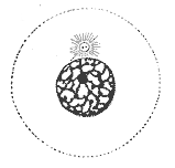
In cell biology a centriole is a cylindrical organelle composed mainly of a protein called tubulin. Centrioles are found in most eukaryotic cells, but are not present in conifers ( Pinophyta), flowering plants ( angiosperms) and most fungi, and are only present in the male gametes of charophytes, bryophytes, seedless vascular plants, cycads, and ''Ginkgo''. A bound pair of centrioles, surrounded by a highly ordered mass of dense material, called the pericentriolar material (PCM), makes up a structure called a centrosome. Centrioles are typically made up of nine sets of short microtubule triplets, arranged in a cylinder. Deviations from this structure include crabs and ''Drosophila melanogaster'' embryos, with nine doublets, and '' Caenorhabditis elegans'' sperm cells and early embryos, with nine singlets. Additional proteins include centrin, cenexin and tektin. The main function of centrioles is to produce cilia during interphase and the aster and the spindle durin ... [...More Info...] [...Related Items...] OR: [Wikipedia] [Google] [Baidu] |
Eukaryotic
The eukaryotes ( ) constitute the Domain (biology), domain of Eukaryota or Eukarya, organisms whose Cell (biology), cells have a membrane-bound cell nucleus, nucleus. All animals, plants, Fungus, fungi, seaweeds, and many unicellular organisms are eukaryotes. They constitute a major group of Outline of life forms, life forms alongside the two groups of prokaryotes: the Bacteria and the Archaea. Eukaryotes represent a small minority of the number of organisms, but given their generally much larger size, their collective global biomass is much larger than that of prokaryotes. The eukaryotes emerged within the archaeal Kingdom (biology), kingdom Asgard (Archaea), Promethearchaeati and its sole phylum Promethearchaeota. This implies that there are only Two-domain system, two domains of life, Bacteria and Archaea, with eukaryotes incorporated among the Archaea. Eukaryotes first emerged during the Paleoproterozoic, likely as Flagellated cell, flagellated cells. The leading evolutiona ... [...More Info...] [...Related Items...] OR: [Wikipedia] [Google] [Baidu] |
Centrosomes
In cell biology, the centrosome (Latin centrum 'center' + Greek sōma 'body') (archaically cytocentre) is an organelle that serves as the main microtubule organizing center (MTOC) of the animal cell, as well as a regulator of cell-cycle progression. The centrosome provides structure for the cell. It is thought to have evolved only in the metazoan lineage of eukaryotic cells. Fungi and plants lack centrosomes and therefore use other structures to organize their microtubules. Although the centrosome has a key role in efficient mitosis in animal cells, it is not essential in certain fly and flatworm species. Centrosomes are composed of two centrioles arranged at right angles to each other, and surrounded by a dense, highly structured mass of protein termed the pericentriolar material (PCM). The PCM contains proteins responsible for microtubule nucleation and anchoring — including γ-tubulin, pericentrin and ninein. In general, each centriole of the centrosome is based on ... [...More Info...] [...Related Items...] OR: [Wikipedia] [Google] [Baidu] |
Blausen 0214 Centrioles
Blausen Medical Communications, Inc. (BMC) is the creator and owner of a library of two- and three-dimensional medical and scientific images and animations, a developer of information technology allowing access to that content, and a business focused on licensing and distributing the content. It was founded by Bruce Blausen in Houston, Texas, in 1991, and is privately held. Background Blausen Medical Communications is a privately held company founded by Bruce Blausen in Houston, Texas in 1991. BMC created and owns a library of medical and scientific images and animations, and has developed information technology tools allowing access to the library; as well, it licenses and otherwise works to distribute the content. As of this date, BMC's animation library comprised approximately 1,500 animations and over 27,000 two- and three-dimensional images designed for point-of-care patient education, which could be accessed by consumers or professional caregivers (primarily via hospital o ... [...More Info...] [...Related Items...] OR: [Wikipedia] [Google] [Baidu] |
Drosophila Melanogaster
''Drosophila melanogaster'' is a species of fly (an insect of the Order (biology), order Diptera) in the family Drosophilidae. The species is often referred to as the fruit fly or lesser fruit fly, or less commonly the "vinegar fly", "pomace fly", or "banana fly". In the wild, ''D. melanogaster'' are attracted to rotting fruit and fermenting beverages, and are often found in orchards, kitchens and pubs. Starting with Charles W. Woodworth's 1901 proposal of the use of this species as a model organism, ''D. melanogaster'' continues to be widely used for biological research in genetics, physiology, microbial pathogenesis, and Life history theory, life history evolution. ''D. melanogaster'' was the first animal to be Fruit flies in space, launched into space in 1947. As of 2017, six Nobel Prizes have been awarded to drosophilists for their work using the insect. ''Drosophila melanogaster'' is typically used in research owing to its rapid life cycle, relatively simple genetics with on ... [...More Info...] [...Related Items...] OR: [Wikipedia] [Google] [Baidu] |
Edouard Van Beneden
Édouard Joseph Louis Marie Van Beneden (5 March 1846 in Leuven – 28 April 1910 in Liège) was a Belgian embryologist, cytologist and marine biologist. He was professor of zoology at the University of Liège. He contributed to cytogenetics by his works on the roundworm '' Ascaris''. In this work he discovered how chromosomes organized meiosis (the production of gametes). He is son of Pierre-Joseph Van Beneden, a zoologist Zoology ( , ) is the scientific study of animals. Its studies include the structure, embryology, classification, habits, and distribution of all animals, both living and extinct, and how they interact with their ecosystems. Zoology is one ... and paleontologist. Van Beneden elucidated, together with Walther Flemming and Eduard Strasburger, the essential facts of mitosis, where, in contrast to meiosis, there is a qualitative and quantitative equality of chromosome distribution to daughter cells. (See karyotype). Publications * ''Rech ... [...More Info...] [...Related Items...] OR: [Wikipedia] [Google] [Baidu] |
Walther Flemming
Walther Flemming (21 April 1843 – 4 August 1905) was a German biologist and a founder of cytogenetics. He was born in Sachsenberg (now part of Schwerin) as the fifth child and only son of the psychiatrist Carl Friedrich Flemming (1799–1880) and his second wife, Auguste Winter. He graduated from the ''Gymnasium der Residenzstadt'', where one of his colleagues and lifelong friends was writer Heinrich Seidel. Career Flemming trained in medicine at the University of Prague, graduating in 1868. Afterwards, he served in 1870–71 as a military physician in the Franco-Prussian War. From 1873 to 1876 he worked as a teacher at the University of Prague. In 1876 he accepted a post as a professor of anatomy at the University of Kiel. He became the director of the Anatomical Institute and stayed there until his death. With the use of aniline dyes he was able to find a structure which strongly absorbed basophilic dyes, which he named chromatin. He identified that chromatin was corr ... [...More Info...] [...Related Items...] OR: [Wikipedia] [Google] [Baidu] |
Spindle Apparatus
In cell biology, the spindle apparatus is the cytoskeletal structure of eukaryotic cells that forms during cell division to separate sister chromatids between daughter Cell (biology), cells. It is referred to as the mitotic spindle during mitosis, a process that produces genetically identical daughter cells, or the meiotic spindle during meiosis, a process that produces gametes with half the number of chromosomes of the parent cell. Besides chromosomes, the spindle apparatus is composed of hundreds of proteins. Microtubules comprise the most abundant components of the machinery. Spindle structure Attachment of microtubules to chromosomes is mediated by kinetochores, which actively monitor spindle assembly checkpoint, spindle formation and prevent premature anaphase onset. Microtubule polymerization and depolymerization dynamic drive chromosome congression. Depolymerization of microtubules generates tension at kinetochores; bipolar attachment of sister kinetochores to microtubu ... [...More Info...] [...Related Items...] OR: [Wikipedia] [Google] [Baidu] |
Aster (cell Biology)
An aster is a cellular structure shaped like a star, consisting of a centrosome and its associated microtubules during the early stages of mitosis in an animal cell. Asters do not form during mitosis in plants. Astral rays, composed of microtubules, radiate from the centrosphere and look like a cloud. Astral rays are one variant of microtubule which comes out of the centrosome; others include kinetochore microtubules and polar microtubules. During mitosis, there are five stages of cell division: Prophase, Prometaphase, Metaphase, Anaphase, and Telophase. During prophase, two aster-covered centrosomes migrate to opposite sides of the nucleus in preparation of mitotic spindle formation. During prometaphase there is fragmentation of the nuclear envelope and formation of the mitotic spindles. During metaphase, the kinetochore microtubules extending from each centrosome connect to the centromeres of the chromosomes. Next, during anaphase, the kinetochore microtubules pull the ... [...More Info...] [...Related Items...] OR: [Wikipedia] [Google] [Baidu] |
Interphase
Interphase is the active portion of the cell cycle that includes the G1, S, and G2 phases, where the cell grows, replicates its DNA, and prepares for mitosis, respectively. Interphase was formerly called the "resting phase," but the cell in interphase is not simply dormant. Calling it so would be misleading since a cell in interphase is very busy synthesizing proteins, transcribing DNA into RNA, engulfing extracellular material, and processing signals, to name just a few activities. The cell is quiescent only in G0. Interphase is the phase of the cell cycle in which a typical cell spends most of its life. Interphase is the "daily living" or metabolic phase of the cell, in which the cell obtains nutrients and metabolizes them, grows, replicates its DNA in preparation for mitosis, and conducts other "normal" cell functions. A common misconception is that interphase is the first stage of mitosis, but since mitosis is the division of the nucleus, prophase is actually ... [...More Info...] [...Related Items...] OR: [Wikipedia] [Google] [Baidu] |
Cilium
The cilium (: cilia; ; in Medieval Latin and in anatomy, ''cilium'') is a short hair-like membrane protrusion from many types of eukaryotic cell. (Cilia are absent in bacteria and archaea.) The cilium has the shape of a slender threadlike projection that extends from the surface of the much larger cell body. Eukaryotic flagella found on sperm cells and many protozoans have a similar structure to motile cilia that enables swimming through liquids; they are longer than cilia and have a different undulating motion. There are two major classes of cilia: ''motile'' and ''non-motile'' cilia, each with two subtypes, giving four types in all. A cell will typically have one primary cilium or many motile cilia. The structure of the cilium core, called the axoneme, determines the cilium class. Most motile cilia have a central pair of single microtubules surrounded by nine pairs of double microtubules called a 9+2 axoneme. Most non-motile cilia have a 9+0 axoneme that lacks the centr ... [...More Info...] [...Related Items...] OR: [Wikipedia] [Google] [Baidu] |
Tektin
Tektins are cytoskeletal proteins found in cilia and flagella as structural components of outer doublet microtubules. They are also present in centrioles and basal bodies. They are polymeric in nature, and form filaments.MA Pirner and RW LinckTektins are heterodimeric polymers in flagellar microtubules with axial periodicities matching the tubulin lattice J. Biol. Chem., Vol. 269, Issue 50, 31800-31806, Dec, 1994 They include TEKT1, TEKT2, TEKT3, TEKT4, TEKT5. Structure Tektin filaments are 2 to 3 nm diameter with two alpha helical segments. They have the consensus amino acid sequence of RPNVELCRD. Different types of tektins, designated as A (53 kDa), B (51 kDa), C (47 kDa) form dimers, trimers and oligomers in various combinations and are also associated with tubulin in the microtubule. Tektins A and B form heteropolymeric protofilaments whereas tektin C forms homodimers. Tektin filaments are present in a supercoiled state. This structure of tektins suggests that they ... [...More Info...] [...Related Items...] OR: [Wikipedia] [Google] [Baidu] |


