|
Caridoid Escape Reaction
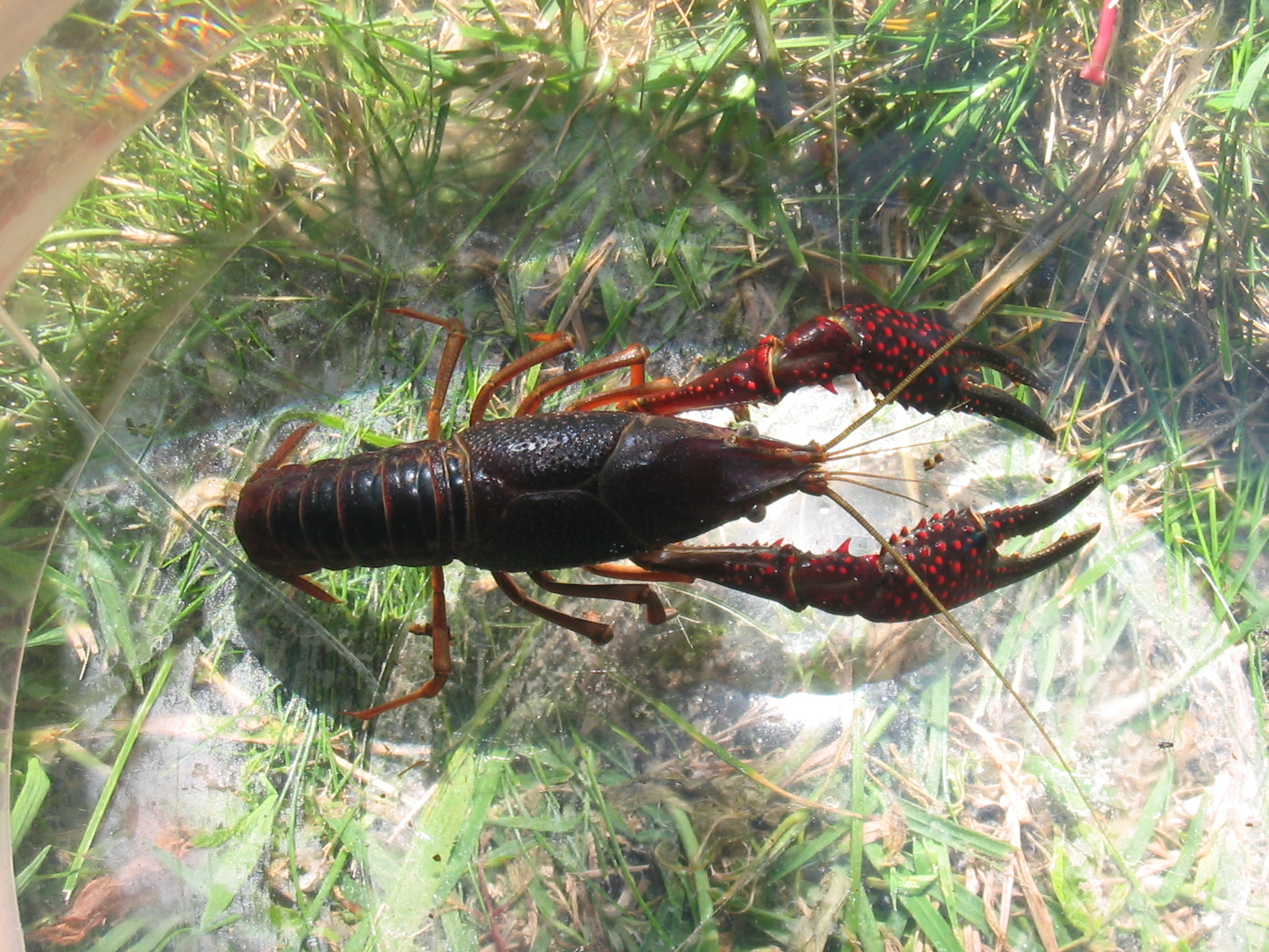
The caridoid escape reaction, also known as lobstering or tail-flipping, is an innate escape behavior in marine and freshwater eucarid crustaceans such as lobsters, krill, shrimp and crayfish. The reaction, most extensively researched in crayfish, allows crustaceans to escape predators through rapid abdominal flexions that produce powerful thrusts that make the crustacean quickly swim backwards through the water and away from danger. The type of response depends on the part of the crustacean stimulated, but this behavior is complex and is regulated both spatially and temporally through the interactions of several neurons. Discovery of the first command neuron-mediated behavior In 1946, C. A. G. Wiersma first described the tail-flip escape in the crayfish '' Procambarus clarkii'' and noted that the giant interneurons present in the tail were responsible for the reaction. The aforementioned neuronal fibres consist of a pair of lateral giant interneurons and a pair of medi ... [...More Info...] [...Related Items...] OR: [Wikipedia] [Google] [Baidu] [Amazon] |
Medial Giant Interneuron
The medial giant interneuron (MG) is an interneuron in the abdominal nerve cord of crayfish. It is part of the system that controls the caridoid escape reaction of crayfish, clawed lobsters, and other decapod crustaceans. Crayfish have a pair of medial giants running the length of the entire animal, and are the largest neurons in the animal. When a crayfish is given a sudden visual or tactile stimulus to the front part of the animal, the MG activates fast flexor motor neurons that cause the abdomen to flex, resulting in the crayfish moving directly backward, away from the source of the stimulation. This connection was first demonstrated by C. A. G. Wiersma in the red swamp crayfish, '' Procambarus clarkii''. The medial giant interneurons are less well studied than the lateral giant neurons, which trigger a similar escape behavior. See also *Squid giant axon The squid giant axon is the very large (up to 1.5 mm in diameter; typically around 0.5 mm) axon that controls pa ... [...More Info...] [...Related Items...] OR: [Wikipedia] [Google] [Baidu] [Amazon] |
Motor Nerve
A motor nerve, or efferent nerve, is a nerve that contains exclusively efferent nerve fibers and transmits motor signals from the central nervous system (CNS) to the effector organs (muscles and glands), as opposed to sensory nerves, which transfer signals from sensory receptors in the periphery to the CNS. This is different from the motor neuron, which includes a cell body and branching of dendrites, while the nerve is made up of a bundle of axons. In the strict sense, a "motor nerve" can refer exclusively to the connection to muscles, excluding other organs. The vast majority of nerves contain both sensory and motor fibers and are therefore called mixed nerves. Structure and function Motor nerve fibers Transduction (physiology), transduce signals from the CNS to peripheral neurons of proximal muscle tissue. Motor nerve axon terminals innervate Skeletal muscle, skeletal and Smooth muscle tissue, smooth muscle, as they are heavily involved in muscle control. Motor nerves tend t ... [...More Info...] [...Related Items...] OR: [Wikipedia] [Google] [Baidu] [Amazon] |
Ganglia
A ganglion (: ganglia) is a group of neuron cell bodies in the peripheral nervous system. In the somatic nervous system, this includes dorsal root ganglia and trigeminal ganglia among a few others. In the autonomic nervous system, there are both sympathetic and parasympathetic ganglia which contain the cell bodies of postganglionic sympathetic and parasympathetic neurons respectively. A pseudoganglion looks like a ganglion, but only has nerve fibers and has no nerve cell bodies. Structure Ganglia are primarily made up of somata and dendritic structures, which are bundled or connected. Ganglia often interconnect with other ganglia to form a complex system of ganglia known as a plexus. Ganglia provide relay points and intermediary connections between different neurological structures in the body, such as the peripheral and central nervous systems. Among vertebrates there are three major groups of ganglia: * Dorsal root ganglia (also known as the spinal ganglia) cont ... [...More Info...] [...Related Items...] OR: [Wikipedia] [Google] [Baidu] [Amazon] |
Muscle
Muscle is a soft tissue, one of the four basic types of animal tissue. There are three types of muscle tissue in vertebrates: skeletal muscle, cardiac muscle, and smooth muscle. Muscle tissue gives skeletal muscles the ability to muscle contraction, contract. Muscle tissue contains special Muscle contraction, contractile proteins called actin and myosin which interact to cause movement. Among many other muscle proteins, present are two regulatory proteins, troponin and tropomyosin. Muscle is formed during embryonic development, in a process known as myogenesis. Skeletal muscle tissue is striated consisting of elongated, multinucleate muscle cells called muscle fibers, and is responsible for movements of the body. Other tissues in skeletal muscle include tendons and perimysium. Smooth and cardiac muscle contract involuntarily, without conscious intervention. These muscle types may be activated both through the interaction of the central nervous system as well as by innervation ... [...More Info...] [...Related Items...] OR: [Wikipedia] [Google] [Baidu] [Amazon] |
Extensor
In anatomy, extension is a movement of a joint that increases the angle between two bones or body surfaces at a joint. Extension usually results in straightening of the bones or body surfaces involved. For example, extension is produced by extending the flexed (bent) elbow. Straightening of the arm would require extension at the elbow joint. If the head is tilted all the way back, the neck is said to be extended. Extensor muscles Upper limb *of arm at shoulder **Axilla and shoulder ***Latissimus dorsi *** Posterior fibres of deltoid *** Teres major *of forearm at elbow **Posterior compartment of the arm ***Triceps brachii *** Anconeus *of hand at wrist **Posterior compartment of the forearm *** Extensor carpi radialis longus ***Extensor carpi radialis brevis *** Extensor carpi ulnaris *** Extensor digitorum *of phalanges, at all joints **Posterior compartment of the forearm *** Extensor digitorum *** Extensor digiti minimi (little finger only) ***Extensor indicis (index finger ... [...More Info...] [...Related Items...] OR: [Wikipedia] [Google] [Baidu] [Amazon] |
Flexor
In anatomy, flexor is a muscle that contracts to perform flexion (from the Latin verb ''flectere'', to bend), a movement that decreases the angle between the bones converging at a joint. For example, one's elbow joint flexes when one brings their hand closer to the shoulder, thus decreasing the angle between the upper arm and the forearm. Flexors Upper limb *of the humerus bone (the bone in the upper arm) at the shoulder ** Pectoralis major ** Anterior deltoid ** Coracobrachialis ** Biceps brachii * of the forearm at the elbow ** Brachialis ** Brachioradialis ** Biceps brachii *of carpus (the carpal bones) at the wrist ** flexor carpi radialis ** flexor carpi ulnaris ** palmaris longus *of the hand ** flexor pollicis longus muscle ** flexor pollicis brevis muscle ** flexor digitorum profundus muscle ** flexor digitorum superficialis muscle Lower limb Hip The hip flexors are (in descending order of importance to the action of flexing the hip joint):Platzer (20 ... [...More Info...] [...Related Items...] OR: [Wikipedia] [Google] [Baidu] [Amazon] |
Exoskeleton
An exoskeleton () . is a skeleton that is on the exterior of an animal in the form of hardened integument, which both supports the body's shape and protects the internal organs, in contrast to an internal endoskeleton (e.g. human skeleton, that of a human) which is enclosed underneath other soft tissues. Some large, hard and non-flexible protective exoskeletons are known as mollusc shell, shell or armour (anatomy), armour. Examples of exoskeletons in animals include the arthropod exoskeleton, cuticle skeletons shared by arthropods (insects, chelicerates, myriapods and crustaceans) and tardigrades, as well as the corallite, skeletal cups formed by hardened secretion of stony corals, the test (biology), test/tunic of sea squirts and sea urchins, and the prominent mollusc shell shared by snails, bivalvia, clams, tusk shells, chitons and nautilus. Some vertebrate animals, such as the turtle, have both an endoskeleton and a turtle shell, protective exoskeleton. Role Exoskeletons c ... [...More Info...] [...Related Items...] OR: [Wikipedia] [Google] [Baidu] [Amazon] |
Decapoda
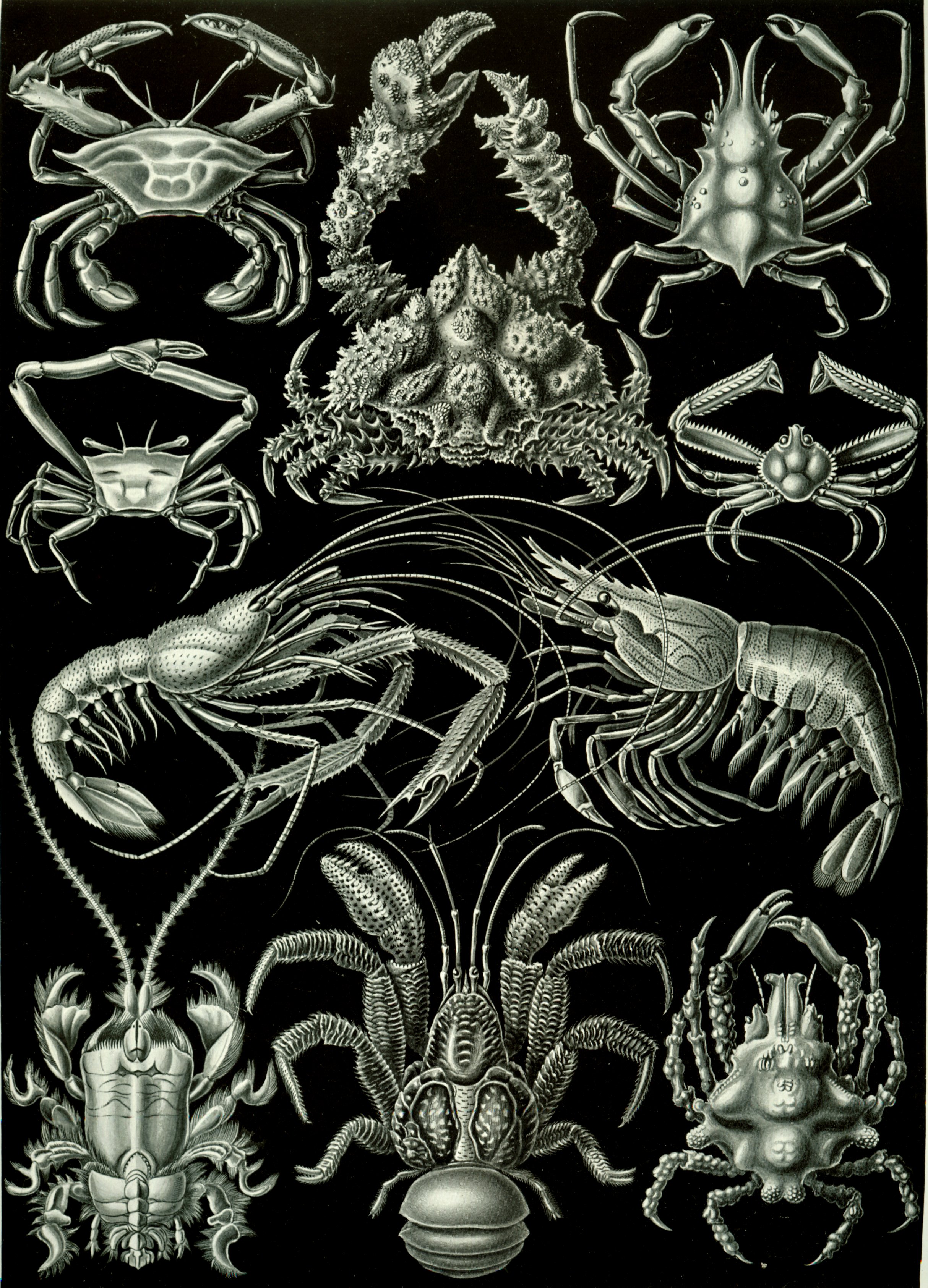
The Decapoda or decapods, from Ancient Greek δεκάς (''dekás''), meaning "ten", and πούς (''poús''), meaning "foot", is a large order of crustaceans within the class Malacostraca, and includes crabs, lobsters, crayfish, shrimp, and prawns. Most decapods are scavengers. The order is estimated to contain nearly 15,000 extant species in around 2,700 genera, with around 3,300 fossil species. Nearly half of these species are crabs, with the shrimp (about 3,000 species) and Anomura including hermit crabs, king crabs, porcelain crabs, squat lobsters (about 2500 species) making up the bulk of the remainder. The earliest fossils of the group date to the Devonian. Anatomy Decapods can have as many as 38 appendages, arranged in one pair per body segment. As the name Decapoda (from the Greek , ', "ten", and , '' -pod'', "foot") implies, ten of these appendages are considered legs. They are the pereiopods, found on the last five thoracic segments. In many decapods, one ... [...More Info...] [...Related Items...] OR: [Wikipedia] [Google] [Baidu] [Amazon] |
Telson
The telson () is the hindmost division of the body of an arthropod. Depending on the definition, the telson is either considered to be the final segment (biology), segment of the arthropod body, or an additional division that is not a true segment on account of not arising in the embryo from teloblast areas as other segments. It never carries any appendages, but a forked "tail" called the caudal furca may be present. The shape and composition of the telson differs between arthropod groups. Crustaceans In lobsters, Caridea, shrimp and other Decapoda, decapods, the telson, along with the uropods, forms the tail fan. This is used as a paddle in the caridoid escape reaction ("lobstering"), whereby an alarmed animal rapidly flexes its tail, causing it to dart backwards. Krill can reach speeds of over 60 cm per second by this means. The Induction period, trigger time to optical stimulus (physiology), stimulus is, in spite of the low temperatures, only 55 milliseconds. In th ... [...More Info...] [...Related Items...] OR: [Wikipedia] [Google] [Baidu] [Amazon] |
Escape Response
Escape response, escape reaction, or escape behavior is a mechanism by which animals avoid potential predation. It consists of a rapid sequence of movements, or lack of movement, that position the animal in such a way that allows it to hide, freeze, or flee from the supposed predator. Often, an animal's escape response is representative of an instinctual defensive mechanism, though there is evidence that these escape responses may be learned or influenced by experience. The classical escape response follows this generalized, conceptual timeline: threat detection, escape initiation, escape execution, and escape termination or conclusion. Threat detection notifies an animal to a potential predator or otherwise dangerous stimulus, which provokes escape initiation, through neural reflexes or more coordinated cognitive processes. Escape execution refers to the movement or series of movements that will hide the animal from the threat or will allow for the animal to flee. Once the ani ... [...More Info...] [...Related Items...] OR: [Wikipedia] [Google] [Baidu] [Amazon] |
Fixed Action Pattern
"Fixed action pattern" is an Ethology, ethological term describing an instinctive behavioral sequence that is highly stereotyped and species-characteristic. Fixed action patterns are said to be produced by the innate releasing mechanism, a "hard-wired" neural network, in response to a Fixed action pattern#Sign stimulus, sign/key stimulus or releaser. Once released, a fixed action pattern runs to completion. This term is often associated with Konrad Lorenz, who is the founder of the concept. Lorenz identified six characteristics of fixed action patterns. These characteristics state that fixed action patterns are stereotyped, complex, species-characteristic, released, triggered, and independent of experience. Fixed action patterns have been observed in many species, but most notably in fish and birds. Classic studies by Konrad Lorenz and Nikolaas Tinbergen, Niko Tinbergen involve male stickleback mating behavior and greylag goose egg-retrieval behavior. Fixed action patterns have be ... [...More Info...] [...Related Items...] OR: [Wikipedia] [Google] [Baidu] [Amazon] |


