|
Canthotomy
Canthotomy (also called lateral canthotomy and canthotomy with cantholysis) is a surgical procedure where the lateral canthus, or corner, of the eye is cut to relieve the fluid pressure inside or behind the eye, known as intraocular pressure (IOC). The procedure is typically done in emergency situations when the intraocular pressure becomes too high, which can damage the optic nerve and lead to blindness if left untreated. The most common cause of elevated intraocular pressure is orbital compartment syndrome (OCS) caused by trauma, retrobulbar hemorrhage, infections, tumors, or prolonged hypoxemia. Absolute contraindications to canthotomy include globe rupture. Complications include bleeding, infections, cosmetic deformities, and functional impairment of eyelids. Lateral canthotomy further specifies that the lateral canthus is being cut. Canthotomy with cantholysis includes cutting the lateral palpebral ligament, also known as the canthal tendon. History The first case of ... [...More Info...] [...Related Items...] OR: [Wikipedia] [Google] [Baidu] [Amazon] |
Canthus
The canthus (: canthi, palpebral commissures) is either corner of the eye where the upper and lower eyelids meet. More specifically, the inner and outer canthi are, respectively, the medial and lateral ends/angles of the palpebral fissure. The bicanthal plane is the transversal plane linking both canthi and defines the upper boundary of the midface. Etymology The word ' is the Latinized form of the Ancient Greek ('), meaning 'corner of the eye'. Population distribution The eyes of East Asian and some Southeast Asian people tend to have the inner canthus veiled by the epicanthus. In the Caucasian or double eyelid, the inner corner tends to be exposed completely. Commissures * The ''lateral palpebral commissure'' (commissura palpebrarum lateralis; external canthus) is more acute than the medial, and the eyelids here lie in close contact with the bulb of the eye. * The ''medial palpebral commissure'' (commissura palpebrarum medialis; internal canthus) is prolonged for a s ... [...More Info...] [...Related Items...] OR: [Wikipedia] [Google] [Baidu] [Amazon] |
Retrobulbar Hemorrhage
Retrobulbar bleeding, also known as retrobulbar hemorrhage, is when bleeding occurs behind the eye. Symptoms may include pain, bruising around the eye, the proptosis, eye bulging outwards, vomiting, and vision loss. Retrobulbar bleeding can occur as a result of trauma to the eye, surgery to the eye, anticoagulation, blood thinners, or an arteriovenous malformation. In those with significant symptoms lateral canthotomy with cantholysis is indicated. This is recommended to be carried out within two hours. The condition is rare. References Eye injury Gross pathology {{Injury-stub} ... [...More Info...] [...Related Items...] OR: [Wikipedia] [Google] [Baidu] [Amazon] |
Ophthalmology
Ophthalmology (, ) is the branch of medicine that deals with the diagnosis, treatment, and surgery of eye diseases and disorders. An ophthalmologist is a physician who undergoes subspecialty training in medical and surgical eye care. Following a medical degree, a doctor specialising in ophthalmology must pursue additional postgraduate residency training specific to that field. In the United States, following graduation from medical school, one must complete a four-year residency in ophthalmology to become an ophthalmologist. Following residency, additional specialty training (or fellowship) may be sought in a particular aspect of eye pathology. Ophthalmologists prescribe medications to treat ailments, such as eye diseases, implement laser therapy, and perform surgery when needed. Ophthalmologists provide both primary and specialty eye care—medical and surgical. Most ophthalmologists participate in academic research on eye diseases at some point in their training and many inc ... [...More Info...] [...Related Items...] OR: [Wikipedia] [Google] [Baidu] [Amazon] |
Surgical Drain
A surgical drain is a tube used to remove pus, blood or other fluids from a wound, body cavity, or organ. They are commonly placed by surgeons or interventional radiologists after procedures or some types of injuries, but they can also be used as an intervention for decompression. There are several types of drains, and selection of which to use often depends on the placement site and how long the drain is needed. Use and Management Drains help to remove contents, usually fluids, from inside the body. This is beneficial since fluid accumulation may cause distension and pressure, which can lead to pain. For example, nasogastric (NG) tubes inserted through the nose and into the stomach can help remove stomach contents for patients who have a blockage further along in their gastrointestinal tract. After surgery, drains can be placed to remove blood, lymph, or other fluids that accumulate in the wound bed. This helps to promote wound healing and allows healthcare providers to monit ... [...More Info...] [...Related Items...] OR: [Wikipedia] [Google] [Baidu] [Amazon] |
Iatrogenic
Iatrogenesis is the causation of a disease, a harmful complication, or other ill effect by any medical activity, including diagnosis, intervention, error, or negligence." Iatrogenic", ''Merriam-Webster.com'', Merriam-Webster, Inc., accessed 27 Jun 2020. First used in this sense in 1924, the term was introduced to sociology in 1976 by Ivan Illich, alleging that industrialized societies impair quality of life by overmedicalizing life."iatrogenesis" ''A Dictionary of Sociology'', . updated 31 May 2020. Iatrogenesis may thus include mental suffering via medical beliefs or a practitioner's statements. Some iatrogen ... [...More Info...] [...Related Items...] OR: [Wikipedia] [Google] [Baidu] [Amazon] |
Enophthalmos
Enophthalmos is a posterior displacement of the eyeball within the orbit. It is due to either enlargement of the bony orbit and/or reduction of the orbital content, this in relation to each other. It should not be confused with its opposite, exophthalmos, which is the anterior displacement of the eye. It may be a congenital anomaly, or be acquired as a result of trauma (such as in a blowout fracture of the orbit), Horner's syndrome (apparent enophthalmos due to ptosis), Marfan syndrome, Duane's syndrome, silent sinus syndrome or phthisis bulbi Phthisis bulbi is a shrunken, non-functional eye. It may result from severe eye disease, inflammation or injury, or it may represent a complication of eye surgery. Treatment options include insertion of a prosthesis, which may be preceded by enucl .... References Further reading * External links Disorders of eyelid, lacrimal system and orbit {{med-sign-stub ... [...More Info...] [...Related Items...] OR: [Wikipedia] [Google] [Baidu] [Amazon] |
Subconjunctival Hemorrhage
Subconjunctival bleeding, also known as subconjunctival hemorrhage or subconjunctival haemorrhage, is bleeding from a small blood vessel over the whites of the eye. It results in a red spot in the white of the eye. There is generally little to no pain and vision is not affected. Generally only one eye is affected. Causes can include coughing, vomiting, heavy lifting, straining during acute constipation or the act of "bearing down" during childbirth, as these activities can increase the blood pressure in the vascular systems supplying the conjunctiva. Other causes include blunt or penetrating trauma to the eye. Risk factors include hypertension, diabetes, old age, and blood thinners. Subconjunctival bleeding occurs in about 2% of newborns following a vaginal delivery. The blood accumulates between the conjunctiva and the episclera. Diagnosis is generally based on the appearance of the conjunctiva. The condition is relatively common, and both sexes are affected equally. Sponta ... [...More Info...] [...Related Items...] OR: [Wikipedia] [Google] [Baidu] [Amazon] |
Iris (anatomy)
The iris (: irides or irises) is a thin, annular structure in the eye in most mammals and birds that is responsible for controlling the diameter and size of the pupil, and thus the amount of light reaching the retina. In optical terms, the pupil is the eye's aperture, while the iris is the diaphragm (optics), diaphragm. Eye color is defined by the iris. Etymology The word "iris" is derived from the Greek word for "rainbow", also Iris (mythology), its goddess plus messenger of the gods in the ''Iliad'', because of the many eye color, colours of this eye part. Structure The iris consists of two layers: the front pigmented Wikt:fibrovascular, fibrovascular layer known as a stroma of iris, stroma and, behind the stroma, pigmented epithelial cells. The stroma is connected to a sphincter muscle (sphincter pupillae), which contracts the pupil in a circular motion, and a set of dilator muscles (dilator pupillae), which pull the iris radially to enlarge the pupil, pulling it in folds. ... [...More Info...] [...Related Items...] OR: [Wikipedia] [Google] [Baidu] [Amazon] |
Pupil
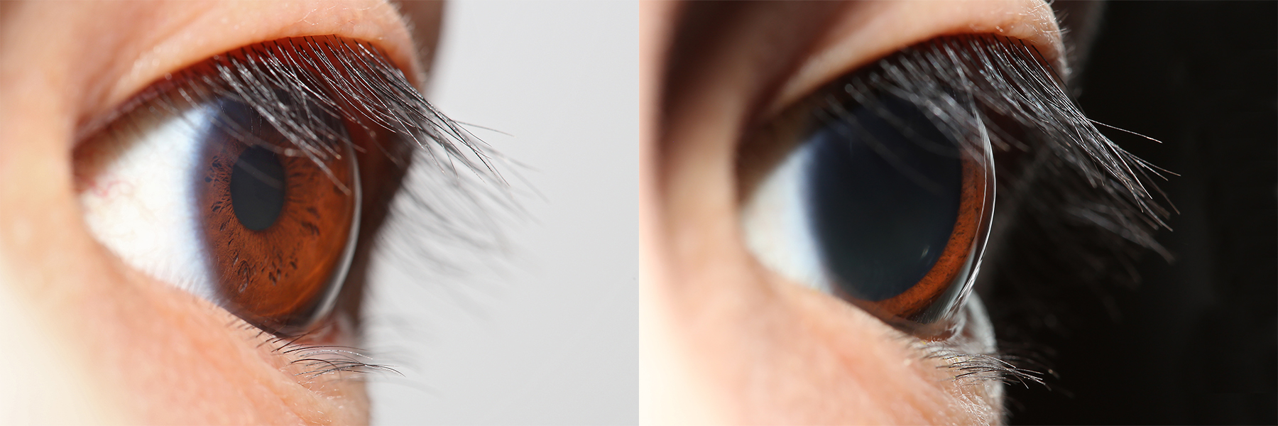
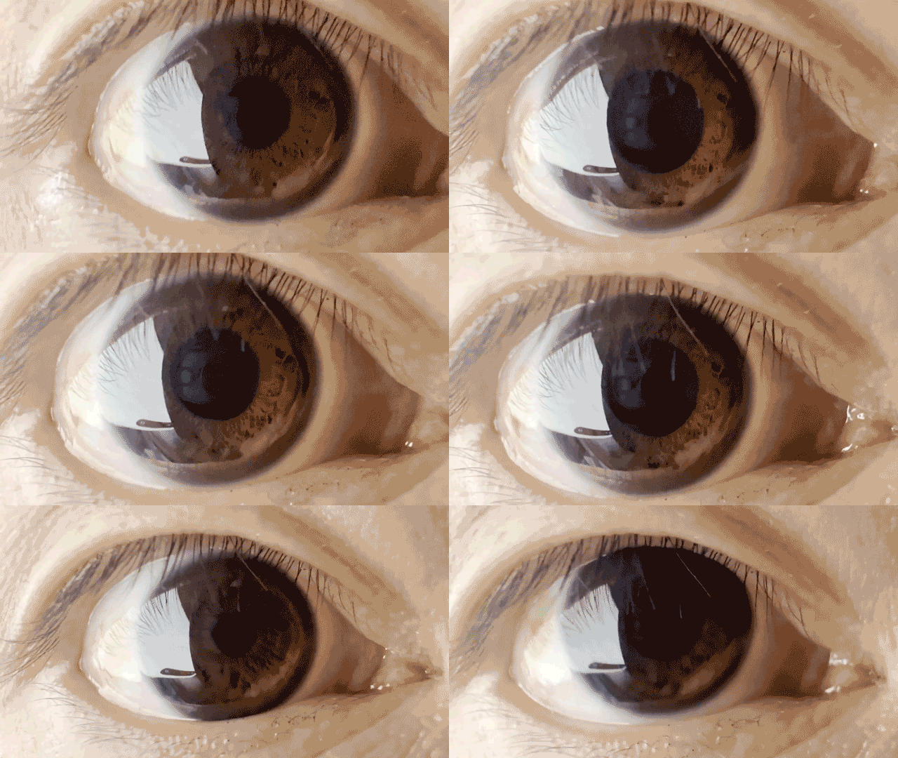
The pupil is a hole located in the center of the iris of the eye that allows light to strike the retina.Cassin, B. and Solomon, S. (1990) ''Dictionary of Eye Terminology''. Gainesville, Florida: Triad Publishing Company. It appears black because light rays entering the pupil are either absorbed by the tissues inside the eye directly, or absorbed after diffuse reflections within the eye that mostly miss exiting the narrow pupil. The size of the pupil is controlled by the iris, and varies depending on many factors, the most significant being the amount of light in the environment. The term "pupil" was coined by Gerard of Cremona. In humans, the pupil is circular, but its shape varies between species; some cats, reptiles, and foxes have vertical slit pupils, goats and sheep have horizontally oriented pupils, and some catfish have annular types. In optical terms, the anatomical pupil is the eye's aperture and the iris is the aperture stop. The image of the pupil as seen from o ... [...More Info...] [...Related Items...] OR: [Wikipedia] [Google] [Baidu] [Amazon] |
Extraocular Muscles
The extraocular muscles, or extrinsic ocular muscles, are the seven extrinsic muscles of the eye in human eye, humans and other animals. Six of the extraocular muscles, the four recti muscles, and the superior oblique muscle, superior and inferior oblique muscles, control Eye movement, movement of the eye. The other muscle, the Levator palpebrae superioris muscle, levator palpebrae superioris, controls eyelid elevation and depression, elevation. The actions of the six muscles responsible for eye movement depend on the position of the eye at the time of muscle contraction. The ciliary muscle, pupillary sphincter muscle and pupillary dilator muscle sometimes are called intrinsic ocular muscles or intraocular muscles. Structure Since only a small part of the eye called the Fovea centralis, fovea provides sharp vision, the eye must move to follow a target. Eye movements must be precise and fast. This is seen in scenarios like reading, where the reader must shift gaze constantly. Alt ... [...More Info...] [...Related Items...] OR: [Wikipedia] [Google] [Baidu] [Amazon] |


