|
Bumper Fracture
A bumper fracture is a fracture of the lateral tibial plateau caused by the bumper of a car coming into contact with the outer side of the knee when a person is standing. Specifically, it is caused by a forced valgus applied to the knee. This causes the lateral part of the distal femur and the lateral tibial plateau to come into contact, compressing the tibial plateau and causing the tibia to fracture. The name of the injury is because it was described as being caused by the impact of a car bumper on the lateral side of the knee while the foot is planted on the ground, although this mechanism is only seen in about 25% of tibial plateau fractures. Fracture of the neck of the fibula may also be found, and associated injury to the medial collateral ligament or cruciate ligament Cruciate ligaments (also cruciform ligaments) are pairs of ligaments arranged like a letter X. They occur in several joints of the body, such as the knee joint and the atlanto-axial joint. In a fashi ... [...More Info...] [...Related Items...] OR: [Wikipedia] [Google] [Baidu] |
Bone Fracture
A bone fracture (abbreviated FRX or Fx, Fx, or #) is a medical condition in which there is a partial or complete break in the continuity of any bone in the body. In more severe cases, the bone may be broken into several fragments, known as a ''comminuted fracture''. A bone fracture may be the result of high force impact or stress, or a minimal trauma injury as a result of certain medical conditions that weaken the bones, such as osteoporosis, osteopenia, bone cancer, or osteogenesis imperfecta, where the fracture is then properly termed a pathologic fracture. Signs and symptoms Although bone tissue contains no pain receptors, a bone fracture is painful for several reasons: * Breaking in the continuity of the periosteum, with or without similar discontinuity in endosteum, as both contain multiple pain receptors. * Edema and hematoma of nearby soft tissues caused by ruptured bone marrow evokes pressure pain. * Involuntary muscle spasms trying to hold bone fragments in pla ... [...More Info...] [...Related Items...] OR: [Wikipedia] [Google] [Baidu] |
Human Anatomical Terms
Anatomical terminology is a form of scientific terminology used by anatomists, zoologists, and health professionals such as doctors. Anatomical terminology uses many unique terms, suffixes, and prefixes deriving from Ancient Greek and Latin. These terms can be confusing to those unfamiliar with them, but can be more precise, reducing ambiguity and errors. Also, since these anatomical terms are not used in everyday conversation, their meanings are less likely to change, and less likely to be misinterpreted. To illustrate how inexact day-to-day language can be: a scar "above the wrist" could be located on the forearm two or three inches away from the hand or at the base of the hand; and could be on the palm-side or back-side of the arm. By using precise anatomical terminology such ambiguity is eliminated. An international standard for anatomical terminology, '' Terminologia Anatomica'' has been created. Word formation Anatomical terminology has quite regular morphology: the ... [...More Info...] [...Related Items...] OR: [Wikipedia] [Google] [Baidu] |
Tibia
The tibia (; ), also known as the shinbone or shankbone, is the larger, stronger, and anterior (frontal) of the two bones in the leg below the knee in vertebrates (the other being the fibula, behind and to the outside of the tibia); it connects the knee with the ankle. The tibia is found on the medial side of the leg next to the fibula and closer to the median plane. The tibia is connected to the fibula by the interosseous membrane of leg, forming a type of fibrous joint called a syndesmosis with very little movement. The tibia is named for the flute '' tibia''. It is the second largest bone in the human body, after the femur. The leg bones are the strongest long bones as they support the rest of the body. Structure In human anatomy, the tibia is the second largest bone next to the femur. As in other vertebrates the tibia is one of two bones in the lower leg, the other being the fibula, and is a component of the knee and ankle joints. The ossification or formation of the ... [...More Info...] [...Related Items...] OR: [Wikipedia] [Google] [Baidu] |
Valgus Deformity
A valgus deformity is a condition in which the bone segment distal to a joint is angled outward, that is, angled laterally, away from the body's midline. The opposite deformation, where the twist or angulation is directed medially, toward the center of the body, is called varus. Common causes of valgus knee ( genu valgum or "knock-knee") in adults include arthritis of the knee and traumatic injuries. Knee arthritis with valgus knee Rheumatoid knee commonly presents as valgus knee. Osteoarthritis knee may also sometimes present with valgus deformity though varus deformity is common. Total knee arthroplasty (TKA) to correct valgus deformity is surgically difficult and requires specialized implants called constrained condylar knees. Examples * Ankle: ''talipes valgus'' (from Latin ''talus'' = ankle and ''pes'' = foot) – outward turning of the heel, resulting in a 'flat foot' presentation. * Elbows: ''cubitus valgus'' (from Latin ''cubitus'' = elbow) – forearm is angled aw ... [...More Info...] [...Related Items...] OR: [Wikipedia] [Google] [Baidu] |
Knee
In humans and other primates, the knee joins the thigh with the leg and consists of two joints: one between the femur and tibia (tibiofemoral joint), and one between the femur and patella (patellofemoral joint). It is the largest joint in the human body. The knee is a modified hinge joint, which permits flexion and extension as well as slight internal and external rotation. The knee is vulnerable to injury and to the development of osteoarthritis. It is often termed a ''compound joint'' having tibiofemoral and patellofemoral components. (The fibular collateral ligament is often considered with tibiofemoral components.) Structure The knee is a modified hinge joint, a type of synovial joint, which is composed of three functional compartments: the patellofemoral articulation, consisting of the patella, or "kneecap", and the patellar groove on the front of the femur through which it slides; and the medial and lateral tibiofemoral articulations linking the femur, or thigh ... [...More Info...] [...Related Items...] OR: [Wikipedia] [Google] [Baidu] |
Femur
The femur (; ), or thigh bone, is the proximal bone of the hindlimb in tetrapod vertebrates. The head of the femur articulates with the acetabulum in the pelvic bone forming the hip joint, while the distal part of the femur articulates with the tibia (shinbone) and patella (kneecap), forming the knee joint. By most measures the two (left and right) femurs are the strongest bones of the body, and in humans, the largest and thickest. Structure The femur is the only bone in the upper leg. The two femurs converge medially toward the knees, where they articulate with the proximal ends of the tibiae. The angle of convergence of the femora is a major factor in determining the femoral-tibial angle. Human females have thicker pelvic bones, causing their femora to converge more than in males. In the condition ''genu valgum'' (knock knee) the femurs converge so much that the knees touch one another. The opposite extreme is ''genu varum'' (bow-leggedness). In the general popu ... [...More Info...] [...Related Items...] OR: [Wikipedia] [Google] [Baidu] |
Bumper (automobile)
A bumper is a structure attached to or integrated with the front and rear ends of a motor vehicle, to absorb impact in a minor collision, ideally minimizing repair costs. Stiff metal bumpers appeared on automobiles as early as 1904 that had a mainly ornamental function. Numerous developments, improvements in materials and technologies, as well as greater focus on functionality for protecting vehicle components and improving safety have changed bumpers over the years. Bumpers ideally minimize height mismatches between vehicles and protect pedestrians from injury. Regulatory measures have been enacted to reduce vehicle repair costs and, more recently, impact on pedestrians. History Bumpers were at first just rigid metal bars. George Albert Lyon invented the earliest car bumper. The first bumper appeared on a vehicle in 1897, and it was installed by Nesselsdorfer Wagenbau-Fabriksgesellschaft, a Czech carmaker. The construction of these bumpers was not reliable as they featured on ... [...More Info...] [...Related Items...] OR: [Wikipedia] [Google] [Baidu] |
EMedicine
eMedicine is an online clinical medical knowledge base founded in 1996 by doctors Scott Plantz and Jonathan Adler, and computer engineer Jeffrey Berezin. The eMedicine website consists of approximately 6,800 medical topic review articles, each of which is associated with a clinical subspecialty "textbook". The knowledge base includes over 25,000 clinically multimedia files. Each article is authored by board certified specialists in the subspecialty to which the article belongs and undergoes three levels of physician peer-review, plus review by a Doctor of Pharmacy. The article's authors are identified with their current faculty appointments. Each article is updated yearly, or more frequently as changes in practice occur, and the date is published on the article. eMedicine.com was sold to WebMD in January, 2006 and is available as the Medscape Reference. History Plantz, Adler and Berezin evolved the concept for eMedicine.com in 1996 and deployed the initial site via Boston Medi ... [...More Info...] [...Related Items...] OR: [Wikipedia] [Google] [Baidu] |
Fibula
The fibula or calf bone is a leg bone on the lateral side of the tibia, to which it is connected above and below. It is the smaller of the two bones and, in proportion to its length, the most slender of all the long bones. Its upper extremity is small, placed toward the back of the head of the tibia, below the knee joint and excluded from the formation of this joint. Its lower extremity inclines a little forward, so as to be on a plane anterior to that of the upper end; it projects below the tibia and forms the lateral part of the ankle joint. Structure The bone has the following components: * Lateral malleolus * Interosseous membrane connecting the fibula to the tibia, forming a syndesmosis joint * The superior tibiofibular articulation is an arthrodial joint between the lateral condyle of the tibia and the head of the fibula. * The inferior tibiofibular articulation (tibiofibular syndesmosis) is formed by the rough, convex surface of the medial side of the lower end of th ... [...More Info...] [...Related Items...] OR: [Wikipedia] [Google] [Baidu] |
Medial Collateral Ligament
The medial collateral ligament (MCL), or tibial collateral ligament (TCL), is one of the four major ligaments of the knee. It is on the medial (inner) side of the knee joint in humans and other primates. Its primary function is to resist outward turning forces on the knee. Structure It is a broad, flat, membranous band, situated slightly posterior on the medial side of the knee joint. It is attached proximally to the medial epicondyle of the femur immediately below the adductor tubercle; below to the medial condyle of the tibia and medial surface of its body. It resists forces that would push the knee medially, which would otherwise produce valgus deformity. The fibers of the posterior part of the ligament are short and incline backward as they descend; they are inserted into the tibia above the groove for the semimembranosus muscle. The anterior part of the ligament is a flattened band, about 10 centimeters long, which inclines forward as it descends. It is inserted into ... [...More Info...] [...Related Items...] OR: [Wikipedia] [Google] [Baidu] |
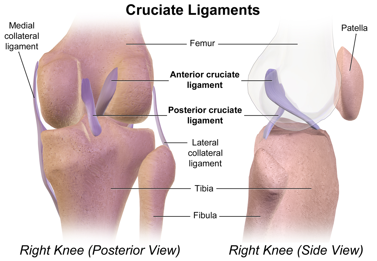
Cruciate Ligament
Cruciate ligaments (also cruciform ligaments) are pairs of ligaments arranged like a letter X. They occur in several joints of the body, such as the knee joint and the atlanto-axial joint. In a fashion similar to the cords in a toy Jacob's ladder, the crossed ligaments stabilize the joint while allowing a very large range of motion. Knee Structure Cruciate ligaments occur in the knee of humans and other bipedal animals and the corresponding stifle of quadrupedal animals, and in the neck, fingers, and foot. * The cruciate ligaments of the knee are the anterior cruciate ligament (ACL) and the posterior cruciate ligament (PCL). These ligaments are two strong, rounded bands that extend from the head of the tibia to the intercondyloid notch of the femur. The ACL is lateral and the PCL is medial. They cross each other like the limbs of an X. They are named for their insertion into the tibia: the ACL attaches to the anterior aspect of the intercondylar area, the PCL to the po ... [...More Info...] [...Related Items...] OR: [Wikipedia] [Google] [Baidu] |


_deLuxe_Two_door_1958_Bumper.jpg)

