|
Breech Birth
A breech birth is when a baby is born bottom first instead of head first, as is normal. Around 3–5% of pregnant women at term (37–40 weeks pregnant) have a breech baby. Due to their higher than average rate of possible complications for the baby, breech births are generally considered higher risk. Breech births also occur in many other mammals such as dogs and horses, see veterinary obstetrics. Most babies in the breech position are delivered via caesarean section because it is seen as safer than being born vaginally. Doctors and midwives in the developing world often lack many of the skills required to safely assist women giving birth to a breech baby vaginally. Also, delivering all breech babies by caesarean section in developing countries is difficult to implement as there are not always resources available to provide this service. OB-GYNs do not recommend home births if a breech birth is expected, even when attended by a medical professional. Cause With regard to th ... [...More Info...] [...Related Items...] OR: [Wikipedia] [Google] [Baidu] |
William Smellie (obstetrician)
William Smellie (5 February 1697 – 5 March 1763) was a Scottish obstetrician and medical instructor who practiced and taught primarily in London. One of the first prominent male midwives in Britain, he designed an improved version of the obstetrical forceps, established safer delivery practices, and through his teaching and writing helped make obstetrics more scientifically based. He is often called the "father of British midwifery". Early life and education Smellie was born on 5 February 1697 in the town of Lesmahagow, Scotland. He was the only child of Sara Kennedy (1657–1727) and Archibald Smellie (1663/4–1735), a merchant and burgess of the town. Smellie practiced medicine before getting a license, opening an apothecary in 1720 in Lanark. It was not a particularly lucrative venture, as he also sold cloth as a side business to supplement his income, but he began reading medical books and teaching himself obstetrics at this time. By 1728, he was married to Eupham Borlan ... [...More Info...] [...Related Items...] OR: [Wikipedia] [Google] [Baidu] |
Potter Anomaly
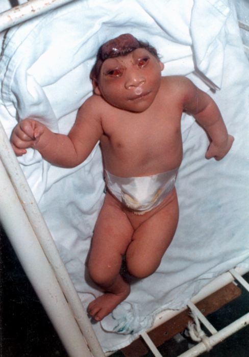
Potter sequence is the atypical physical appearance of a baby due to oligohydramnios experienced when in the uterus. It includes clubbed feet, pulmonary hypoplasia and cranial anomalies related to the oligohydramnios. Oligohydramnios is the decrease in amniotic fluid volume sufficient to cause deformations in morphogenesis of the baby. Oligohydramnios is the cause of Potter sequence but there are many things that can lead to oligohydramnios. It can be caused by renal diseases such as bilateral renal agenesis (BRA), atresia of the ureter or urethra causing obstruction of the urinary tract, polycystic or multicystic kidney diseases, renal hypoplasia, amniotic rupture, toxemia, or uteroplacental insufficiency from maternal hypertension. The term ''Potter sequence'' was initially intended to only refer to cases caused by BRA; however, it is now commonly used by many clinicians and researchers to refer to any case that presents with oligohydramnios or anhydramnios regardless of t ... [...More Info...] [...Related Items...] OR: [Wikipedia] [Google] [Baidu] |
Amelia (birth Defect)
Amelia is the birth defect of lacking one or more limbs. It can also result in a shrunken or deformed limb. The term may be modified to indicate the number of legs or arms missing at birth, such as tetra-amelia for the absence of all four limbs. A related term is meromelia, which is the partial absence of a limb or limbs. The term is from Greek ἀ- "lack of" plus μέλος (plural: μέλεα or μέλη) "limb" Symptoms The diagnosis of tetra-amelia syndrome is established clinically and can be made on routine prenatal ultrasonography. WNT3 is the only gene known to be associated with tetra-amelia syndrome. Molecular genetic testing on a clinical basis can be used to diagnose the incidence of the syndrome. The mutation detection frequency is unknown as only a limited number of families have been studied. Affected infants are often stillborn or die shortly after birth. Description Amelia may be present as an isolated defect, but it is often associated with major malformation ... [...More Info...] [...Related Items...] OR: [Wikipedia] [Google] [Baidu] |
Achondrogenesis
Achondrogenesis is a number of disorders that are the most severe form of congenital chondrodysplasia (malformation of bones and cartilage). These conditions are characterized by a small body, short limbs, and other skeletal abnormalities. As a result of their serious health problems, infants with achondrogenesis are usually born prematurely, are stillborn, or die shortly after birth from respiratory failure. Some infants, however, have lived for a while with intensive medical support. Researchers have described at least three forms of achondrogenesis, designated as Achondrogenesis type 1A, achondrogenesis type 1B and achondrogenesis type 2. These types are distinguished by their signs and symptoms, inheritance pattern, and genetic cause. Other types of achondrogenesis may exist, but they have not been characterized or their cause is unknown. Achondrogenesis type 1A is caused by a defect in the microtubules of the Golgi apparatus. In mice, a nonsense mutation in the thyroid hor ... [...More Info...] [...Related Items...] OR: [Wikipedia] [Google] [Baidu] |
Amyoplasia
Amyoplasia is a condition characterized by a generalized lack in the newborn of muscular development and growth, with contracture and deformity at most joints. It is the most common form of arthrogryposis. It is characterized by the four limbs being involved, and by the replacement of skeletal muscle by dense fibrous and adipose tissue. Studies involving amyoplasia have revealed similar findings of the muscle tissue due to various causes including that seen in sacral agenesis and amyotrophic lateral sclerosis. So amyoplasia may also include an intermediate common pathway, rather than the primary cause of the contractors. Signs and symptoms Amyoplasia results when a fetus is unable to move sufficiently in the womb. Mothers of children with the disorder often report that their baby was abnormally still during the pregnancy. The lack of movement in utero (also known as fetal akinesia) allows extra connective tissue to form around the joints and, therefore, the joints become fixed. Th ... [...More Info...] [...Related Items...] OR: [Wikipedia] [Google] [Baidu] |
Osteogenesis Imperfecta
Osteogenesis imperfecta (; OI), colloquially known as brittle bone disease, is a group of genetic disorders that all result in bones that break easily. The range of symptoms—on the skeleton as well as on the body's other organs—may be mild to severe. Symptoms found in various types of OI include whites of the eye (sclerae) that are blue instead, short stature, loose joints, hearing loss, breathing problems and problems with the teeth ( dentinogenesis imperfecta). Potentially life-threatening complications, all of which become more common in more severe OI, include: tearing ( dissection) of the major arteries, such as the aorta; pulmonary valve insufficiency secondary to distortion of the ribcage; and basilar invagination. The underlying mechanism is usually a problem with connective tissue due to a lack of, or poorly formed, type I collagen. In more than 90% of cases, OI occurs due to mutations in the '' COL1A1'' or ''COL1A2'' genes. These mutations may be ... [...More Info...] [...Related Items...] OR: [Wikipedia] [Google] [Baidu] |
Congenital Hydrocephalus
Hydrocephalus is a condition in which an accumulation of cerebrospinal fluid (CSF) occurs within the brain. This typically causes increased pressure inside the skull. Older people may have headaches, double vision, poor balance, urinary incontinence, personality changes, or mental impairment. In babies, it may be seen as a rapid increase in head size. Other symptoms may include vomiting, sleepiness, seizures, and downward pointing of the eyes. Hydrocephalus can occur due to birth defects or be acquired later in life. Associated birth defects include neural tube defects and those that result in aqueductal stenosis. Other causes include meningitis, brain tumors, traumatic brain injury, intraventricular hemorrhage, and subarachnoid hemorrhage. The four types of hydrocephalus are communicating, noncommunicating, ''ex vacuo'', and normal pressure. Diagnosis is typically made by physical examination and medical imaging. Hydrocephalus is typically treated by the surgical pla ... [...More Info...] [...Related Items...] OR: [Wikipedia] [Google] [Baidu] |
Spina Bifida
Spina bifida (Latin for 'split spine'; SB) is a birth defect in which there is incomplete closing of the spine and the membranes around the spinal cord during early development in pregnancy. There are three main types: spina bifida occulta, meningocele and myelomeningocele. Meningocele and myelomeningocele may be grouped as spina bifida cystica. The most common location is the lower back, but in rare cases it may be in the middle back or neck. Occulta has no or only mild signs, which may include a hairy patch, dimple, dark spot or swelling on the back at the site of the gap in the spine. Meningocele typically causes mild problems, with a sac of fluid present at the gap in the spine. Myelomeningocele, also known as open spina bifida, is the most severe form. Problems associated with this form include poor ability to walk, impaired bladder or bowel control, accumulation of fluid in the brain (hydrocephalus), a tethered spinal cord and latex allergy. Learning problems are relat ... [...More Info...] [...Related Items...] OR: [Wikipedia] [Google] [Baidu] |
Anencephalus
Anencephaly is the absence of a major portion of the brain, skull, and scalp that occurs during embryonic development. It is a cephalic disorder that results from a neural tube defect that occurs when the rostral (head) end of the neural tube fails to close, usually between the 23rd and 26th day following conception. Strictly speaking, the Greek term translates as "without a brain" (or totally lacking the inside part of the head), but it is accepted that children born with this disorder usually only lack a telencephalon, the largest part of the brain consisting mainly of the cerebral hemispheres, including the neocortex, which is responsible for cognition. The remaining structure is usually covered only by a thin layer of membrane—skin, bone, meninges, etc., are all lacking. With very few exceptions, infants with this disorder do not survive longer than a few hours or days after birth. Signs and symptoms The National Institute of Neurological Disorders and Stroke (NINDS) descr ... [...More Info...] [...Related Items...] OR: [Wikipedia] [Google] [Baidu] |
21 Trisomy Syndrome
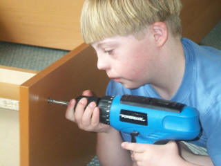
Down syndrome or Down's syndrome, also known as trisomy 21, is a genetic disorder caused by the presence of all or part of a third copy of chromosome 21. It is usually associated with physical growth delays, mild to moderate intellectual disability, and characteristic facial features. The average IQ of a young adult with Down syndrome is 50, equivalent to the mental ability of an eight- or nine-year-old child, but this can vary widely. The parents of the affected individual are usually genetically normal. The probability increases from less than 0.1% in 20-year-old mothers to 3% in those of age 45. The extra chromosome is believed to occur by chance, with no known behavioral activity or environmental factor that changes the probability. Down syndrome can be identified during pregnancy by prenatal screening followed by diagnostic testing or after birth by direct observation and genetic testing. Since the introduction of screening, Down syndrome pregnancies are often abor ... [...More Info...] [...Related Items...] OR: [Wikipedia] [Google] [Baidu] |
18 Trisomy Syndrome
Edwards syndrome, also known as trisomy 18, is a genetic disorder caused by the presence of a third copy of all or part of chromosome 18. Many parts of the body are affected. Babies are often born small and have heart defects. Other features include a small head, small jaw, clenched fists with overlapping fingers, and severe intellectual disability. Most cases of Edwards syndrome occur due to problems during the formation of the reproductive cells or during early development. The rate of disease increases with the mother's age. Rarely, cases may be inherited from a person's parents. Occasionally, not all cells have the extra chromosome, known as mosaic trisomy, and symptoms in these cases may be less severe. An ultrasound during pregnancy can increase suspicion for the condition, which can be confirmed by amniocentesis. Treatment is supportive. After having one child with the condition, the risk of having a second is typically around one percent. It is the second-most com ... [...More Info...] [...Related Items...] OR: [Wikipedia] [Google] [Baidu] |


