|
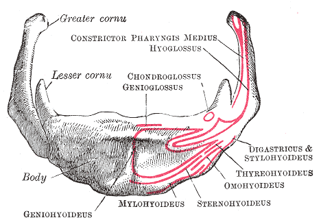
Body Of Hyoid Bone
The hyoid bone (lingual bone or tongue-bone) () is a horseshoe-shaped bone situated in the anterior midline of the neck between the chin and the thyroid cartilage. At rest, it lies between the base of the mandible and the third cervical vertebra. Unlike other bones, the hyoid is only distantly articulated to other bones by muscles or ligaments. It is the only bone in the human body that is not connected to any other bones nearby. The hyoid is anchored by muscles from the anterior, posterior and inferior directions, and aids in tongue movement and swallowing. The hyoid bone provides attachment to the muscles of the floor of the mouth and the tongue above, the larynx below, and the epiglottis and pharynx behind. Its name is derived . Structure The hyoid bone is classed as an irregular bone and consists of a central part called the body, and two pairs of horns, the greater and lesser horns. Body The body of the hyoid bone is the central part of the hyoid bone. *At the fron ... [...More Info...] [...Related Items...] OR: [Wikipedia] [Google] [Baidu] |
Branchial Arch
Branchial arches, or gill arches, are a series of bony "loops" present in fish, which support the gills. As gills are the primitive condition of vertebrates, all vertebrate embryos develop pharyngeal arches, though the eventual fate of these arches varies between taxa. In jawed fish, the first arch develops into the jaws, the second into the hyomandibular complex, with the posterior arches supporting gills. In amphibians and reptiles, many elements are lost including the gill arches, resulting in only the oral jaws and a hyoid apparatus remaining. In mammals and birds, the hyoid is still more simplified. All basal vertebrates breathe with gills. The gills are carried right behind the head, bordering the posterior margins of a series of openings from the esophagus to the exterior. Each gill is supported by a cartilaginous or bony gill arch. Bony fish have four pairs of arches, cartilaginous fish have five to seven pairs, and primitive jawless fish have seven. The vertebrate ... [...More Info...] [...Related Items...] OR: [Wikipedia] [Google] [Baidu] |
Geniohyoid Muscle
The geniohyoid muscle is a narrow muscle situated superior to the medial border of the mylohyoid muscle. It is named for its passage from the chin ("genio-" is a standard prefix for "chin") to the hyoid bone. Structure It arises from the inferior mental spine, on the back of the mandibular symphysis, and runs backward and slightly downward, to be inserted into the anterior surface of the body of the hyoid bone. It lies in contact with its fellow of the opposite side. It thus belongs to the suprahyoid muscles. The muscle is supplied by branches of the lingual artery. Innervation The geniohyoid muscle is innervated by fibres from the first cervical spinal nerve travelling alongside the hypoglossal nerve. Although the first three cervical nerves give rise to the ansa cervicalis, the geniohyoid muscle is said to be innervated by the first cervical nerve, as some of its efferent fibers do not contribute to ansa cervicalis. Variations It may be blended with the one on opposite side ... [...More Info...] [...Related Items...] OR: [Wikipedia] [Google] [Baidu] |
Lateral Thyrohyoid Ligament
The lateral thyrohyoid ligament (lateral hyothyroid ligament) is a round elastic cord, which forms the posterior border of the thyrohyoid membrane and passes between the tip of the superior cornu of the thyroid cartilage and the extremity of the greater cornu of the hyoid bone. The internal branch of the superior laryngeal nerve The superior laryngeal nerve is a branch of the vagus nerve. It arises from the middle of the inferior ganglion of vagus nerve and in its course receives a branch from the superior cervical ganglion of the sympathetic nervous system. The superi ... typical lies lateral to this ligament. Triticeal cartilage A small cartilaginous nodule (cartilago triticea), sometimes bony, is frequently found in the lateral thyrohyoid ligament. References External links * - "Larynx, anterior view" * - "Larynx, lateral view" Human head and neck Ligaments {{Portal bar, Anatomy ... [...More Info...] [...Related Items...] OR: [Wikipedia] [Google] [Baidu] |
Tubercle (anatomy)
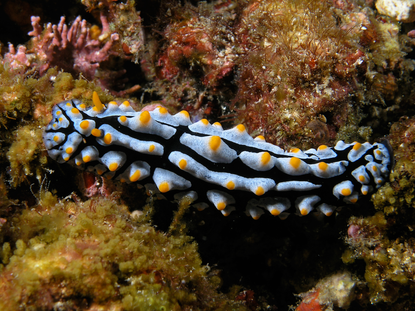
In anatomy, a tubercle (literally 'small tuber', Latin for 'lump') is any round nodule, small eminence, or warty outgrowth found on external or internal organs of a plant or an animal. In plants A tubercle is generally a wart-like projection, but it has slightly different meaning depending on which family of plants or animals it is used to refer to. In the case of certain orchids and cacti, it denotes a round nodule, small eminence, or warty outgrowth found on the lip. They are also known as podaria (singular ''podarium''). When referring to some members of the pea family, it is used to refer to the wart-like excrescences that are found on the roots. In fungi In mycology, a tubercle is used to refer to a mass of hyphae from which a mushroom is made. In animals When it is used in relation to certain dorid nudibranchs such as ''Peltodoris nobilis'', it means the nodules on the dorsum of the animal. The tubercles in nudibranchs can present themselves in different ways: e ... [...More Info...] [...Related Items...] OR: [Wikipedia] [Google] [Baidu] |
Thyrohyoid Muscle
The thyrohyoid muscle is a small skeletal muscle on the neck. It originates from the lamina of the thyroid cartilage, and inserts into the greater cornu of the hyoid bone. It is supplied by the hypoglossal nerve, and a branch of the ventral rami of the cervical plexus, spinal nerve C1, which travels with the hypoglossal nerve. The thyrohyoid muscle depresses the hyoid bone and elevates the larynx. By controlling the position and shape of the larynx, it aids in making sound. Structure The thyrohyoid muscle is a quadrilateral muscle in shape. It appears like an upward continuation of the sternothyroid muscle. It belongs to the infrahyoid muscles group. It lies in the carotid triangle. It arises from the oblique line on the lamina of the thyroid cartilage. It is inserted into the lower border of the greater cornu of the hyoid bone. Nerve supply The thyrohyoid muscle is supplied by the hypoglossal nerve (XII). It is the only infrahyoid muscle that is not supplied by the ansa c ... [...More Info...] [...Related Items...] OR: [Wikipedia] [Google] [Baidu] |
Genioglossus Muscle
The genioglossus is one of the paired extrinsic muscles of the tongue. The genioglossus is the major muscle responsible for protruding (or sticking out) the tongue. Structure Genioglossus is the fan-shaped extrinsic tongue muscle that forms the majority of the body of the tongue. It arises from the mental spine of the mandible and its insertions are the hyoid bone and the bottom of the tongue. The genioglossus is innervated by the hypoglossal nerve, as are all muscles of the tongue except for the palatoglossus. Blood is supplied to the sublingual branch of the lingual artery, a branch of the external carotid artery. The canine genioglossus muscle has been divided into horizontal and oblique compartments. Function The left and right genioglossus muscles protrude the tongue and deviate it towards the opposite side. When acting together, the muscles depress the center of the tongue at its back. Clinical significance Contraction of the genioglossus stabilizes and enlarges the porti ... [...More Info...] [...Related Items...] OR: [Wikipedia] [Google] [Baidu] |
Aponeurosis
An aponeurosis (; plural: ''aponeuroses'') is a type or a variant of the deep fascia, in the form of a sheet of pearly-white fibrous tissue that attaches sheet-like muscles needing a wide area of attachment. Their primary function is to join muscles and the body parts they act upon, whether bone or other muscles. They have a shiny, whitish-silvery color, are histologically similar to tendons, and are very sparingly supplied with blood vessels and nerves. When dissected, aponeuroses are papery and peel off by sections. The primary regions with thick aponeuroses are in the ventral abdominal region, the dorsal lumbar region, the ventriculus in birds, and the palmar (palms) and plantar (soles) regions. Anatomy Anterior abdominal aponeuroses The anterior abdominal aponeuroses are located just superficial to the rectus abdominis muscle. It has for its borders the external oblique, pectoralis muscles, and the latissimus dorsi. Posterior lumbar aponeuroses The posterior lumbar aponeu ... [...More Info...] [...Related Items...] OR: [Wikipedia] [Google] [Baidu] |
Bursa (anatomy)
( grc-gre, Προῦσα, Proûsa, Latin: Prusa, ota, بورسه, Arabic:بورصة) is a city in northwestern Turkey and the administrative center of Bursa Province. The fourth-most populous city in Turkey and second-most populous in the Marmara Region, Bursa is one of the industrial centers of the country. Most of Turkey's automotive production takes place in Bursa. As of 2019, the Metropolitan Province was home to 3,056,120 inhabitants, 2,161,990 of whom lived in the 3 city urban districts (Osmangazi, Yildirim and Nilufer) plus Gursu and Kestel, largely conurbated. Bursa was the first major and second overall capital of the Ottoman State between 1335 and 1363. The city was referred to as (, meaning "God's Gift" in Ottoman Turkish, a name of Persian origin) during the Ottoman period, while a more recent nickname is ("") in reference to the parks and gardens located across its urban fabric, as well as to the vast and richly varied forests of the surrounding region ... [...More Info...] [...Related Items...] OR: [Wikipedia] [Google] [Baidu] |
Loose Areolar Tissue
Loose connective tissue, sometimes called areolar tissue, is a cellular connective tissue with thin and relatively sparse collagen fibers. Its ground substance occupies more volume than the fibers do. It has a viscous to gel-like consistency and plays an important role in the diffusion of oxygen and nutrients from the capillaries that course through this connective tissue as well as in the diffusion of carbon dioxide and metabolic wastes back to the vessels. Moreover, loose connective tissue is primarily located beneath the epithelia that cover the body surfaces and line the internal surfaces of the body. It is also associated with the epithelium of glands and surrounds the smallest blood vessels. This tissue is thus the initial site where pathogenic agents, such as bacteria that have breached an epithelial surface, are challenged and destroyed by cells of the immune system. In the past, the designations areolar tissue, adipose tissue, and reticular tissue have been listed ... [...More Info...] [...Related Items...] OR: [Wikipedia] [Google] [Baidu] |
Hyothyroid Membrane
The thyrohyoid membrane (or hyothyroid membrane) is a broad, fibro-elastic sheet of the larynx. It connects the upper border of the thyroid cartilage to the hyoid bone. Structure The thyrohyoid membrane is attached below to the upper border of the thyroid cartilage and to the front of its superior cornu, and above to the upper margin of the posterior surface of the body and greater cornu of the hyoid bone. It passes behind the posterior surface of the body of the hyoid. It is separated from the hyoid bone by a mucous bursa, which allows for the upward movement of the larynx during swallowing. Its middle thicker part is termed the median thyrohyoid ligament. Its lateral thinner portions are pierced by the superior laryngeal vessels and the internal branch of the superior laryngeal nerve. Its anterior surface is in relation with the thyrohyoid muscle, sternohyoid muscle, and omohyoid muscles, and with the body of the hyoid bone. It is pierced by the superior laryngeal nerve. It ... [...More Info...] [...Related Items...] OR: [Wikipedia] [Google] [Baidu] |
Omohyoid Muscle
The omohyoid muscle is a muscle that depresses the hyoid. It is located in the front of the neck, and consists of two bellies separated by an intermediate tendon. The omohyoid muscle is proximally attached to the scapula and distally attached to the hyoid bone, stabilising it. Its superior belly serves as the most lateral member of the infrahyoid muscles, located lateral to both the sternothyroid muscles and the thyrohyoid muscles.Illustrated Anatomy of the Head and Neck, Fehrenbach and Herring, Elsevier, 2012, page 102 Structure The omohyoid muscle arises from the upper border of the scapula, inserting into the lower border of the body of the hyoid bone. It has two separate bellies, superior and inferior: * The ''inferior belly'' forms a flat, narrow fasciculus, which inclines forward and slightly upward across the lower part of the neck, being bound down to the clavicle by a fibrous expansion; it then passes behind the sternocleidomastoid, becomes tendinous and changes its ... [...More Info...] [...Related Items...] OR: [Wikipedia] [Google] [Baidu] |

.jpg)


