|
Bright-field Microscopy
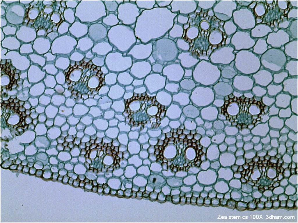
Bright-field microscopy (BF) is the simplest of all the optical microscopy illumination techniques. Sample illumination is transmitted (i.e., illuminated from below and observed from above) white light, and contrast in the sample is caused by attenuation of the transmitted light in dense areas of the sample. Bright-field microscopy is the simplest of a range of techniques used for illumination of samples in light microscopes, and its simplicity makes it a popular technique. The typical appearance of a bright-field microscopy image is a dark sample on a bright background, hence the name. History of microscopy Compound microscopes first appeared in Europe around 1620. The actual inventor of the compound microscope is unknown although many claims have been made over the years. These include a dubious claim that Dutch spectacle-maker Zacharias Janssen invented the compound microscope and the telescope as early as 1590. Another claim is that Janssen's competitor Hans Lippersh ... [...More Info...] [...Related Items...] OR: [Wikipedia] [Google] [Baidu] |
Tulane University
The Tulane University of Louisiana (commonly referred to as Tulane University) is a private research university in New Orleans, Louisiana, United States. Founded as the Medical College of Louisiana in 1834 by a cohort of medical doctors, it became a comprehensive public university in the University of Louisiana in 1847. The institution became private under the endowments of Paul Tulane and Josephine Louise Newcomb in 1884 and 1887. The Tulane University School of Law and Tulane University Medical School are, respectively, the 12th oldest law school and 15th oldest medical school in the United States. Tulane has been a member of the Association of American Universities since 1958 and is classified among "R1: Doctoral Universities – Very high research activity". Alumni include 12 governors of Louisiana; 1 Chief Justice of the United States; members of the United States Congress, including a Speaker of the House; 2 Surgeons General of the United States; 23 Marshall S ... [...More Info...] [...Related Items...] OR: [Wikipedia] [Google] [Baidu] |
Staining
Staining is a technique used to enhance contrast in samples, generally at the Microscope, microscopic level. Stains and dyes are frequently used in histology (microscopic study of biological tissue (biology), tissues), in cytology (microscopic study of cell (biology), cells), and in the medical fields of histopathology, hematology, and cytopathology that focus on the study and diagnoses of diseases at the microscopic level. Stains may be used to define biological tissues (highlighting, for example, muscle fibers or connective tissue), cell (biology), cell populations (classifying different blood cells), or organelles within individual cells. In biochemistry, it involves adding a class-specific (DNA, proteins, lipids, carbohydrates) dye to a substrate to qualify or quantify the presence of a specific compound. Staining and fluorescent tagging can serve similar purposes. Biological staining is also used to mark cells in flow cytometry, and to flag proteins or nucleic acids in gel ... [...More Info...] [...Related Items...] OR: [Wikipedia] [Google] [Baidu] |
Contrast (vision)
Contrast is the difference in luminance or color that makes an object (or its representation in an image or display) visible against a background of different luminance or color. The human visual system is more sensitive to contrast than to absolute luminance; thus, we can perceive the world similarly despite significant changes in illumination throughout the day or across different locations. The maximum contrast of an image is termed the contrast ratio or dynamic range. In images where the contrast ratio approaches the maximum possible for the medium, there is a ''conservation of contrast''. In such cases, increasing contrast in certain parts of the image will necessarily result in a decrease in contrast elsewhere. Brightening an image increases contrast in darker areas but decreases it in brighter areas; conversely, darkening the image will have the opposite effect. Bleach bypass reduces contrast in the darkest and brightest parts of an image while enhancing luminance contr ... [...More Info...] [...Related Items...] OR: [Wikipedia] [Google] [Baidu] |
Optical Resolution
Optical resolution describes the ability of an imaging system to resolve detail, in the object that is being imaged. An imaging system may have many individual components, including one or more lenses, and/or recording and display components. Each of these contributes (given suitable design, and adequate alignment) to the optical resolution of the system; the environment in which the imaging is done often is a further important factor. Lateral resolution Resolution depends on the distance between two distinguishable radiating points. The sections below describe the theoretical estimates of resolution, but the real values may differ. The results below are based on mathematical models of Airy discs, which assumes an adequate level of contrast. In low-contrast systems, the resolution may be much lower than predicted by the theory outlined below. Real optical systems are complex, and practical difficulties often increase the distance between distinguishable point sources. The ... [...More Info...] [...Related Items...] OR: [Wikipedia] [Google] [Baidu] |
Visible Light
Light, visible light, or visible radiation is electromagnetic radiation that can be perceived by the human eye. Visible light spans the visible spectrum and is usually defined as having wavelengths in the range of 400–700 nanometres (nm), corresponding to frequencies of 750–420 terahertz. The visible band sits adjacent to the infrared (with longer wavelengths and lower frequencies) and the ultraviolet (with shorter wavelengths and higher frequencies), called collectively '' optical radiation''. In physics, the term "light" may refer more broadly to electromagnetic radiation of any wavelength, whether visible or not. In this sense, gamma rays, X-rays, microwaves and radio waves are also light. The primary properties of light are intensity, propagation direction, frequency or wavelength spectrum, and polarization. Its speed in vacuum, , is one of the fundamental constants of nature. All electromagnetic radiation exhibits some properties of both particles and waves ... [...More Info...] [...Related Items...] OR: [Wikipedia] [Google] [Baidu] |
Wavelength
In physics and mathematics, wavelength or spatial period of a wave or periodic function is the distance over which the wave's shape repeats. In other words, it is the distance between consecutive corresponding points of the same ''phase (waves), phase'' on the wave, such as two adjacent crests, troughs, or zero crossings. Wavelength is a characteristic of both traveling waves and standing waves, as well as other spatial wave patterns. The multiplicative inverse, inverse of the wavelength is called the ''spatial frequency''. Wavelength is commonly designated by the Greek letter lambda (''λ''). For a modulated wave, ''wavelength'' may refer to the carrier wavelength of the signal. The term ''wavelength'' may also apply to the repeating envelope (mathematics), envelope of modulated waves or waves formed by Interference (wave propagation), interference of several sinusoids. Assuming a sinusoidal wave moving at a fixed phase velocity, wave speed, wavelength is inversely proportion ... [...More Info...] [...Related Items...] OR: [Wikipedia] [Google] [Baidu] |
Optical Resolution
Optical resolution describes the ability of an imaging system to resolve detail, in the object that is being imaged. An imaging system may have many individual components, including one or more lenses, and/or recording and display components. Each of these contributes (given suitable design, and adequate alignment) to the optical resolution of the system; the environment in which the imaging is done often is a further important factor. Lateral resolution Resolution depends on the distance between two distinguishable radiating points. The sections below describe the theoretical estimates of resolution, but the real values may differ. The results below are based on mathematical models of Airy discs, which assumes an adequate level of contrast. In low-contrast systems, the resolution may be much lower than predicted by the theory outlined below. Real optical systems are complex, and practical difficulties often increase the distance between distinguishable point sources. The ... [...More Info...] [...Related Items...] OR: [Wikipedia] [Google] [Baidu] |
Magnification
Magnification is the process of enlarging the apparent size, not physical size, of something. This enlargement is quantified by a size ratio called optical magnification. When this number is less than one, it refers to a reduction in size, sometimes called ''de-magnification''. Typically, magnification is related to scaling up visuals or images to be able to see more detail, increasing resolution, using microscope, printing techniques, or digital processing. In all cases, the magnification of the image does not change the perspective of the image. Examples of magnification Some optical instruments provide visual aid by magnifying small or distant subjects. * A magnifying glass, which uses a positive (convex) lens to make things look bigger by allowing the user to hold them closer to their eye. * A telescope, which uses its large objective lens or primary mirror to create an image of a distant object and then allows the user to examine the image closely with a smaller ... [...More Info...] [...Related Items...] OR: [Wikipedia] [Google] [Baidu] |
Köhler Illumination
Köhler illumination is a method of specimen illumination used for transmitted and reflected light (trans- and epi-illuminated) optical microscopy. Köhler illumination acts to generate an even illumination of the sample and ensures that an image of the illumination source (for example a halogen lamp filament) is not visible in the resulting image. Köhler illumination is the predominant technique for sample illumination in modern scientific light microscopy. It requires additional optical elements which are more expensive and may not be present in more basic light microscopes. History and motivation Prior to Köhler illumination critical illumination was the predominant technique for sample illumination. Critical illumination has the major limitation that the image of the light source (typically a light bulb) falls in the same plane as the image of the specimen, i.e., the bulb filament is visible in the final image. The image of the light source is often referred to as the fil ... [...More Info...] [...Related Items...] OR: [Wikipedia] [Google] [Baidu] |
Critical Illumination
Critical illumination or Nelsonian illumination is a method of Laboratory specimen, specimen illumination (lighting), illumination used for transmitted and reflected light (trans- and epi-illuminated) optical microscopy. Critical illumination focuses an image of a light source on to the specimen for bright illumination. Critical illumination generally has problems with evenness of illumination as an image of the illumination source (for example a halogen lamp filament) is visible in the resulting image. Köhler illumination has largely replaced critical illumination in modern scientific light microscopy although it requires additional optics which may not be present in less expensive and simpler light microscopes. Optical principles Critical illumination acts to form an image of the light source on the specimen to illuminate it. [...More Info...] [...Related Items...] OR: [Wikipedia] [Google] [Baidu] |
Ocular Lens
An eyepiece, or ocular lens, is a type of lens that is attached to a variety of optical devices such as Optical telescope, telescopes and microscopes. It is named because it is usually the lens that is closest to the eye when someone looks through an optical device to observe an object or sample. The Objective (optics), objective lens or mirror collects light from an object or sample and brings it to focus creating an image of the object. The eyepiece is placed near the Focus (optics), focal point of the objective to magnify this image to the eyes. (The eyepiece and the eye together make an image of the image created by the objective, on the retina of the eye.) The amount of magnification depends on the focal length of the eyepiece. An eyepiece consists of several "Lens (optics), lens elements" in a housing, with a "barrel" on one end. The barrel is shaped to fit in a special opening of the instrument to which it is attached. The image can be focus (optics), focused by moving th ... [...More Info...] [...Related Items...] OR: [Wikipedia] [Google] [Baidu] |


